XRAY: Chest radiography
1/23
There's no tags or description
Looks like no tags are added yet.
Name | Mastery | Learn | Test | Matching | Spaced | Call with Kai |
|---|
No analytics yet
Send a link to your students to track their progress
24 Terms
What is this patient presenting with?
Pneumothorax
(tip: look for THIN pleural line with absent lung markings beyond its border)

What is this patient presenting with?
Tension pneumothorax
(look out for mediastinal shift suggesting a tension pneumothorax)
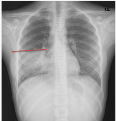
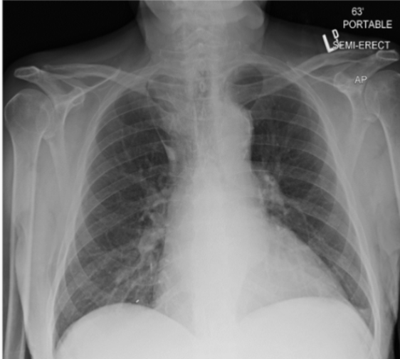
What is the circle indicating?
Deep sulcus sign on supine X-ray

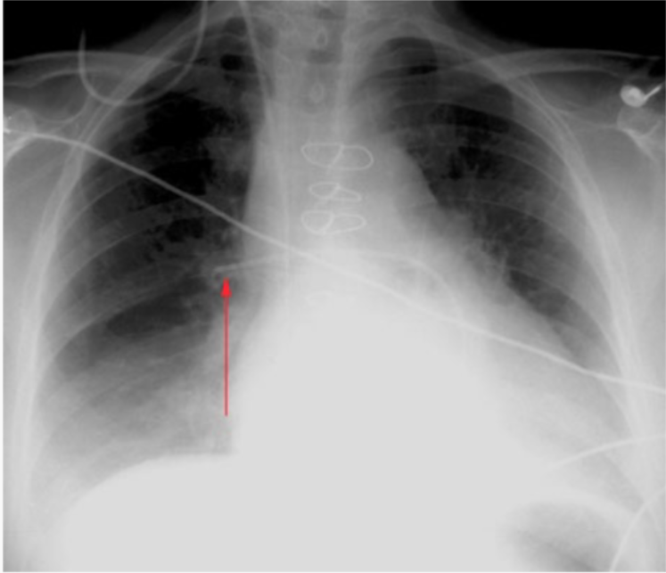
what do the arrows indicate?
pneumomediastinum
What is normal for the following lines and tubes?
Endotracheal tube?
Central lines?
Swan-Ganz catheters?
Feeding tubes?
• Endotracheal Tube:
– With normal neck movement, an ET tube can move down 2 cm
– ET tubes therefore must be at least 2 cm above carina, ideally 4-5 cm above
• Central lines:
– Tip ideally in the superior vena cava
• Swan-Ganz catheters:
– Tip ideally in right or left main pulmonary artery; further than this can cause pulmonary infarction
• Feeding Tube:
– Tip ideally in stomach/proximal duodenum; however any NG/OG tube clearly terminating below the diaphragm is acceptable
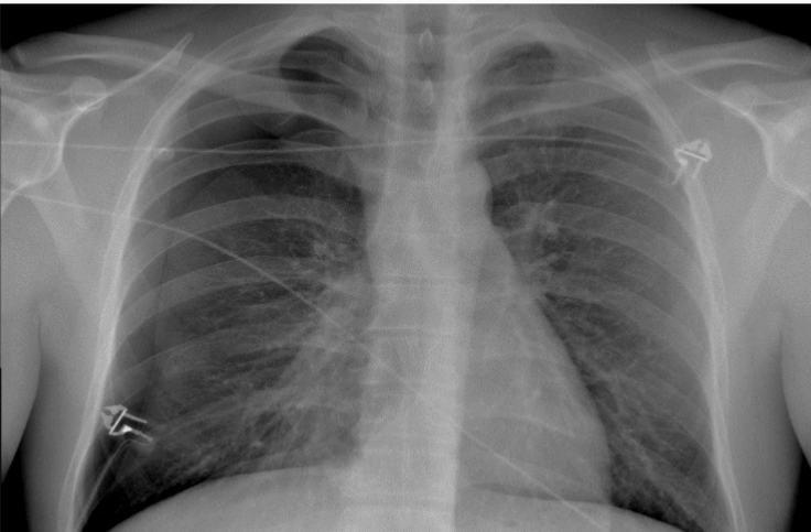
Is this a good placement for a NGT (feeding) tube? Why or why not?
No, it is too shallow in the esophagus, needs to be below the diaphragm for feeding
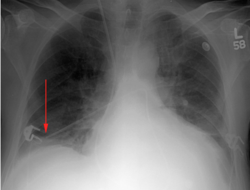
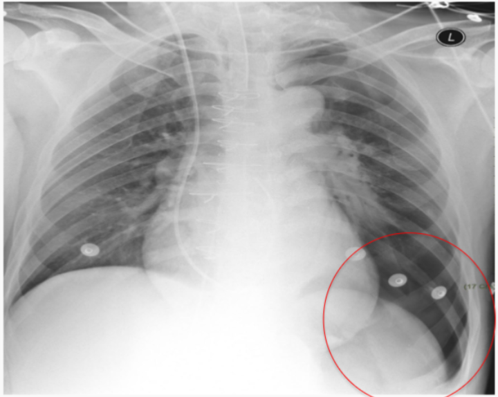
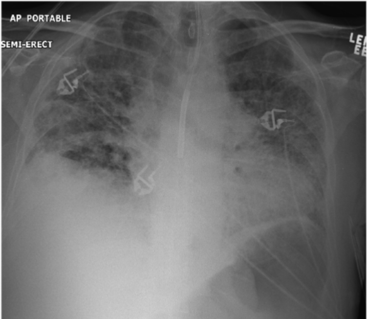
Is this a satisfactory Swan Ganz catheter placement?
No the Swan Ganz is in too far, distal to the main pulmonary artery. A good placement is shown here:
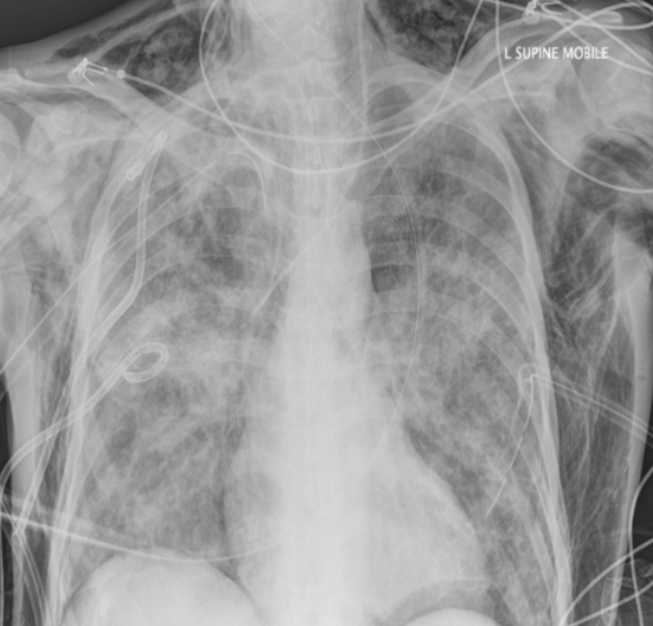
what is happening in this patient?
Severe subcutaneous emphysema (gas leaking into muscle tissues)
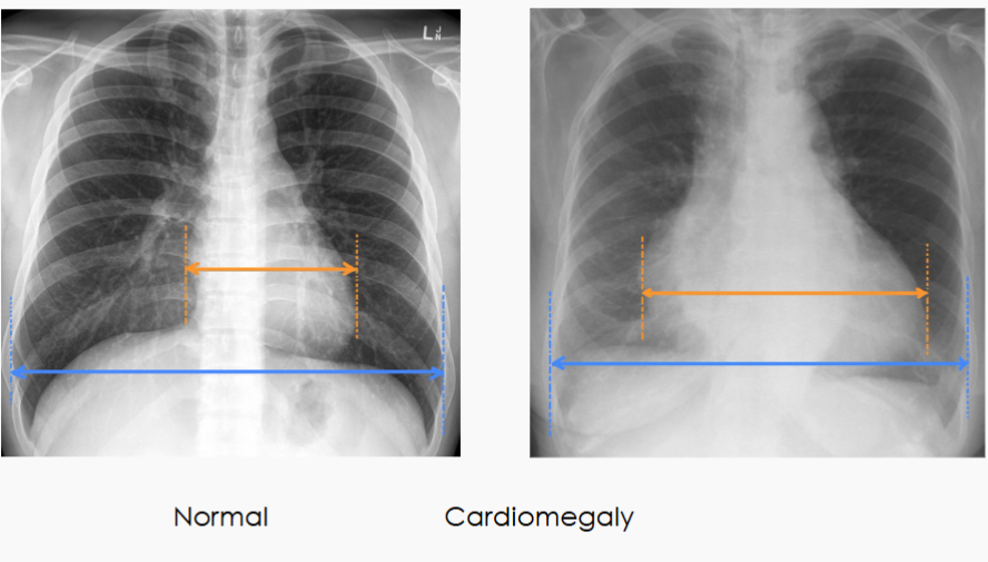
What is a common way to find cardiomegaly on X-ray?
The cardiothoracic ratio (CTR) aids in the detection
of enlargement of the cardiac silhouette (most often cardiomegaly but ALSO pericardial effusion)
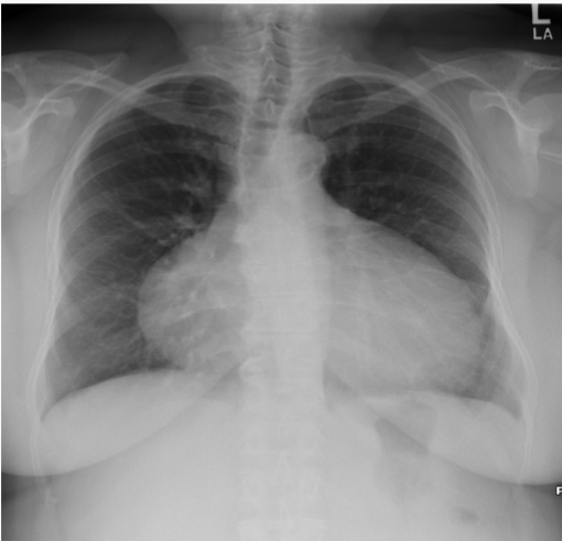
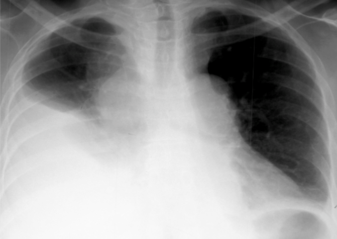
What is this patient presenting with?
Large pericardial effusion-”water bottle sign”
Patterns of atelectasis are very similar to consolidation except:
Volume loss
Left upper lobe collapse
What are some signs of consolidation?
increased opacity
fluffy “air space” opacity
no/negligible volume loss signs
air bronchograms (pathognomonic)
What pattern of consolidation is this?
Left lower lobe pneumonia
Which image represents the “spine sign” and what does this mean?
Right side is spine sign; means that there is something in the lungs (should go bright—>dark, not dark—>bright)
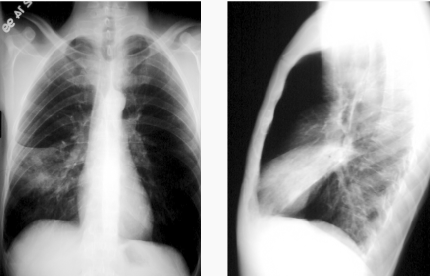
What is this patient presenting with?
Left upper lobe/lingular consolidation
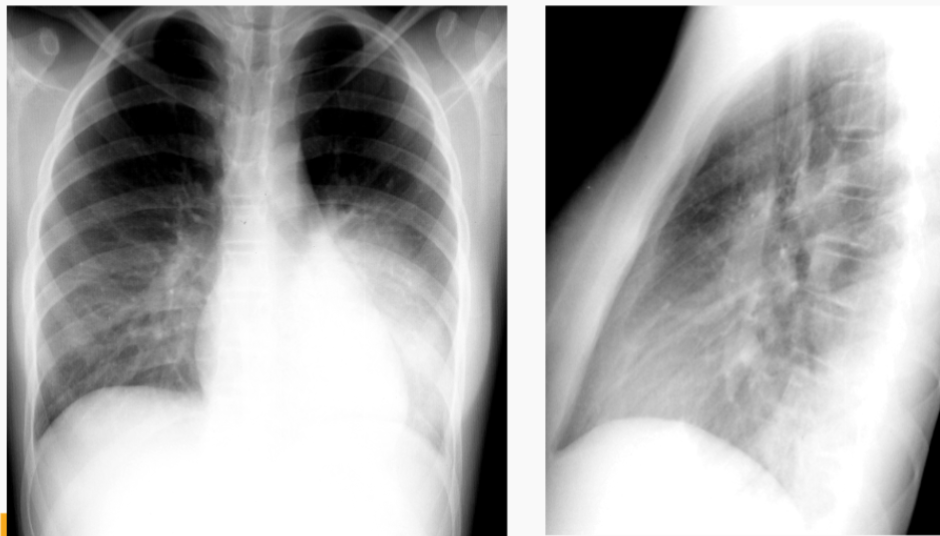
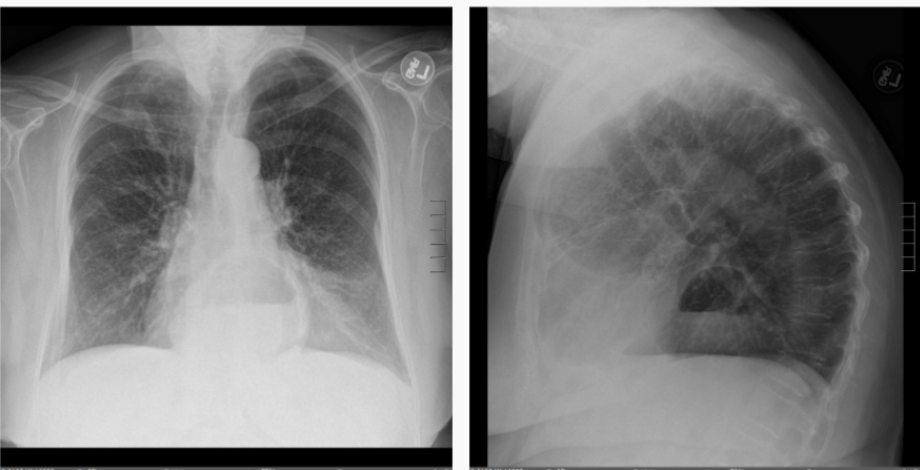
What is this patient presenting with?
Right middle lobe consolidation
What is the silhouette sign?
When there is an increased density in the patient’s right lung that obscures the right heart border
Features on a frontal film for pleural effusions include:
blunting of costophrenic angle
blunting of the cardiophrenic angle
fluid within the horizontal or oblique fissures
What is this patient presenting with?
Large pleural effusion (associated with “meniscus” sign)

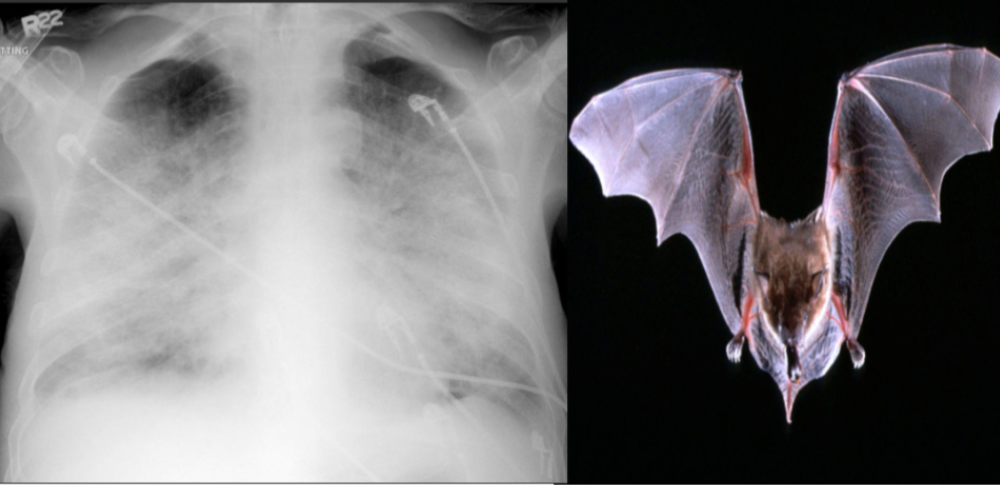
What is this patient presenting with?
Pulmonary edema—classically “Batwing” in appearance
(starts central, spreads outwards)
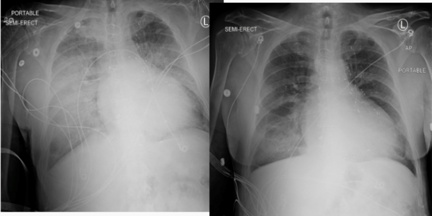
What was the patient’s original dx (left) and what caused improvement (right)?
Pulmonary edema- before (left) and after lasix aka “fluid pill” (right)
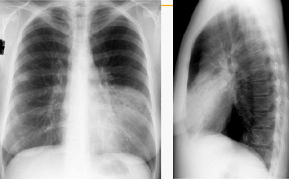
What feature is notable about this patient’s X-ray?
Azygous fissure (teardrop with long tail)
What is this patient presenting with?
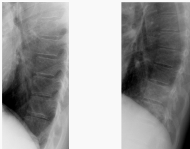
Hiatal hernia (note straight fluid line just behind the heart)

What is this patient presenting with?
Situs inversus (uncommon; picture terrified X-ray tech who mislabeled film, much more common lol)