Cancer Genetics
1/11
There's no tags or description
Looks like no tags are added yet.
Name | Mastery | Learn | Test | Matching | Spaced | Call with Kai |
|---|
No analytics yet
Send a link to your students to track their progress
12 Terms
The Cell Cycle
- tightly controlled
- cellular checkpoints preventing cells from dividing when they should not
- mutations in a cell's DNA change the timing of signals that tell when to grow and divide
- abnormal growth of cells results in a group of disease called cancer
Oncogenes
DNA that codes proteins that promote normal cell growth and division
- mutations can cause these genes to become active at the wrong time or place
Tumor Suppressor Genes
DNA encoding these proteins inhibit cell growth and prevent tumor formation
Tumor
cells keep dividing uncontrollably
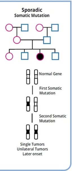
Acquired (somatic) mutations
- exposure to mutagens that affect the DNA
- errors during replication
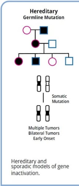
Germline mutation
directly inherited through generations
p53, tumor suppressor protein
- gene located on the short arm of chromosome 17
- mutations to the gene causes the protein to loses its ability to bind to DNA
- p53 that have mutations in specific hot spots promote uncontrolled cell growth and therefore function as oncogenes
- for p53 to play a role in cancer, both alleles need to be altered
Ex: Li-Fraumeni syndrome (LFS)
Li-Fraumeni Syndrome's notable features in family pedigrees
- include a sarcoma patient diagnosed before the age of 45
- at least one immediate relatives with other cancers before the age of 45
- multiple cancers in other family members
Karyotype
chromosome classification
- chromosomes are matched and numbered from largest to smallest, G-banding, and centromere location
- the sex chromosomes are labeled appropriately as either XX in females or XY in males
- X chromosome is much larger than the Y chromosomes, and therefore, X and Y chromosomes are considered nonhomologous
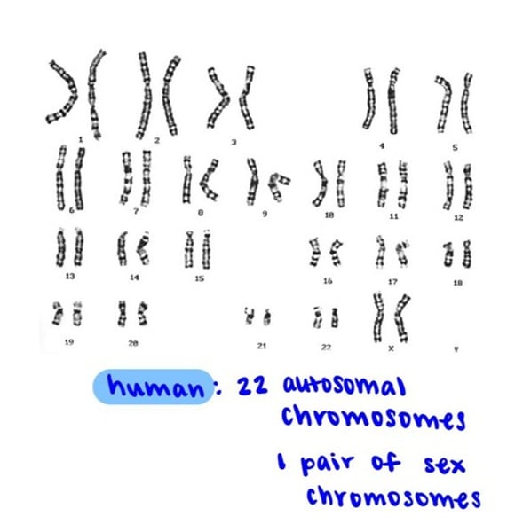
What does Karyotyping detect?
- macroscopic genomic abnormalities
- inversions
- duplications/deletions
- balances and unbalanced translocations
- aneuploidies
G-banding
- also know an Giemsa banding
- a technique used in cytogenetics to produce a visible karyotype by staining condensed chromosomes
More on G-banding
- metaphase chromosomes are treated with trypsin to partially digest the chromosomes
- stained with Giemsa stain
- heterochromatic regions, which tend to be rich with adenine and thymine DNA and relatively gene-poor, stain more darkly in G-banding
- Less condensed chromatin (Euchromatin), which tends to be rich with guanine and cytosine and more transcriptionally active incorporates less Giemsa stain, and these regions appear as light bands in G-banding