Anatomy Exam 3
1/156
There's no tags or description
Looks like no tags are added yet.
Name | Mastery | Learn | Test | Matching | Spaced | Call with Kai |
|---|
No analytics yet
Send a link to your students to track their progress
157 Terms
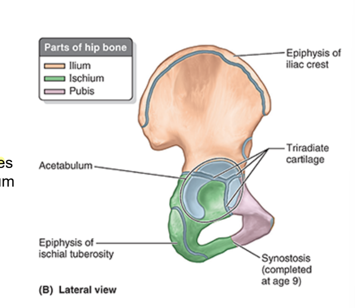
hip bone
fusion of 3 bones
ilium
ischium
pubis
all 3 hip bones contrib to acetabulum
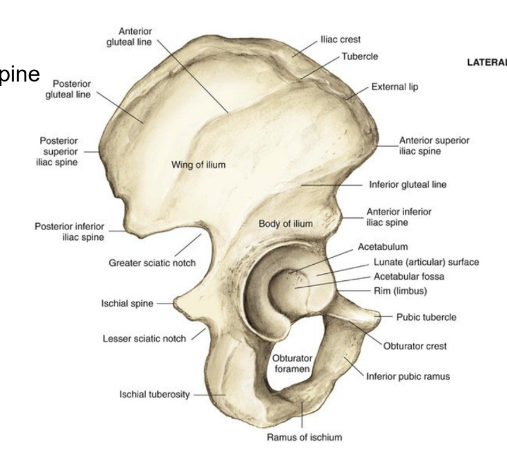
bony landmarks of the hip
iliac crest
ASIS
greater sciatic notch
lesser sciatic notch
acetabulum
ala (wing) of ilium
ischial spine
ischial tuberosity
obturator foramen
pubic tubercle
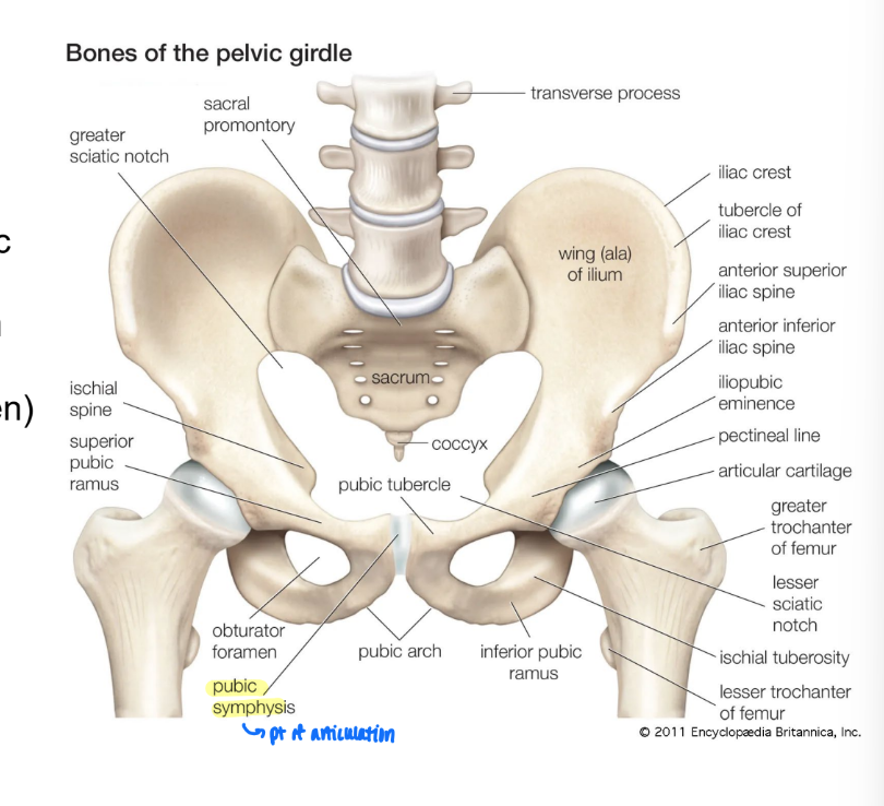
pubic symphysis
pt of articulation between R and L hip bone
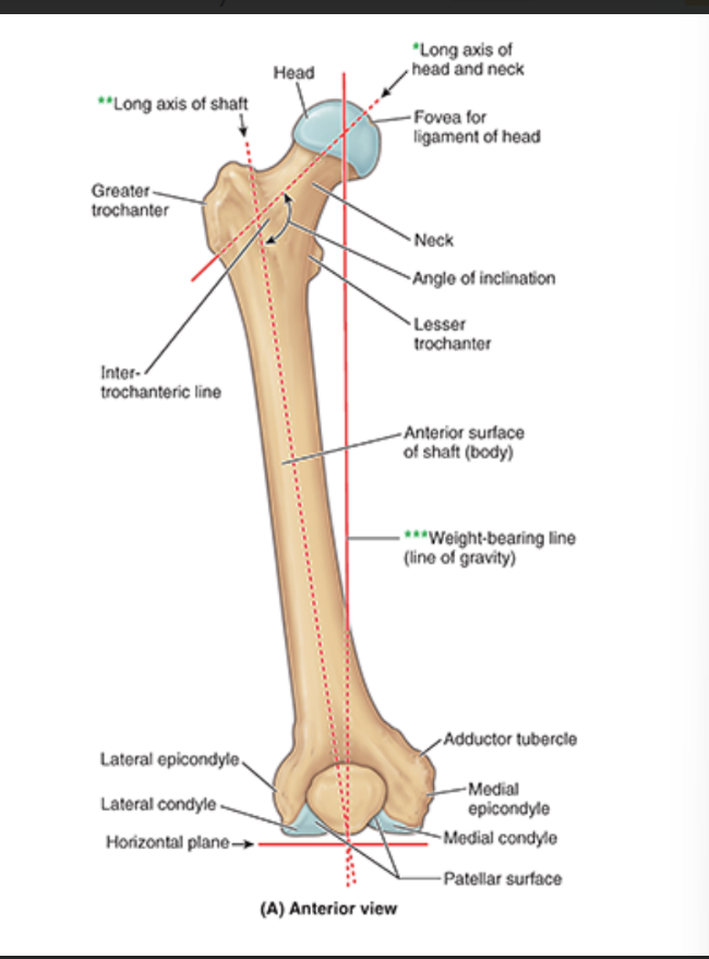
anterior features of the femur
head
fovea for ligament of head
neck
greater trochanter (laterally)
lesser trochanter (medially)
lateral epicondyle
medial epicondyle
adductor tubercle
patellar surfaces
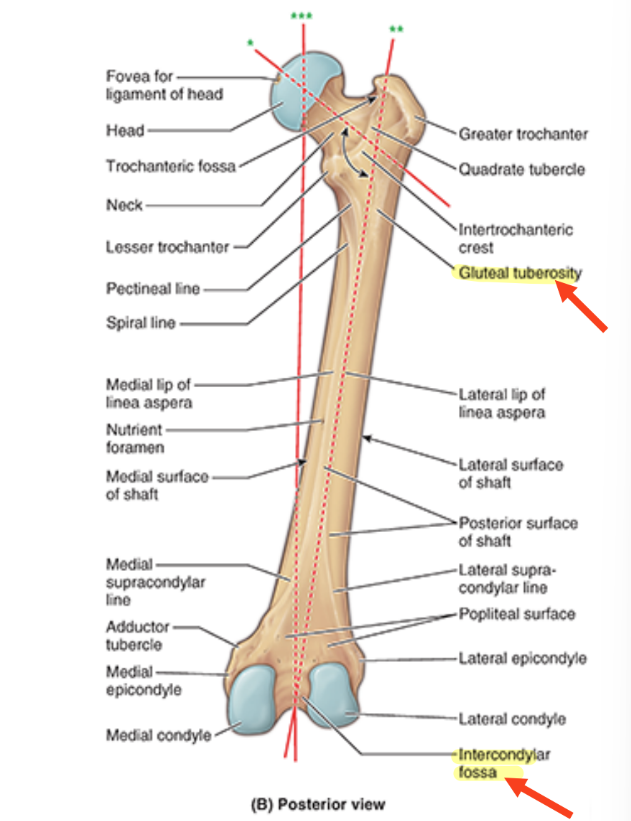
posterior features of the femur
gluteal tuberosity
medial condyle
lateral condyle
intercondylar fossa
linea aspera
longitudinal ridge/crest on middle third of the bone
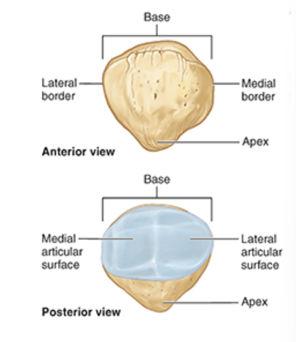
patella
articulates w/ femur at patellar surfaces
base = sup. border
lateral and medial borders
apex = inf. aspect
post. aspect covered w/ articular cartilage for articulation w/ femur
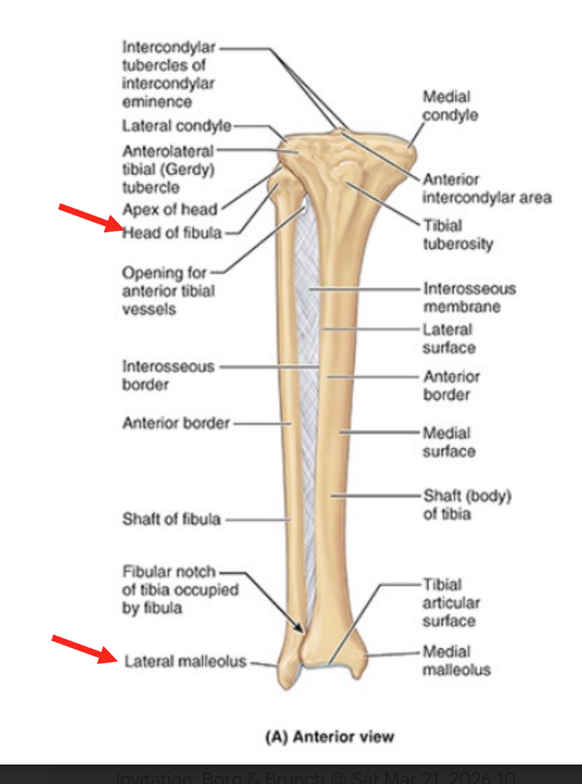
tibia
larger, medial, weight-bearing bone
articulates w/
femur (proximal)
fibula (prox & dist)
talus (distal)
connected to fibula via interosseous membrane on shaft
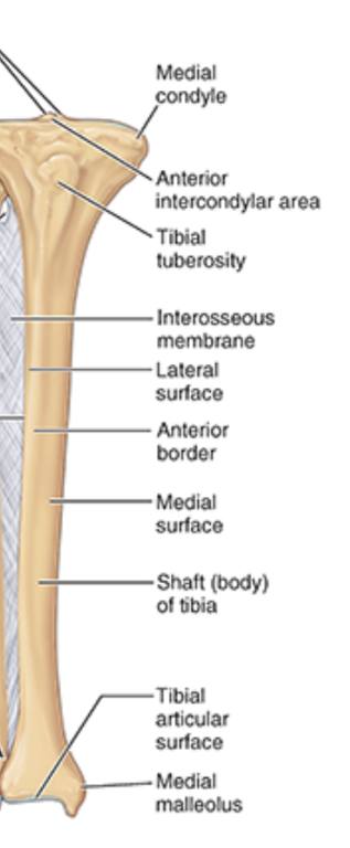
tibia bone markings
medial condyle
lateral condyle
intercondylar) area
medial malleolus
fibular notch
soleal line (posterior)
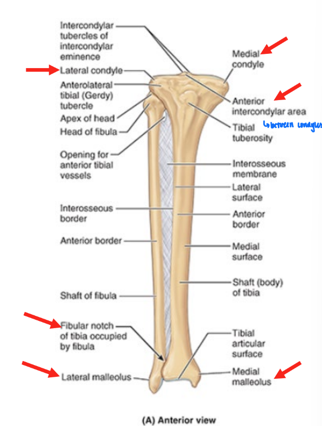
fibula
smaller & lateral bone
not weight bearing
source of attachment
connected to tibia via interosseous membrane on shaft
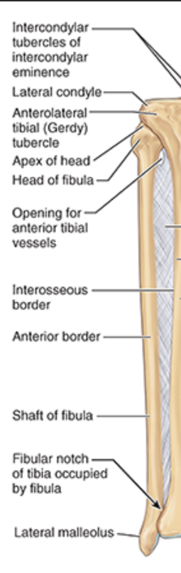
fibula bone markings
head
neck
lateral malleolus
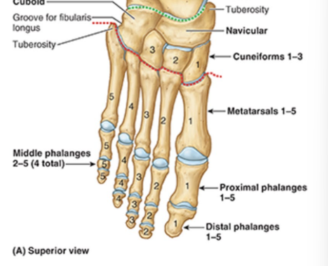
ankle & foot
7 tarsal bones
5 metatarsals
14 phalanges
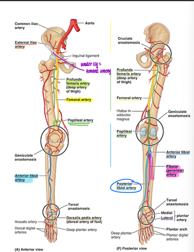
arterial supply of lower limb
femoral artery (continuation of external iliac)
profunda femoris = branch
deep artery of thigh
femoral artery continues = popliteal artery
popliteal artery divides into
ant. & post. tibial artery
post tibial artery → fibular artery
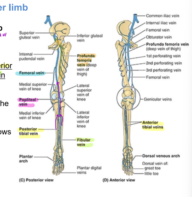
deep venous drainage of the lower limb
same names as arterial supply
ant tibial vein
post tibial vein
fibular vein
= all flow into popliteal vein
which becomes femoral vein
profunda femoris vein flows into femoral
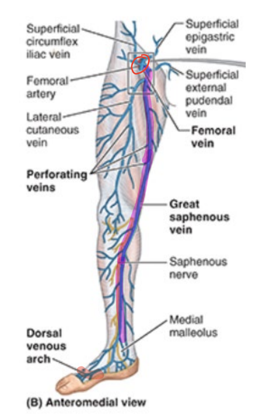
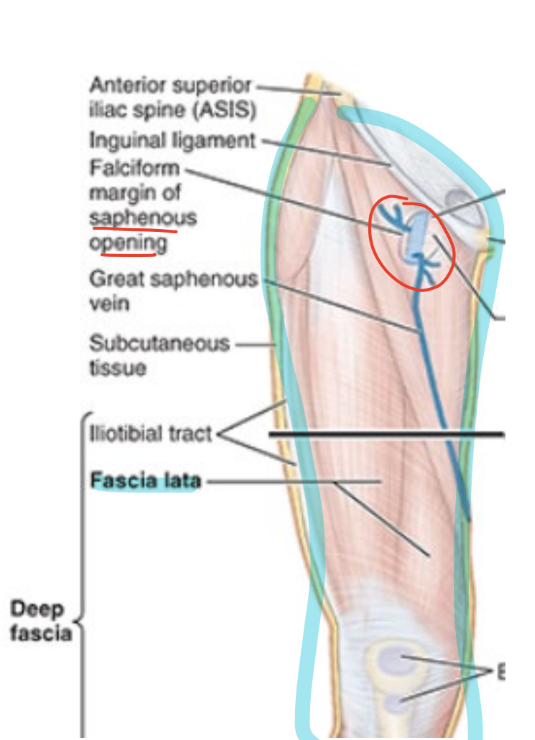
superficial venous drainage of the lower limb: great
superficial to deep fascia
great saphenous vein
medial side of leg
union of dorsal vein of great toe & dorsal venous arch of foot
traverses saphenous opening into fascia lata to empty into femoral vein
used in coronary bypass grafts
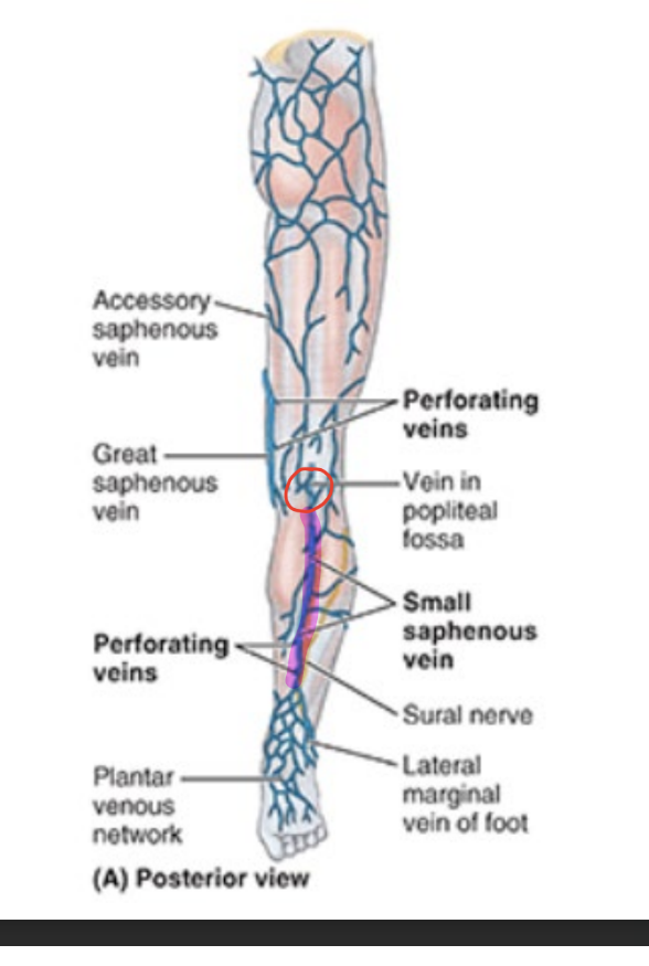
superficial venous drainage of the lower limb: small
superficial to deep fascia
small saphenous vein
arises from lateral side of foot
penetrates deep fascia midway up leg & ascends between heads of gastrocnemius muscle
empties into popliteal vein in popliteal fossa
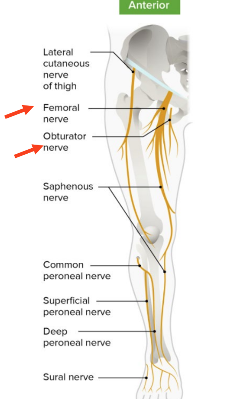
innervation of the lower limb
anterior
femoral nerve
obturator nerve
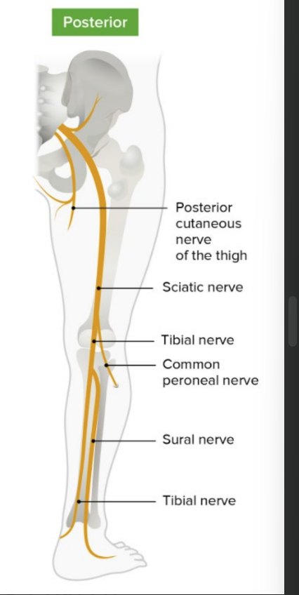
posterior
sciatic nerve
tibial nerve
common fibular (peroneal) nerve
femoral nerve
arises from lumbar plexus
innervates flexors of the hip & extensors of knee
located mainly in ant. compt of thigh

obturator nerve
arises from lumbar plexus
innervates adductors of hip
located in medial comp of thigh
sciatic nerve
arises from sacral plexus
descends into post. compt of thigh
2 comps
tibial nerve
supplies extensor of hip joint & flexors of knee joint
common fibular (peroneal) nerve
supplies short head of biceps muscle on post compt of thigh
anterior and medial thigh: fascia
fascia lata = sleeve-like deep fascia of thigh
saphenous opening = opening in fascia lata inferior to medial part of inguinal ligament
where saphenous and femoral vein unites
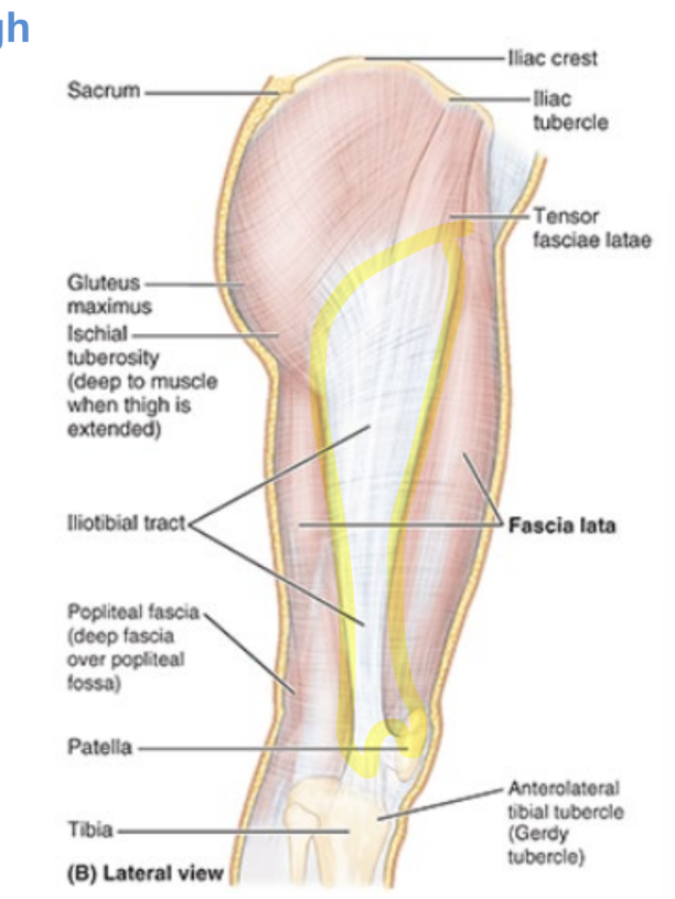
iliotibial tract
IT band
broad band of fibers = shared aponeurosis of tensor fasciae latae & glut maximus muscles
extends from iliac tubercle to anterolateral tubercle of tibia
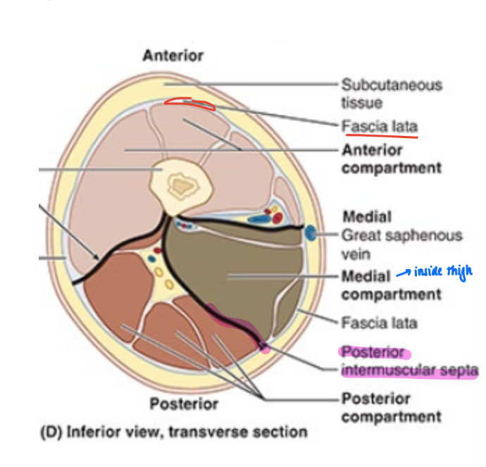
3 compartments of thigh muscles
anterior, medial, & posterior
walls formed by fascia lata & intermuscular septa
medial, lateral, and posterior septa
femoral triangle
subfascial space inf to inguinal ligament

femoral triangle borders
inguinal ligament
adductor longus
sartorius

femoral triangle roof
fascia lata
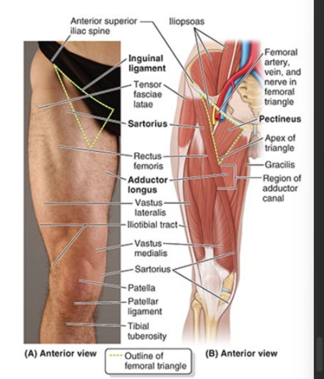
femoral triangle contents
lateral to medial:
femoral nerve
femoral artery
femoral vein
great saphenous & profunda femoris
deep inguinal lymph nodes

innervations of thigh compartments
ant compt = femoral nerve
except psoas part of iliopsoas = L1-L3
medial compt = obturator nerve
except adductor magnus = tibial division of sciatic
post compt = tibial division of sciatic
except short head of biceps femoris = common fibular division
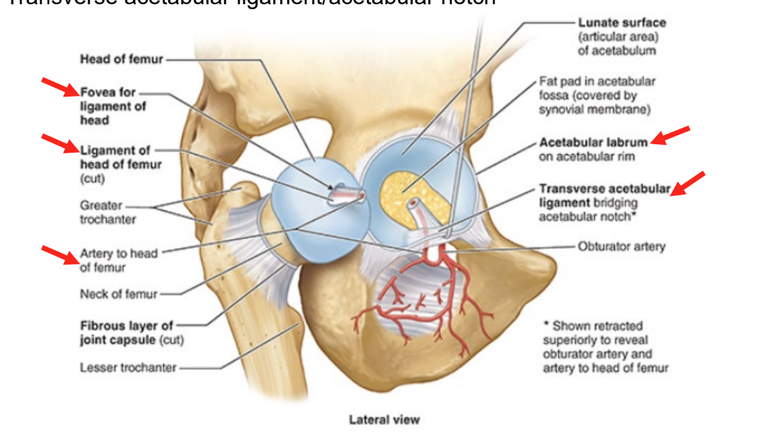
hip joint
acetabular labrum
fovea for ligament of head
ligament of head of femur
artery to head of femur
transverse acetabular ligament / acetabular notch
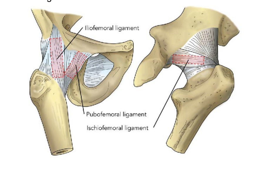
3 major ligaments of hip joint
iliofemoral ligament
pubofemoral ligament
ischiofemoral ligament
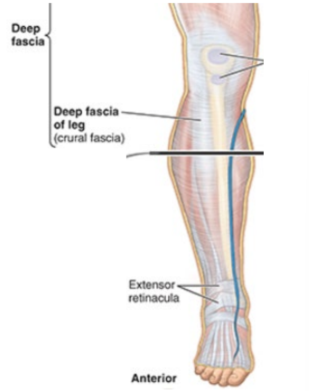
fascia of lower leg
layer of deep fascia covering leg
continuous w/ fascia of thigh region
crural fascia
extensor retinacula (sup and inf)
extensor retinacula
strong bands of fascia that cover tendons passing from ant compt of leg to foot
prevent bowstring of tendons
like wrist tendons
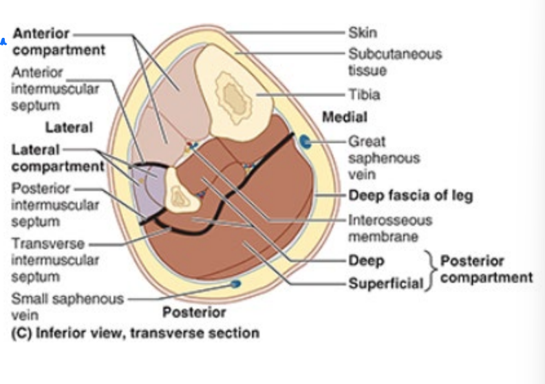
division of compartments of lower leg
intermuscular septa, interosseous memb, bones & crural fascia sep leg in 3 compts
anterior, posterior & lateral
innervation of the lower leg
sciatic nerve
arises from sacral plexus
descends into post. compt of thigh
two comps
tibial nerve
common fibular (peroneal) nerve
arterial supply and venous drainage of leg
femoral artery continues as popliteal artery posteriorly
pop artery divides
ant. tibial artery
post tibial artery
post tibial artery branches into: fibular artery
veins = opposite direction

popliteal fossa boundaries
diamond-shaped space post. to knee
borders:
biceps femoris
semimembranosus
heads of gastrocnemius
roof
popliteal fascia
covers back of knee
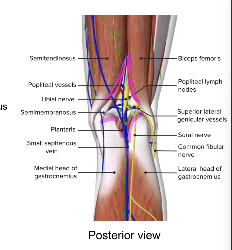
contents of popliteal fossa
passageway for structures from thigh to leg
contents
termination of small saphenous vein
popliteal vein
popliteal artery
tibial nerve
common fibular nerve
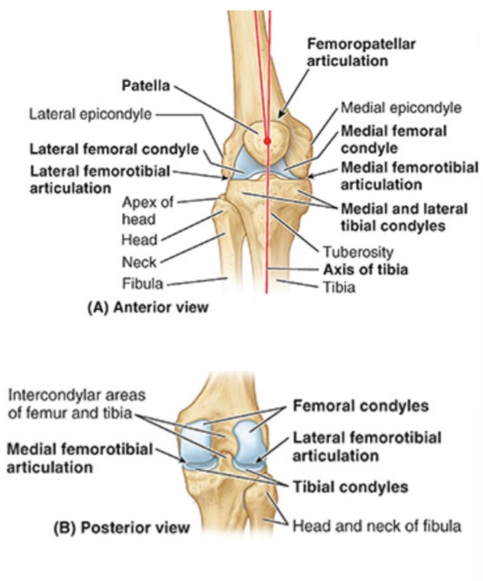
knee joint
2 femorotibial articulations
lateral and medial
between lateral and medial femoral and tibial condyles
1 femoropatellar articulation
fibula does not participate in articulations
menisci
medial and lateral
crescent shaped cartilage on tibia that strengthen joint and act as shock absorbers
keep distal part of femur in place
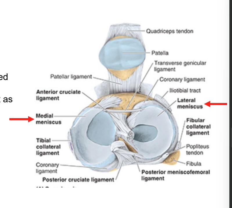
medial collateral ligament
medial epicondyle of femur to superior tibia
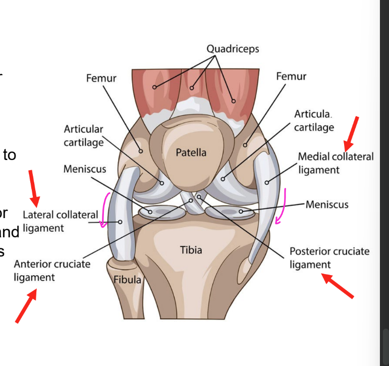
lateral collateral ligament
lateral epicondyle of femur to head of fibula

cruciate ligaments
cruciate = cross one another
anterior & posterior
join femur & tibia
crossing in their paths

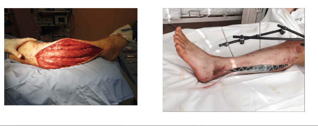
compartment syndrome
devastating lower extremity condition where compt pressure rises to a level that decreases perfusion of leg
blood flow & nerve signaling cut off from inflammation
may lead to irreversible muscle & neurovascular damage
treatment = emergent fasciotomy
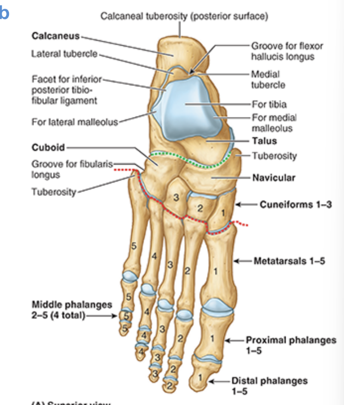
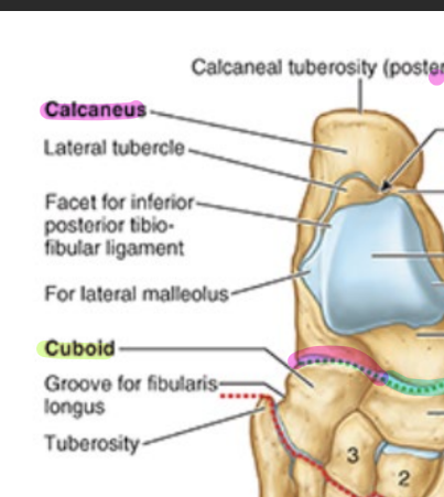
osteology: tarsals
talus
calcaneus
cuboid
navicular
cuneiforms
medial (1st)
intermed (2nd)
lateral (3rd)
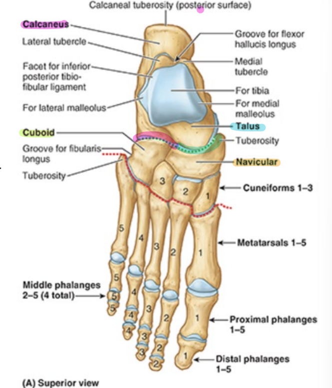
talus
superior surface: trochlea of talus
articulates w/ tibia & fibula
only foot bone that articulates w/ bones in limb
articulates w/ navicular
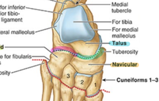
calcaneus
articulates w/ cuboid
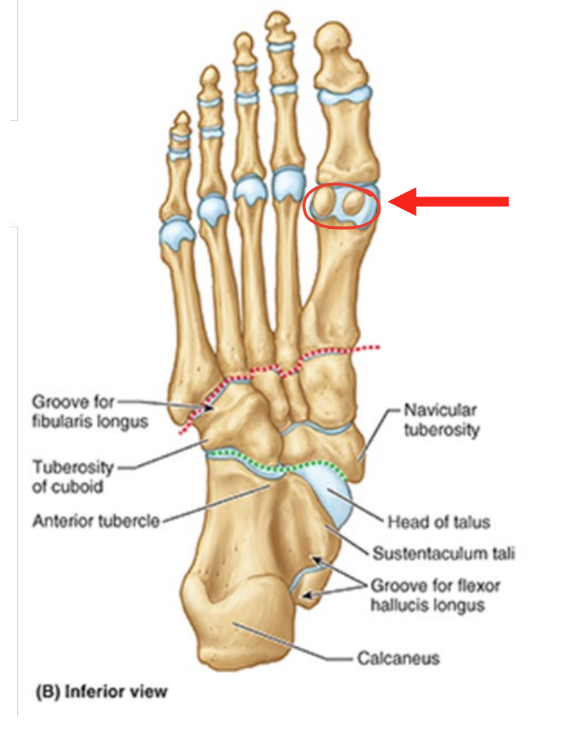
contains groove for flexor hallucis longus tendon
wraps around calcaneus
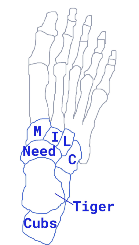
tarsals acronym
Tiger
talus
Cubs
calcaneus
Need
navicular
MILC
medial cune
intermed cune
lateral cune
cuboid
phalanges
hallux = digit 1 = great toe
only proximal and distal phalanges
digits 2-5
proximal, middle, and distal phalanges
sesamoid bones of great toe
two pea-shaped bones located in the ball of the foot
beneath the big toe joint
acts as pulley for tendons
help big toe move normally
provide leverage when big toe pushes off during walking & running
hindfoot
calcaneus
talus

midfoot
cuboid
navicular
cuneiforms (3)
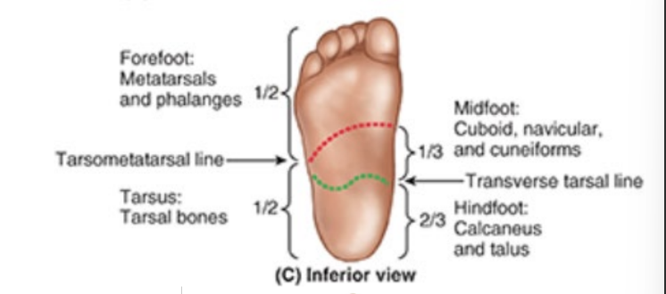
forefoot
metatarsals
phalanges

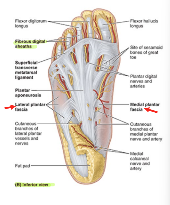
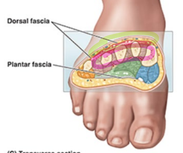
foot fascia
dorsal fascia
contin w/ crural fascia (inf. extensor reticulum)
plantar aponeurosis: thick central part of plantar fascia
extends from calcaneus to phalanges
continuous w/ fibrous digital sheaths that enclose flexor tendons
medial plantar fascia
lateral plantar fascia
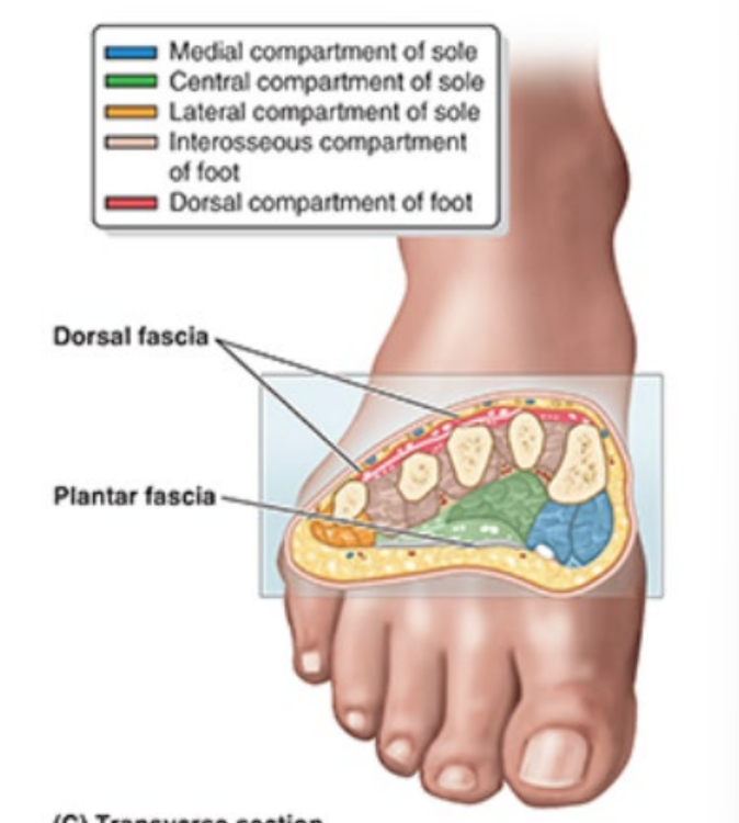
foot compartments
medial
central
lateral
sole = medial, central, lateral
interosseous
dorsal
medial foot compt
part of sole of foot
muscles that control hallux
covered by medial plantar fascia
(pink outline)

central compt of foot
part of sole of foot
muscles controlling lateral four digits
covered by plantar aponeurosis
(blue/green outline)

lateral compt of foot
part of sole of foot
muscles controlling 5th digit
covered by lateral plantar fascia
(orange/red outline)
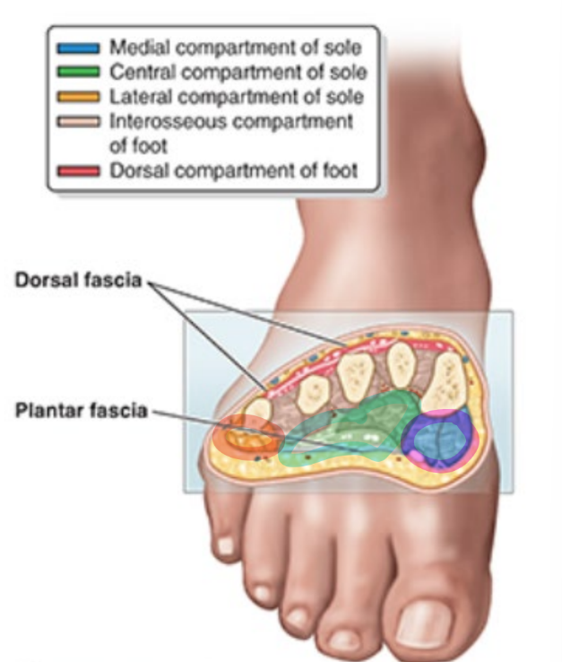
interosseous compt of foot
muscles that contrib to movement of lateral 4 digits
(pink outline)

dorsal compt of foot
muscles contrib to extension of digits
(green outline)
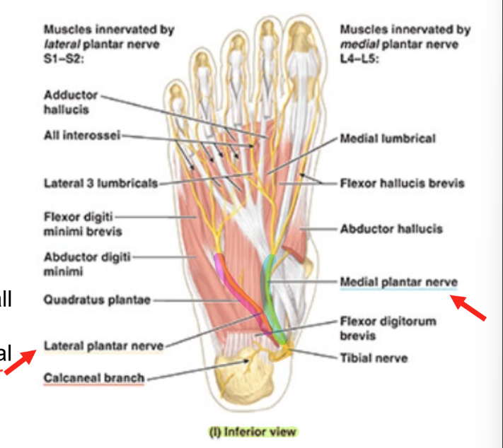
nerves of foot (main)
tibial nerve divides posterior to medial malleolus into
medial plantar nerve
lateral plantar nerve
2 nerves innervate all intrinsic foot muscles except dorsal compt
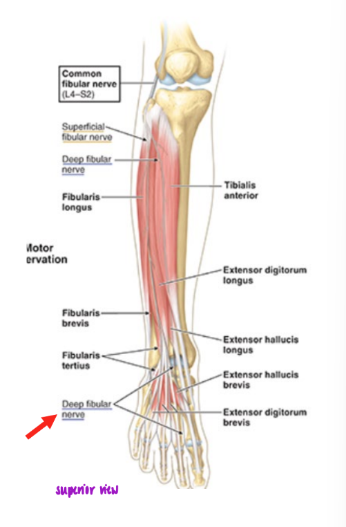
nerves of foot (dorsal)
deep fibular nerve
passes from ant. compt of leg to dorsum of foot
innervates 2 extensor muscles of toes in dorsal compt
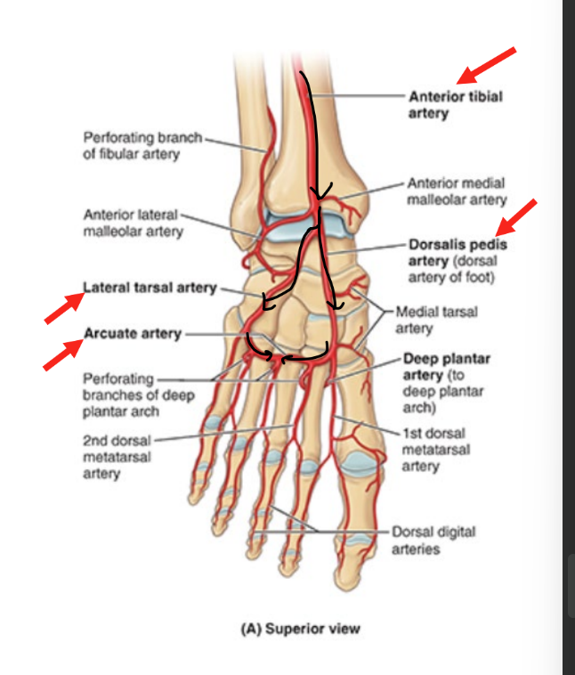
arterial supply of foot (dorsal)
from ant. part of leg
anterior tibial artery continues as dorsalis pedis artery
branches into lateral tarsal artery
runs laterally
both arteries come tg to feed arcuate artery
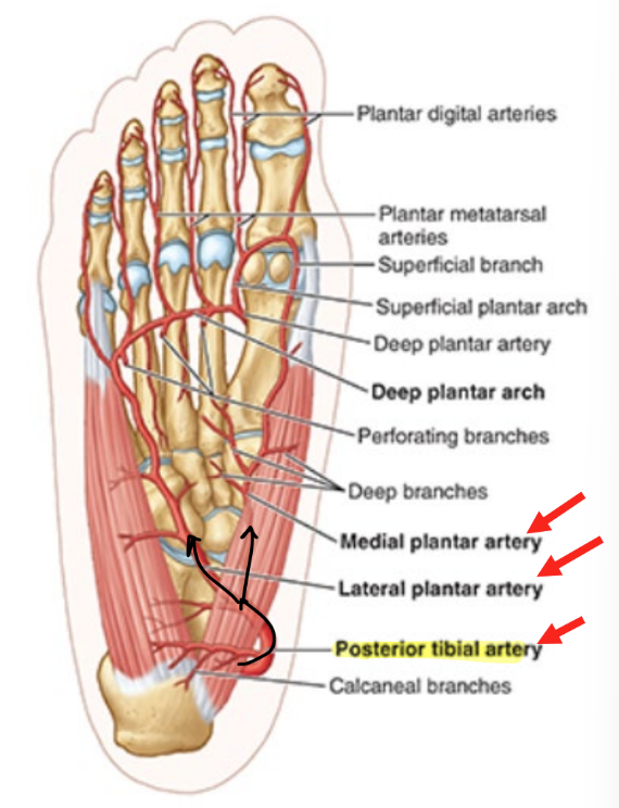
arterial / nervous supply of foot (plantar)
sole of foot supplied by posterior tibial artery
branches into:
medial plantar artery
lateral plantar artery
nerves = corresponding names
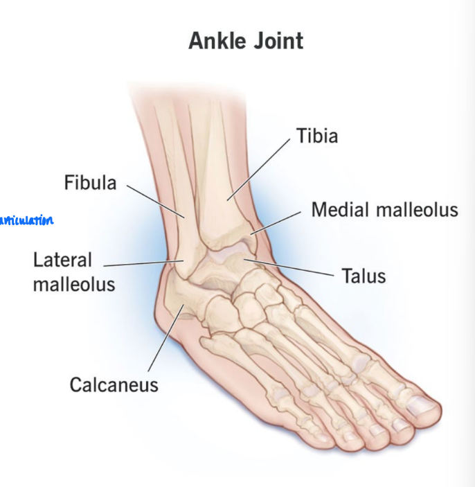
ankle joint
located between distal ends of tibia & fibula & sup. part of talus
only tibia and fibula articulate w/ talus (of leg bones)
trochlea = articulates w/ rounded superior surface of talus
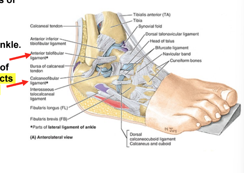
major ligaments of the ankle
medial (deltoid) ligament of ankle
connects tibia to tarsals
lateral ligament of ankle
connects fibula to tarsals
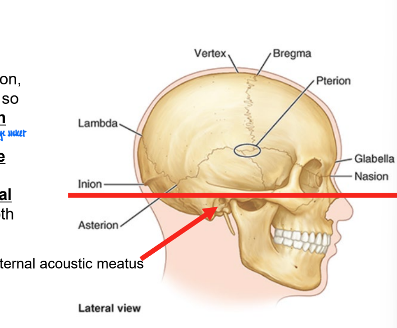
orientation of the cranium
inf margin of the orbit
sup margin of the external acoustic opening of the external acoustic meatus
number of bones in the cranium
contains 22 bones
6 singular bones at midline
8 sets of paired bones that are bilateral
majority are fused at suture joints
immobile joints
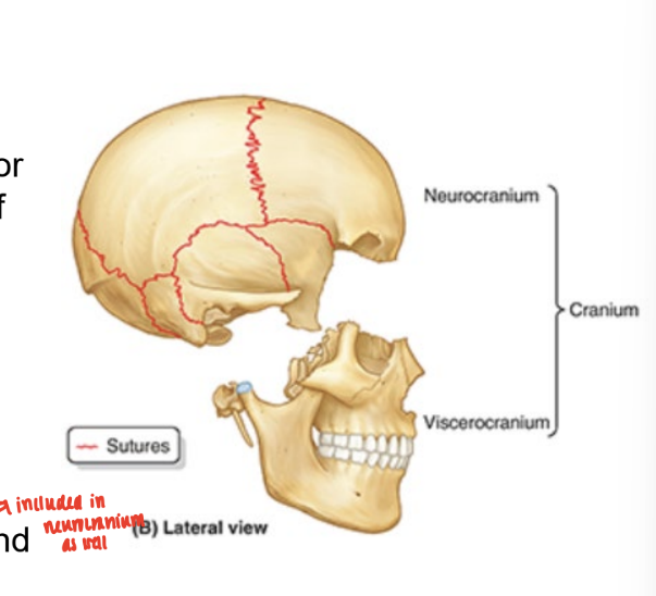
neurocranium
bony case of the brain, membranous coverings, & cranial meninges
formed by series of 8 bones
4 singular bones centered on midline
frontal, ethmoid, sphenoid, occipital
2 sets of bones occurring as bilateral pairs
temporal and parietal
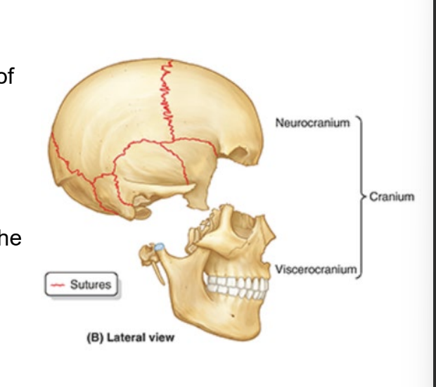
viscerocranium
forms the ant. part of cranium
consists of bones surrounding the mouth (upper & lower jaws), nose/nasal cavity, & most of the orbits
consists of 15 irregular bones
3 singular bones centered on or lying in the midline
mandible, ethmoid, vomer
6 bones as bilateral pairs
mandible, inferior nasal conchae, zygomatic, palatine, nasal, & lacrimal bones
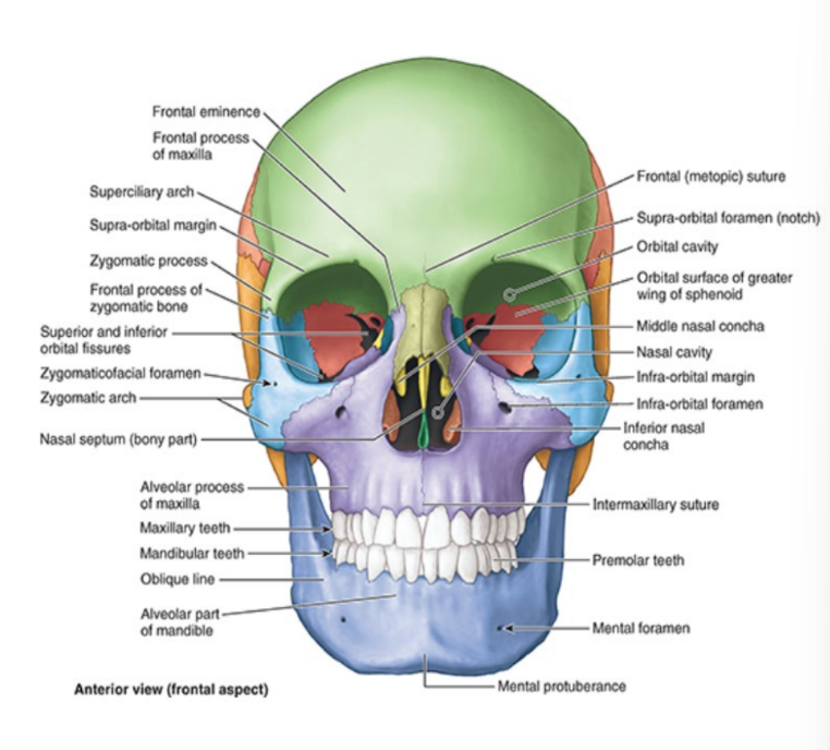
anterior (facial) aspect of cranium
frontal & zygomatic bones
orbits
nasal region
maxillae
mandible
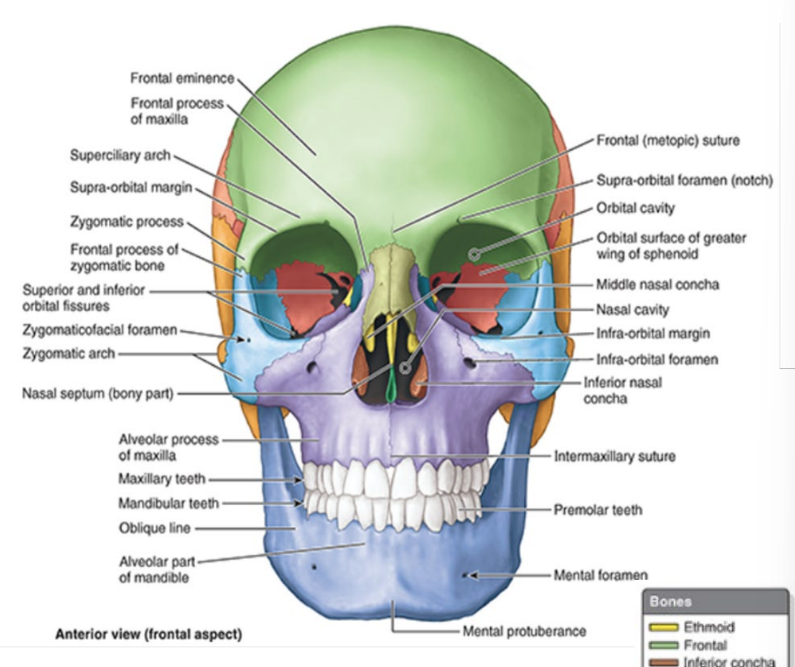
frontal bone
forms skeleton of forehead
articulates inf. w/ nasal & zygomatic bones
intersection of frontal & nasal bones = nasion
bridge of nose

nasion
bridge of nose
zygomatic bones
= cheek bones, malar bones
forms prominences of cheeks
lies on inferolateral sides of orbits
rest on maxillae
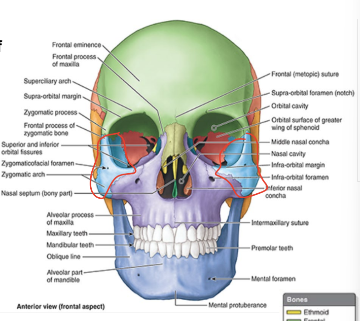
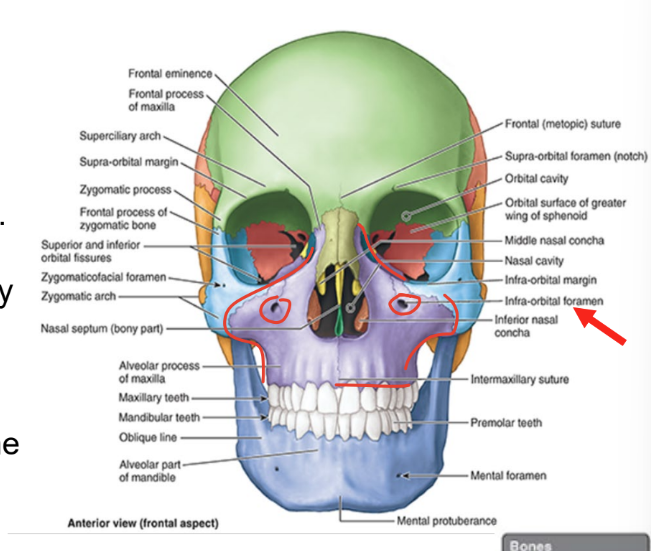
maxillae
form upper jaw
supporting bone for maxillary teeth
maxillae surround most of piriform aperture
forms infra-orbital margins medially
infra-orbital foramen
inf to each orbit for passage of infra-orbital nerve & vessels
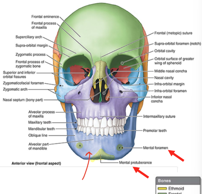
mandible
only moveable bone in skull
U-shaped bone that supports mandibular teeth
consists of horizontal (body) and vertical (ramus) parts
mental foramina
mental nerves & vessels
mental protuberance
forms prominence of chin
triangular bony elevation inf. to mandibular symphysis
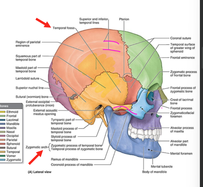
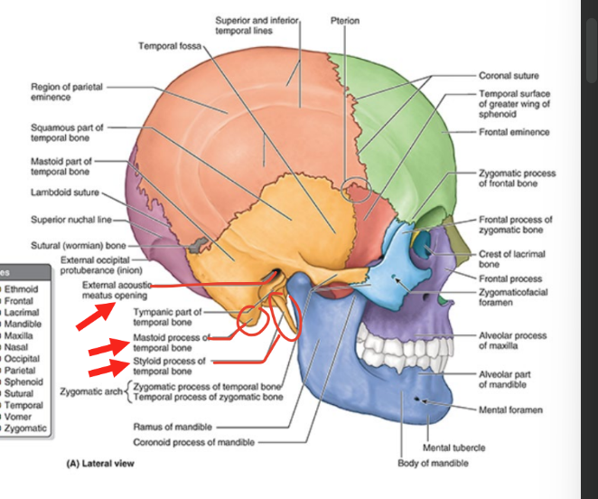
lateral aspect of cranium
includes lateral aspects of frontal, parietal, zygomatic, sphenoid bones & temporal bones
temporal fossa
bounded sup. & post. by sup & inf temporal lines
bounded ant. by frontal & zygomatic bones
bounded inf. by zygomatic arch
zygomatic arch
anterolateral projection of temporal bone & lateral aspect of zygomatic
lateral aspect of cranium (temporal bone)
external acoustic meatus opening
entrance to external acoustic meatus (canal)
leads to tympanic membrane (eardrum)
mastoid process
postero-inf to external acoustic meatus opening
styloid process
anteromedial to mastoid process of temporal bone
slender needle-like, pointed projection
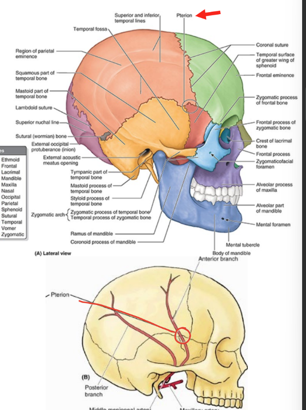
pterion
part of lateral aspect of cranium
ant. part of temporal fossa
3-4 cm sup. to midpoint of zygomatic arch
usually indicated by H-shaped formation of sutures that unite frontal, parietal, sphenoid, & temporal bones
pterion fracture
can be life-threatening
rupture of branches of middle meningeal artery & epidural hematoma
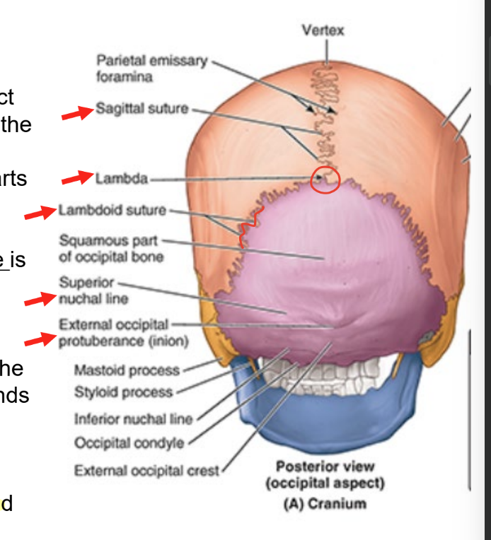
occipital aspect of cranium
post or occipital aspect of cranium
composed of occipital bone, parts of parietal bones, mastoid parts of temporal bones
external occipital protuberance
easily palpable in median plane
sup. nuchal line
marks sup. limit of neck
extends laterally from each side of external protuberance
lambda
jxn of lamboid & sagital sutures
can be palpated
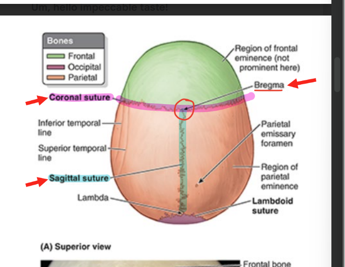
sup. aspect of cranium
coronal suture
separates frontal and parietal bones
sagittal suture
separates parietal bones
bregma
landmark formed by intersection of sagittal & coronal sutures
vertex
most sup part of cranium
external surface of cranial base
cranial base = basicranium
inf portion of neurocranium & viscerocranium
minus mandible
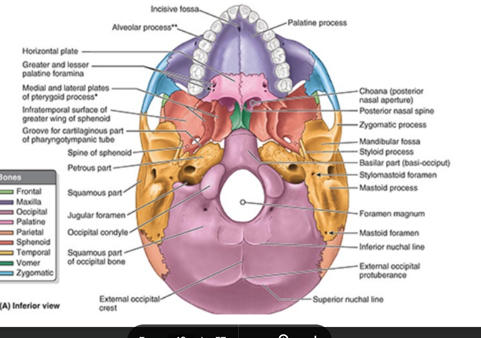
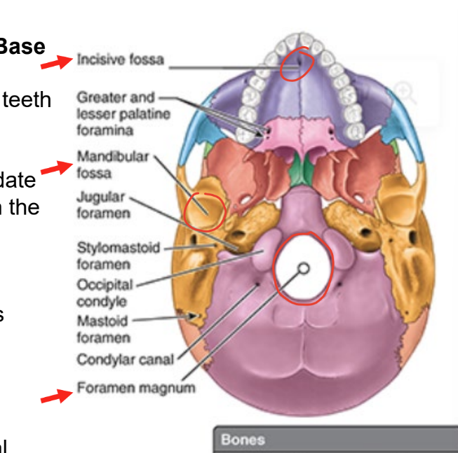
external surface of cranial base (part 1)
incisive fossa
post. to central incisor teeth
mandibular fossae
accommodate mandibular condyles when mouth is closed
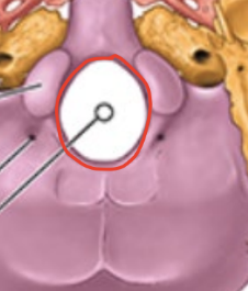
foramen magnum
major structures like spinal cord pass thru
contents of foramen magnum
spinal cord
meninges
vertebral arteries
ant. & post. spinal arteries
external surface of cranial base
occipital condyles
how cranium articulates w/ vert column
jugular foramen
large opening between occipital & temporal bone
entrance to carotid canal
stylomastoid foramen

jugular foramen contents
internal jugular vein
cranial nerves (CN 9-11)

occipital condyles
articulates cranium w/ vert column
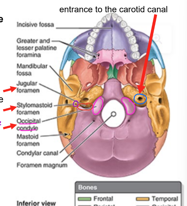
carotid canal contents
internal carotid arteries

stylomastoid foramen contents
facial nerve (CN VII)

internal surface of cranial base
3 large depressions at dif levels
anterior
middle
posterior
form bowl-shaped floor of cranial cavity
space enclosed w/in neurocranium occupied by brain
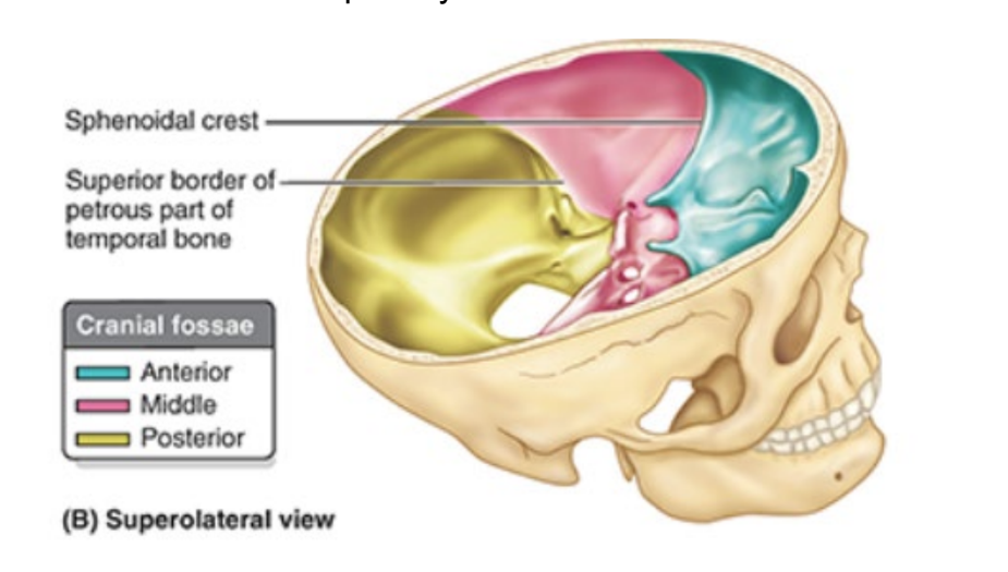
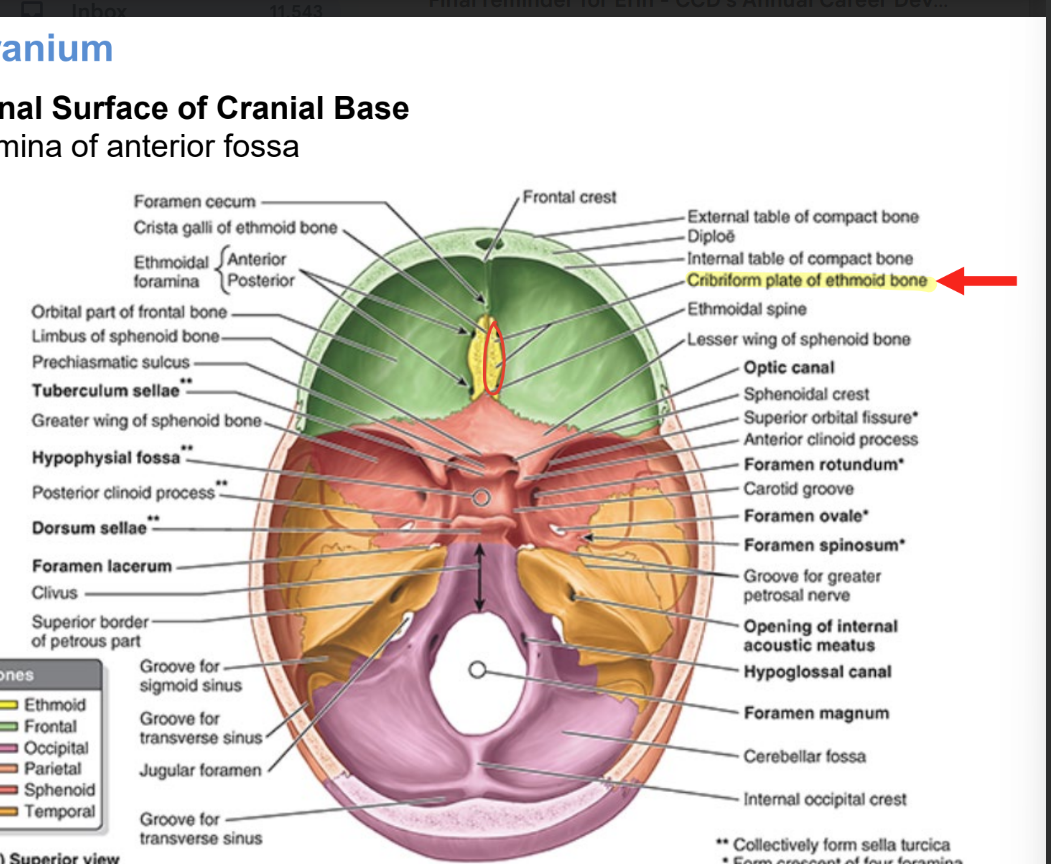
anterior cranial fossa
occupied by inf & ant parts of frontal lobes
cribriform plate of ethmoid bone
has numerous tiny foramina = transmit olfactory nerves (CN 1) branches
from olfactory areas of nasal cavities to olfactory bulbs of brain
middle cranial fossa contents
lateral parts support temporal lobes of brain
hypophysial fossa (pituitary fossa)
foramina
optic canals
foramen lacerum
sup. orbital fissure
foramen rotundum
foramen ovale
foramen spinosum
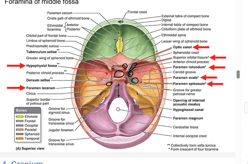
hypophysial fossa (pituitary fossa)
median depression in body of sphenoid
accommodates pituitary gland

optic canals
optic nerves (CN II) & ophthalmic arteries

foramen lacerum
contains internal carotid artery
continuous w/ carotid canal
dif name than exit

superior orbital fissure
ophthalmic nerve (CN V1) → 1st branch of CN V
also CN III, IV, and VI

foramen rotundum
maxillary nerve (CN V2)

foramen ovale
mandibular nerve (CN V3)

foramen spinosum
meningeal branch of CN V3
