ls7c final
1/124
Earn XP
Description and Tags
this class is basically an english class ngl
Name | Mastery | Learn | Test | Matching | Spaced | Call with Kai | Chat |
|---|
No analytics yet
Send a link to your students to track their progress
125 Terms
Identify critical cells involved in auditory and visual sensory transmission to the brain.
auditory: hair cells
visual: rods, cones, bipolar cells, ganglion cells
Describe how hair cells function to transduce mechanical signals to the brain.
mechanical stimulus disturbs kinocilium
K+ channels open and K+ enters the cell
VG Ca2+ channels open and interact with vesicles
vesicles release neurotransmitters into synapse
neurotransmitters bind to ligand-gated channels on afferent neuron
Describe parts of a simple reflex circuit
afferent neurons: receive sensory signals and transmit them to spinal cord
interneurons: relay info in spinal cord from afferent to efferent neurons
efferent neurons: relay signal to muscles
muscles: contract
Predict how photoreceptors change in response to light exposure
in presence of light, rhodopsin dissociates from G-protein, which binds to the effector to turn cGMP into GMP
Predict the effect of toxins or genetic mutations on the function of skeletal muscles
botox prevents muscle contraction
Clostridium tetani causes prolonged muscle contraction
Evaluate the physiological consequences of altering the structure/function of skeletal muscle components (e.g., SR, myosin, troponin)
SR
if SR were always open, Ca2+ presence in the cell would be toxic
if SR were always closed, no muscle contraction could occur
myosin
if myosin could not bind actin or ATP cannot be hydrolyzed, muscle contraction cannot occur
if there is no ATP, muscles stay in rigor
troponin
if troponin cannot bind Ca2+, muscle contraction cannot occur
if troponin gains function, prolonged muscle contraction will occur
Explain how sensory information is encoded in action potentials
intensity of stimulus is proportional to rate of action potentials
Explain how the processes of vision and hearing occur
vision
light hits rod or cone, causing rod or cone to hyperpolarize and stop sending neurotransmitters
without neurotransmitter, mGluR channels on bipolar cells close, causing their VG channels to open and depolarization to occur
in turn, the ganglion cells are depolarized
signals are sent from the optic nerve to the brain for processing
hearing
sound waves travel through endolymph of the cochlea, causing vibrations of the basilar membrane and a mechanical stimulus against the hair cell’s stereocilia to kinocilium
hair cell releases neurotransmitter and signal is processed
Describe the structure and function of a neuromuscular junction
junction between motor neuron and muscle cell
motor neuron releases Ach, binds to channels on muscle cell and allows depolarization of muscle cell
Identify cellular, molecular, and protein components involved in muscle contraction and explain their role
action potential: allows motor neuron’s Ca2+ channels to open, which allows Ach to be released
Ca2+: in the motor neuron, allows Ach release; in muscle cell, binds to troponin to move tropomyosin and expose actin binding sites
Sarcoplasmic Reticulum: stores Ca2+ when muscle is not contracting because Ca2+ is toxic to the cell
voltage-gated channels: motor neuron has VG Ca2+ channels to allow Ca2+ in during action potential
Troponin: binds to Ca2+ and moves tropomyosin
Tropomyosin: covers actin binding sites to prevent muscle contraction in the absence of Ca2+
Actin: “thin” filament of muscle which moves when myosin heads bind to it
Myosin: “thick” filament of muscle which forms cross-bridges with actin to contract the muscle
ATP: binds to myosin after a power stroke, causing it to enter a high-energy state
Relate the structure of skeletal muscle to its function in generating a contractile force
bundles of muscle fibers are individual cells, and bundles of these cells make up fascicles
the large amount of muscle fibers in a muscle allow a high amount of force to be produced during contraction
Describe the cross-bridge cycle in a skeletal muscle
the myosin head’s ATP is hydrolyzed to ADP and Pi as it binds to the exposed binding site
the myosin heads pulls the actin in a power stroke, releasing its ADP and Pi
the power stroke ends and the myosin head is in a low energy state
ATP binds to the myosin head and causes it to enter a high energy state
Compare and contrast the structure and function of slow-twitch versus fast-twitch muscles
slow-twitch
prolonged muscle contraction
many mitochondria
oxidative phosphorylation is main source of ATP
have more myoglobin
are red in color
fast-twitch
short bursts of powerful muscle contraction
can be oxidative or use glycolysis; exercise converts glycolytic cells into oxidative cells
have less myoglobin
have a pale to white color
Explain the relationship between surface area-to-volume ratio and gas exchange efficiency
better surface area-to-volume ratio means more places for gas exchange to occur so gas exchange efficiency is higher
Describe the changes in muscle contraction, volume, and pressure that occur during ventilation
the diaphragm contracts, the volume increases and the pressure in the thoracic cavity decreases causing inhalation; the diaphragm then relaxes, decreasing volume and increasing pressure, leading to exhalation
Relate partial pressure and Boyle’s Law to ventilation and gas exchange
partial pressure is inversely related to volume, driving ventilation
gases flow from high partial pressure to low partial pressure, driving gas exchange
Explain how the nervous system regulates breathing and how this relates to homeostasis
chemoreceptors in the medulla sense pH: if it is too low, breathing rate increases to bring more oxygen to the blood until it rises to normal
Describe how hemoglobin binds oxygen and how this relates to gas exchange
hemoglobin binds 4 oxygen molecules to carry them throughout the blood
Predict how changes in elevation or external pressure will affect ventilation and gas exchange
higher elevation/lower external pressure shifts curve to the right
Describe the properties of blood and blood vessels
blood is 55% plasma, which has water, ions and proteins; 45% cells and platelets
blood vessels allow blood to flow from high to low pressure gradients; they have three layers: adventitia, tunica media, and tunica intima
Discuss the various functions of the cells, proteins, and other components found in human blood
cells
red blood cells
carry oxygen
white blood cells
fight infections
proteins
albumins
create and maintain osmotic pressure
globulins
immune function
fibrinogen
blood coagulation
regulatory proteins
regulate gene expression
clotting factors
convert fibrinogen into fibrin
plasma
transportation, immune function, and contribution to blood volume
platelets
clotting
Trace the flow of blood through the human circulatory system
left ventricle → aorta → arteries → arterioles → capillaries → veins → venae cavae → right atrium → right ventricle → pulmonary artery → lungs → pulmonary vein → left atrium → left ventricle
Describe the changes in blood pressure and muscle contraction that occur during the cardiac cycle
pressure rises during contraction
pressure drops during relaxation
Relate changes in ion movement to the cardiac action potential
phase 0: cardiac cells send Na+ ions through gap junctions to trigger depolarization and action potentials
phase 2: Ca2+ binds to ryanodine receptors on SR, opening SR and causing contraction; K+ flows out, Cl- flows in,
phase 3: K+ pumps remain open to kick out K+ and repolarize cell
phase 4: cardiac cells have only pumps like Na+/K+ pumps working to maintain potential, but cardiac pacemaker cells have slow Na+ influx
Explain heart muscle contraction in response to an action potential
SA node fires
atria contract
AV node
bundle of His
Purkinje fibers
ventricular contraction
Relate the events during the cardiac cycle to those represented on an ECG
SA node contracts = beginning of P wave
atrial contraction = P wave to beginning of Q wave
AV node = end of P wave
atria relax = beginning of Q wave
ventricles contract = beginning of Q wave to beginning of T wave
ventricles relax = beginning of T wave to beginning of Q wave
Discuss how a signal can lead to both short- and long-term responses.
short: most pathways, stuff that relies on phosphorylation
long: anything that activates a transcription factor to alter gene expression
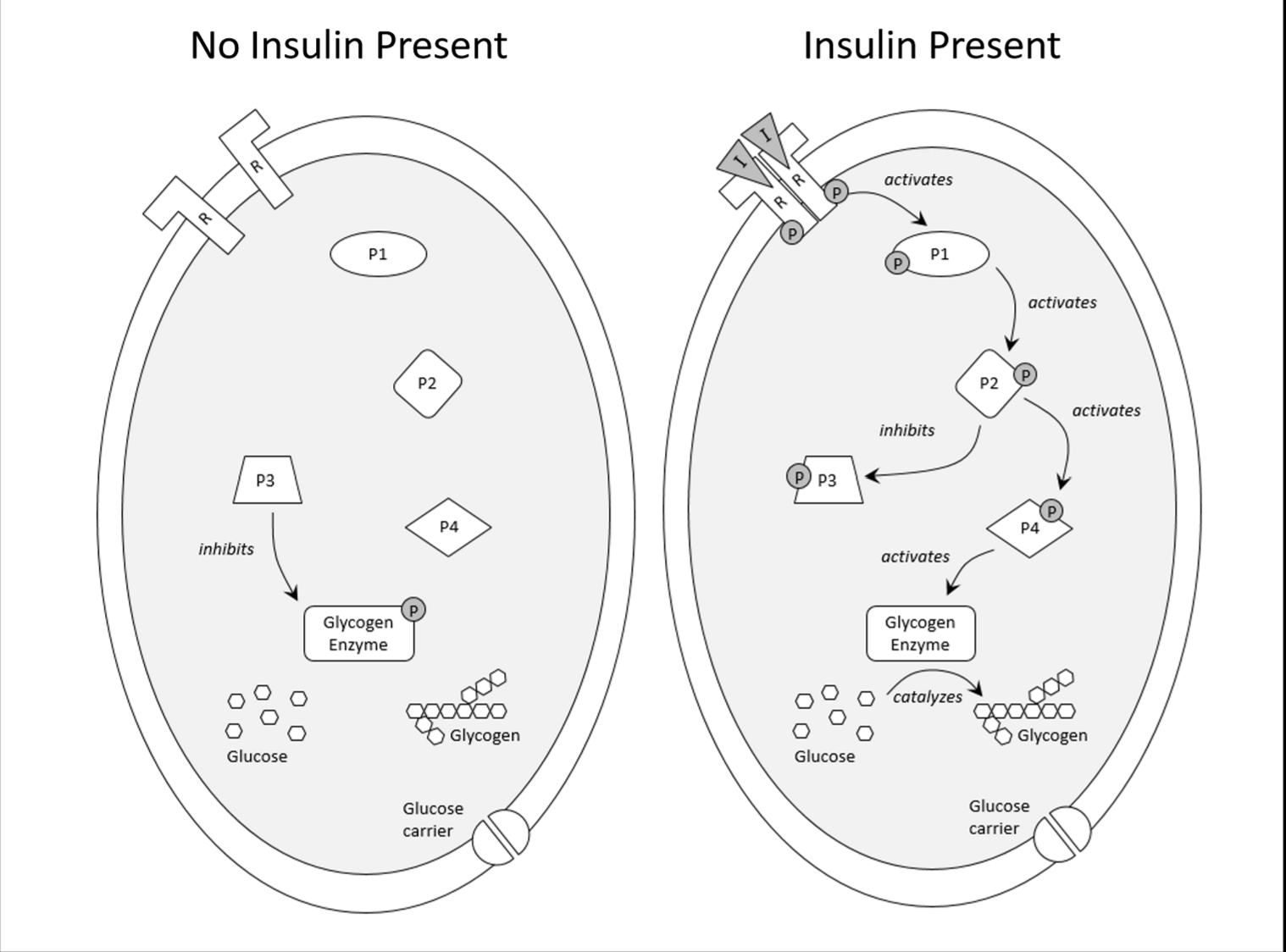
Describe the mechanism of action for a receptor tyrosine kinase pathway.
signal molecule dimerizes RTK
RTK becomes phosphorylated
more proteins are activated, leading to a signaling cascade
Define the role of kinases and phosphatases in cell signaling pathways.
kinases add phosphates
phosphatases take phosphates away
Distinguish the potential for differentiation of totipotent, pluripotent, and multipotent stem cells.
totipotent: can become any cell in the organism
pluripotent: can become any somatic cell
multipotent: can become any cell of a specific tissue type
Explain why diffusion and surface area limit cell size and its implications for large, multicellular organisms.
cells that are too big will have relatively small surface areas
this makes diffusion inefficient
large, multicellular organisms must have lots of cells
Determine if certain proteins in a signaling pathway function as phosphatases, kinases, or neither.
takes a phosphate away from something when active: phosphatase
adds a phosphate to something when active: kinase
does neither: neither
Explain how a signal transduction pathway can be turned off.
ligand dissociates from receptor
Predict the effect of altering part of a signaling transduction pathway.
different product formed
product formed when it isn’t supposed to be
no product formed when it is supposed to be
Interpret data related to different types of cell signaling pathways.
dimers: RTK
squiggly thing with G protein: G protein receptor coupled
Predict cell fate based on cell activation of signaling pathways.
follow the diagram
Describe the different types of cell-cell junctions.
tight junction: seals off layer of cells, divides cells into apical and basal portions, not attached to cytoskeleton
adherens junction: connects cells to epithelial layer and each other, belt-like structure, connected to cytoskeleton, uses cadherins
desmosome: connects cells to each other through button-like points, connected to cytoskeleton, uses cadherins
hemidesmosome: connects cells to basal lamina for anchorage, connected to cytoskeleton, involves integrins and intermediate filaments
gap junction: allows direct cell communication, not attached to cytoskeleton, made of connexins
Define microtubule, microfilament, and intermediate filament.
microtubule: made of tubulin dimers; supports structure, function, and cell division
microfilament: made of actin monomers, supports structure and function
intermediate filament: made of many subunits, provides cell shape and structure
Explain how motor proteins actively move material around the cell.
kinesin: conformational changes, with ATP, to move along microtubules, goes “out” toward + end
dynein: goes “in” toward - end
Explain how cell-cell junctions and the ECM contribute to the cell’s abilities to form tissues and organs.
they connect cells and tissues together
Evaluate how changing components of the cytoskeleton would change cell structure (shape) and/or function (i.e., motility).
cells might not be able to connect to each other
cells might not be anchored
cells might lose communication pathways
Evaluate the effect of modifying cell-cell junctions or ECM components on tissue structure and function.
no tight junctions: movement of stuff through cells is lost
Describe the general structure of a neuron.
cell body connected to many dendrites, also to axon hillock, then to axon covered in myelin sheath which is broken up by nodes of Ranvier, then to axon terminal
Relate the structural features of a neuron (dendrites, axons, etc.) to their functions
dendrites are many = can receive many signals
axon is long = can send signals long distances
myelin sheath = faster signaling
nodes of Ranvier = faster signaling
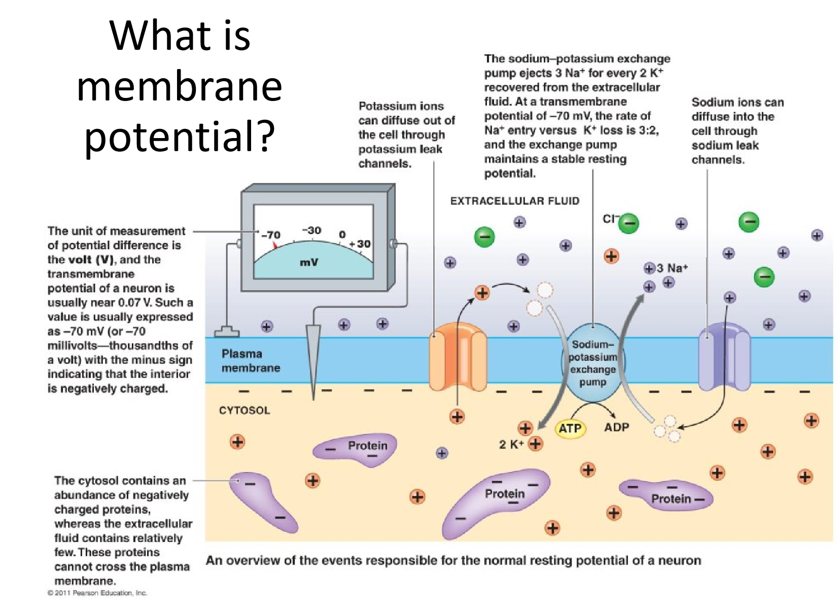
Explain membrane potential and how it arises in both neuronal and non-neuronal cells.
electrochemical gradient produced by active transport
in neurons, Na+/K+ pump
Explain the process by which an action potential is generated and propagated.
enough EPSPs are generated to reach threshold, -55 mV
voltage-gated Na+ channels open
mass influx of Na+ channels open down the axon; depolarization
Compare and contrast ligand-gated and voltage-gated ion channels with respect to their role in signal transduction in a neuron.
ligand-gated are at dendrites and receive neurotransmitters from other neurons
voltage-gated are on the neuron and control action potentials
Explain the process by which two neurons communicate at a synapse.
presynaptic neuron releases neurotransmitters
neurotransmitters bind to ligand-gated receptors on dendrites of postsynaptic neuron
EPSP or IPSP is generated in postsynaptic neuron
Discuss how EPSPs and IPSPs are received and integrated by a postsynaptic neuron.
temporal: many signals at once cause significant effect
spatial: many of the same type at the same time on different dendrites cause effect
they can cancel each other out
Evaluate how multiple signals will be integrated by a postsynaptic neuron that has formed synapses with two or more presynaptic neurons.
see temporal and spatial summation
Predict how a charged molecule will move across a semipermeable membrane in the presence of an electrochemical gradient.
it will attempt to move through a channel with its gradient
Predict how the addition of drugs or the introduction of mutated proteins will alter membrane potential, excitability, and/or signal transmission.
proteins can change resting potential
drugs can act on receptors as agonists or antagonists
Describe the global organization of the human nervous system.
central (brain and spinal cord)
peripheral (everything else)
somatic (voluntary)
autonomic (involuntary)
sympathetic (fight or flight)
parasympathetic (rest and digest)
Relate the major regions of the brain, including the hypothalamus, thalamus, and sensory cortex to their respective functions.
hypothalamus: effects responses in body regulation from brain
thalamus: processes sensory info except smell
sensory cortex: analyzes sensation
Compare and contrast the sympathetic and parasympathetic divisions of the autonomic nervous system.
both are in autonomic divisions
sympathetic: danger mode, increased heart rate, adrenaline
parasympathetic: no danger, decreased heart rate, digestion
Explain how the brain receives, processes, and sends information.
receives: sensory organs, signals from PNS
processes: neuron signaling
sends: more neuron signaling to rest of body; hormones
Evaluate which region of the brain has been damaged in a patient based on a set of symptoms.
Broca’s area: unable to speak well
Wernicke’s area: unable to understand
cerebellum: coordination problems
frontal lobe: personality changes
occipital lobe: vision changes
Predict which branch, sympathetic or parasympathetic, will respond to different stimuli.
dangerous stuff: sympathetic
calming stuff: parasympathetic
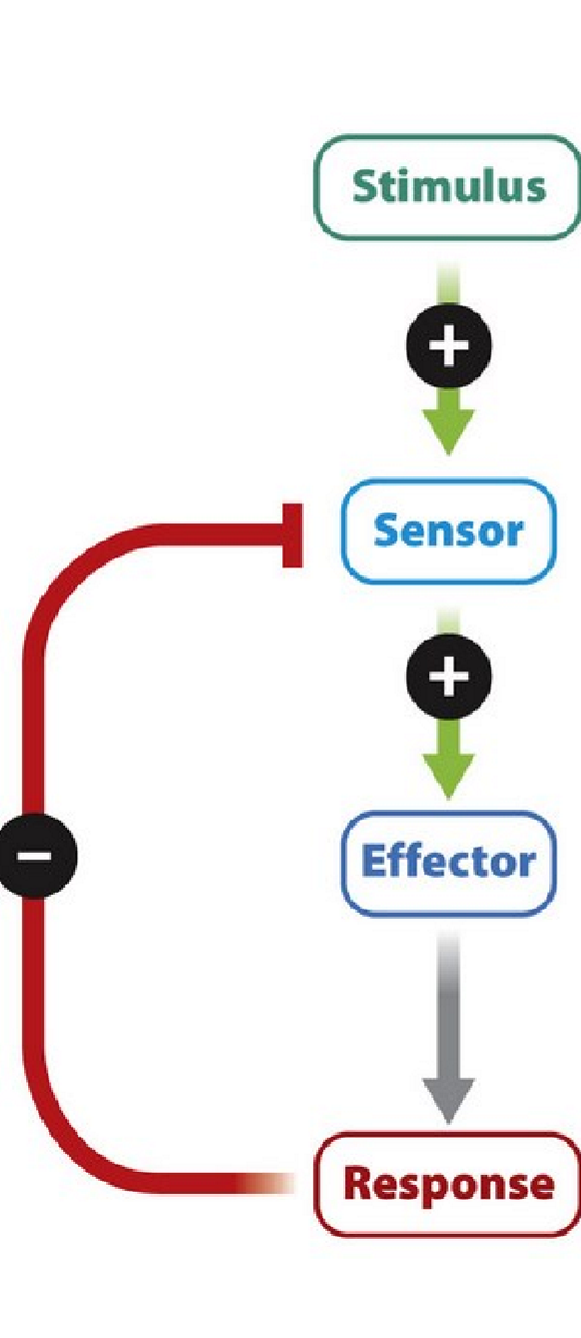
Identify the components of a homeostatic negative feedback system.
stimulus, sensor, effector, response
Explain how each component of a homeostatic negative feedback system contributes to maintaining physiological stability.
sensor senses a stimulus
sensor triggers effector
effector creates response
response stops stimulus
Differentiate between negative feedback and positive feedback, providing examples of each.
negative: response stops stimulus (calcium, blood glucose)
positive: response increases stimulus (childbirth, blood clotting)
Predict how components of a homeostatic system will change when part of the system is perturbed.
sensor may not sense stimulus
effector may not create response or create wrong response
Apply the concept of negative feedback to the process of thermoregulation.
too hot: brain tells blood vessels to dilate and skin to sweat, causing cooldown
too cold: brain tells blood vessels to constrict and muscles to shiver, causing warming up
Relate changes in environmental conditions to changes in physiological or behavioral responses to temperature regulation.
environment affects what temperature brain perceives
causes appropriate response to environment
Relate endocrine function to homeostatic regulation.
endocrine system is used to send signals from brain to body (effector)
Explain when and why hormones are released.
hormones are released in response to signals
hormones cause changes throughout the body
Define the different types of hormones.
peptides are made of chains of amino acids
amines are derived from aromatic amino acids
steroids are derived from cholesterol
Discuss the role that hormones play in the maintenance of homeostasis.
hormones cause changes as effectors
Explain the relationship between the hypothalamus and pituitary.
hypothalamus hormones cause pituitary to release hormones
Determine whether a particular hormone will interact with a cytosolic or membrane-bound receptor.
peptides and amines: membrane-bound
steroids: cytosolic
Predict which hormones have been released from the pituitary to elicit a specific tissue response.
anterior:
ACTH affects blood sugar
FSH causes sperm production or estrogen production
Growth hormone causes growth
LH causes ovulation or testosterone production
prolactin causes milk production
TSH causes thyroid to produce hormones
posterior:
ADH regulates water balance
oxytocin assists in labor and promotes mother-child bonding
Evaluate the consequences of altering a component of a hormone pathway.
too much response produced
not enough produced
Describe different vertebrate reproductive strategies.
ovipary: embryo develops in an egg outside the mother’s body
ovivipary: embryo develops in an egg inside the mother’s body
vivipary: embryo develops in the mother’s body
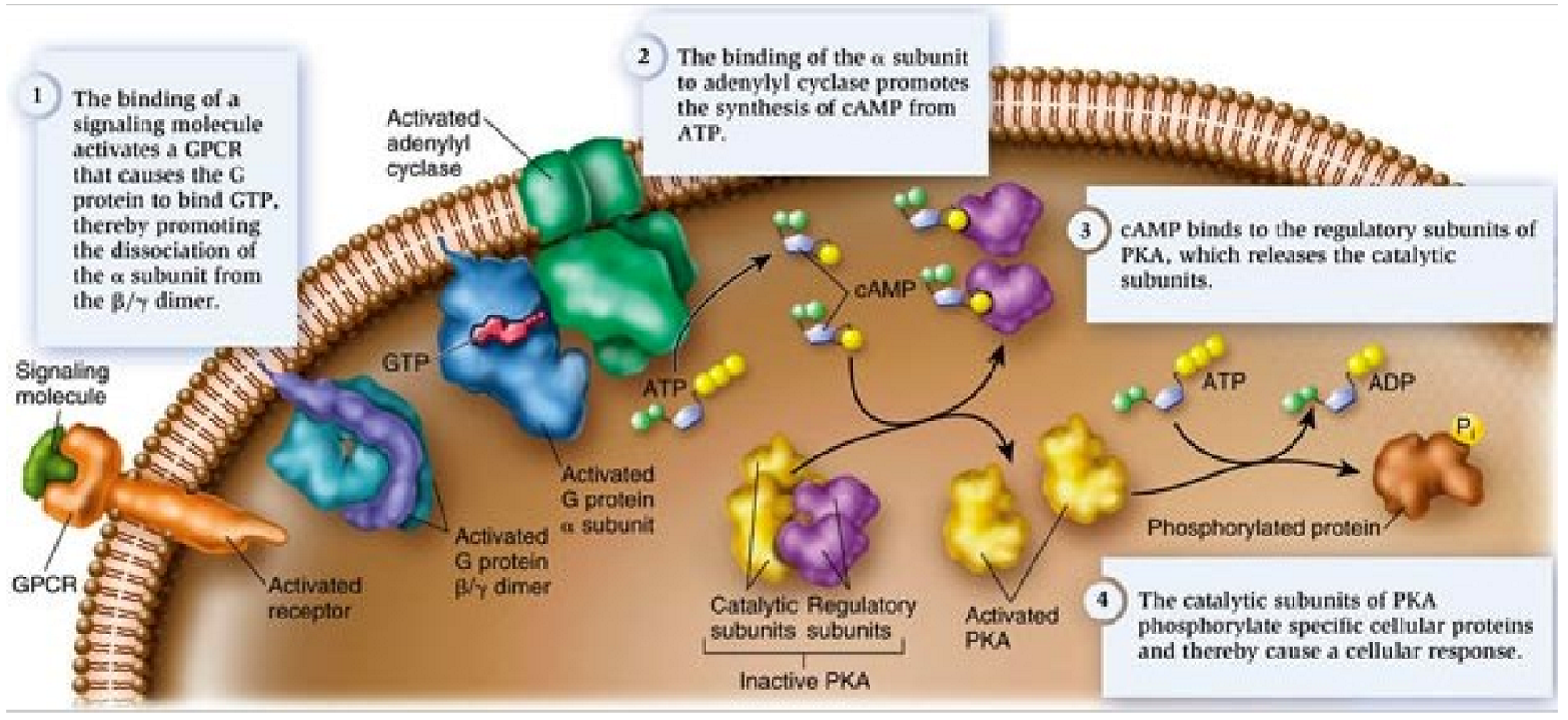
Describe the method of action for a G-protein coupled receptor pathway.
ligand binds to receptor, activating it
G-protein binds to activated receptor
G-proteins GDP is replaced with GTP, attached to the alpha-subunit, which detaches
alpha-subunit with GTP binds to adenylyl cyclase
adenylyl cyclase converts ATP into cAMP
cAMP activates PKA, causing response
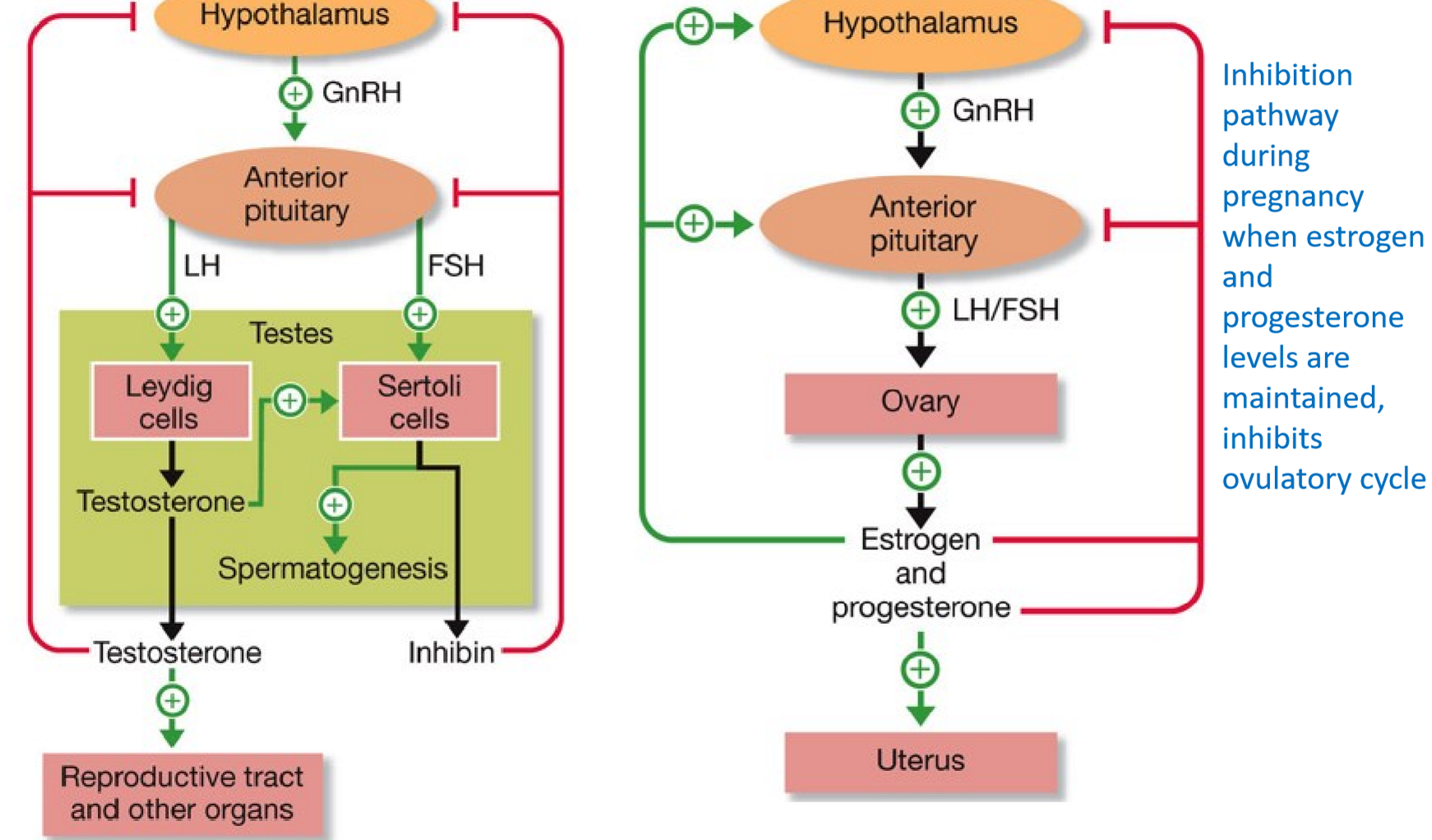
Identify critical components of the endocrine system involved in regulating the human reproductive system.
gonads contain testes/ovary
gametes (sperm or egg)
hormones: testosterone and estrogen or estrogen and progesterone
hormones target Leydig and Sertoli cells or granulosa cells
causes gametes to be released
Discuss the similarities and differences in hormonal control of male and female reproductive systems.
they use some of the same hormones but in different balances and with different responses
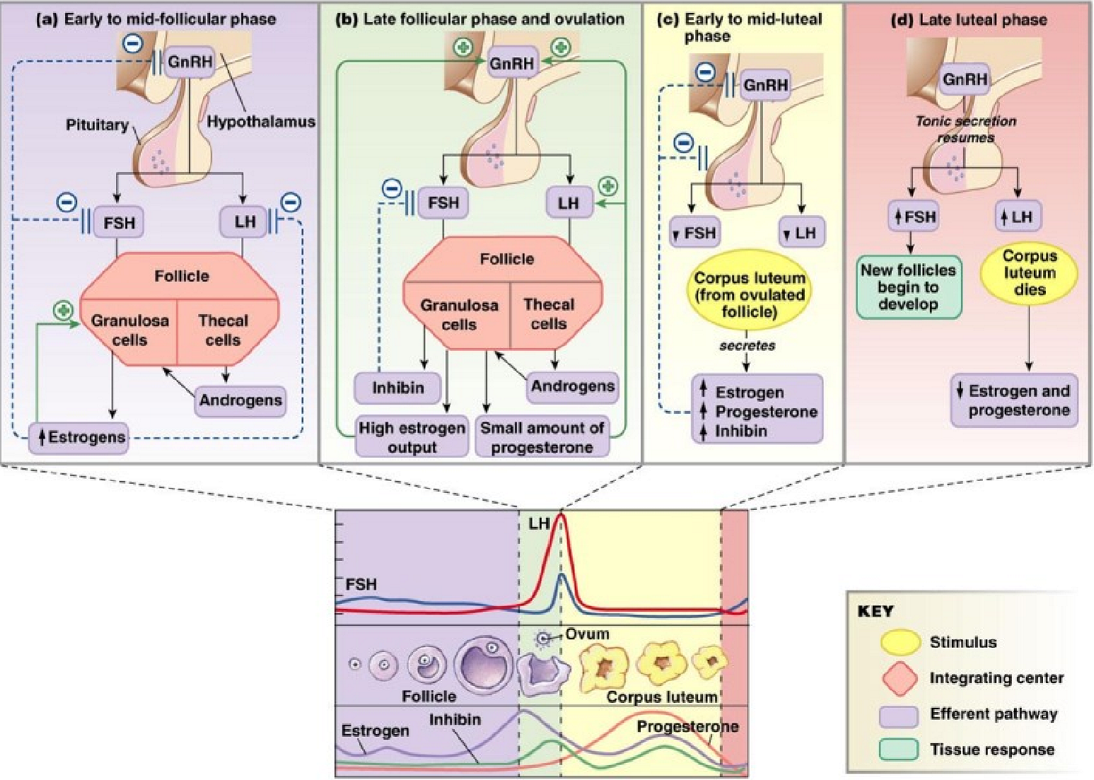
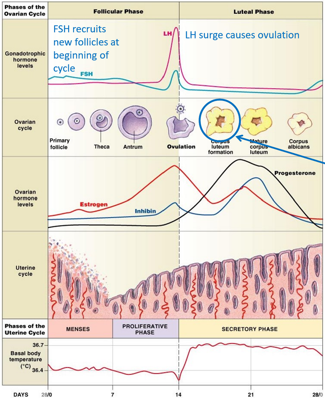
Describe the process of oogenesis and spermatogenesis.
oogenesis:
FSH and LH cause development of secondary follicles around oocyte
after LH surge, secondary oocyte splits off and undergoes meiosis II
ovulation
spermatogenesis
spermatogenic cells reach testes
after meiotic division, spermatids are formed
spermatids differentiate into sperm cells
Predict how changes in release of pituitary sex hormones will alter oogenesis and spermatogenesis.
no hormones = no production
Provide examples of organisms that rely on different modes of reproduction.
yeast: asexual budding
some fish: parthenogenesis
Distinguish between second messengers and other components of signal transduction pathways.
second messengers are intracellular and relay receptor cell signals to effector proteins
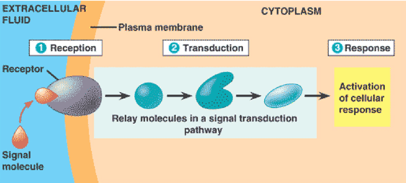
anatomy of a signaling pathway
anatomy of a signaling pathway
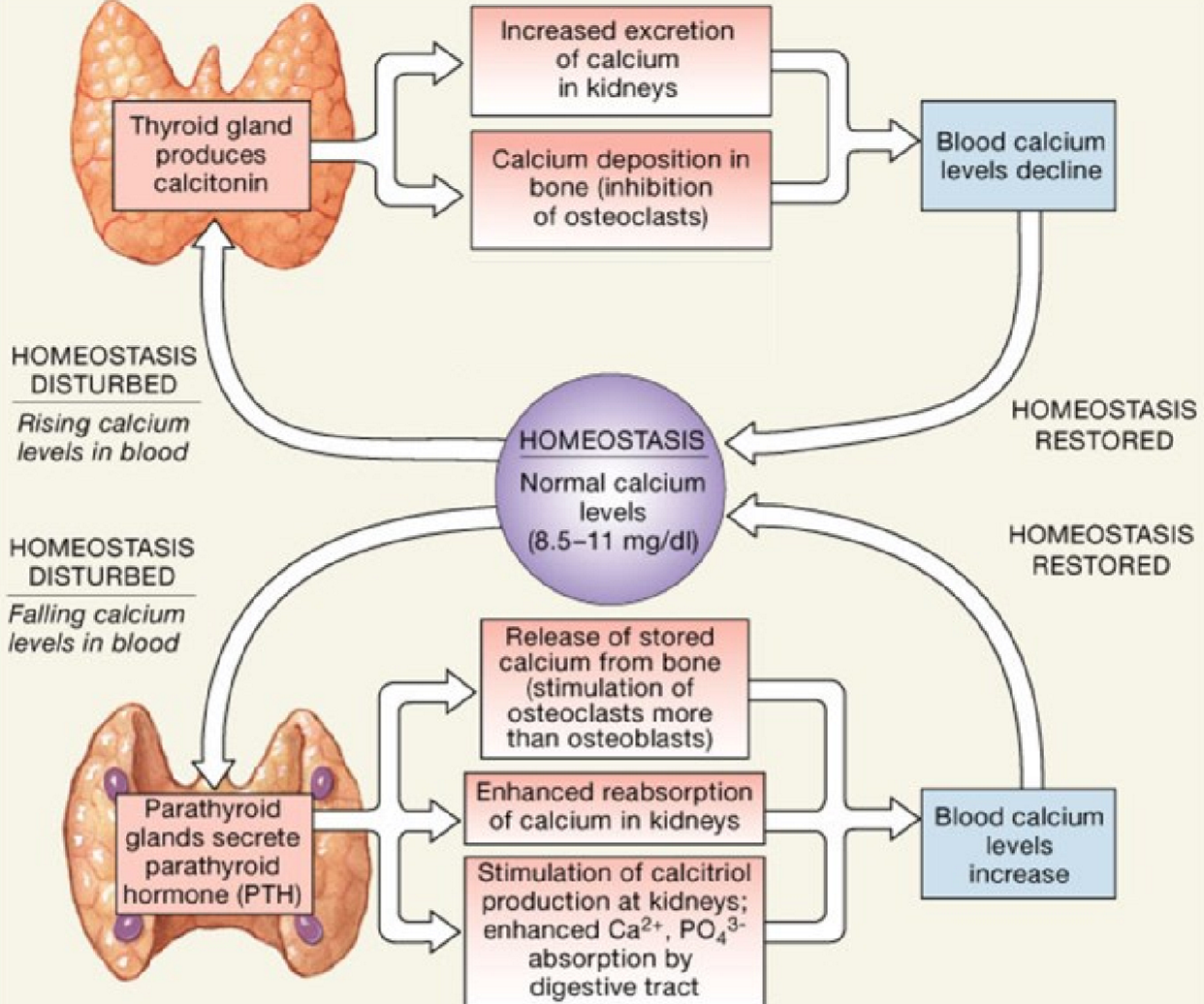
calcitonin and pth
calcitonin and pth
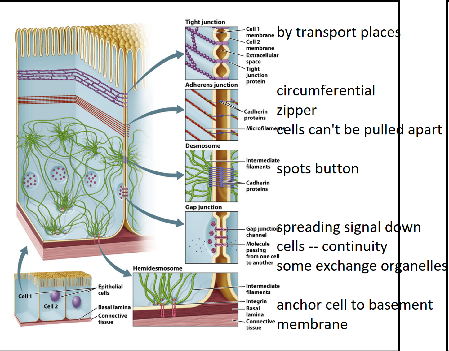
cell junction
cell junction
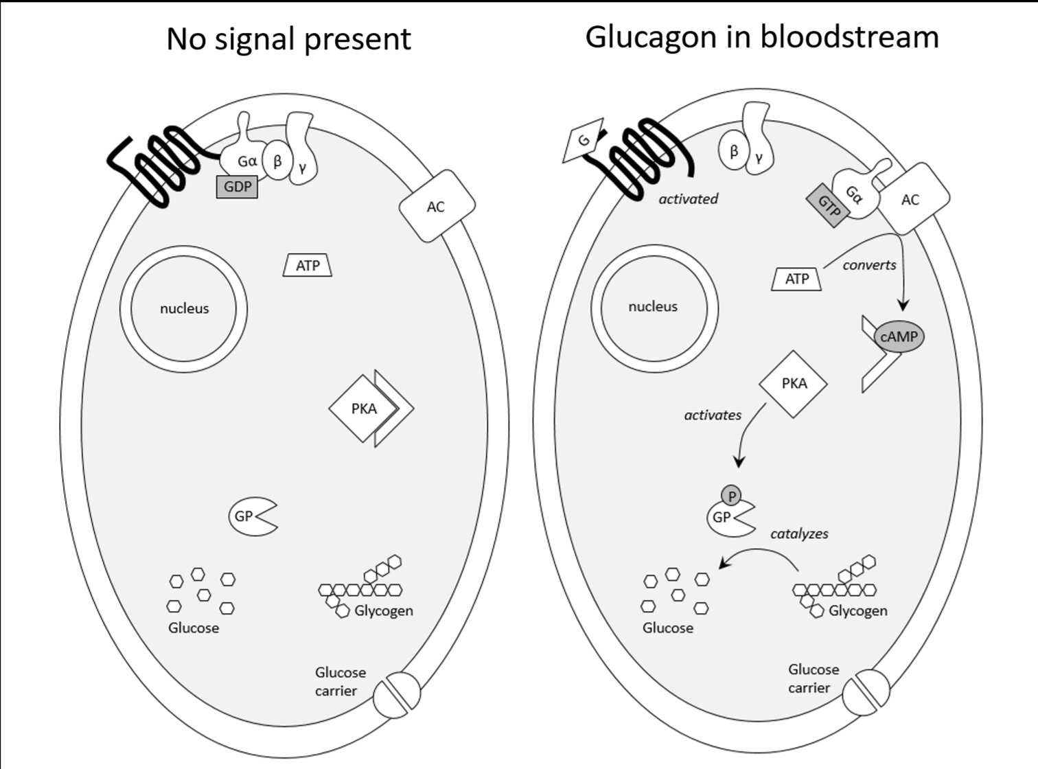
glucagon
glucagon
G-protein coupled receptor
G-protein coupled receptor
homeostasis
homeostasis
insulin
insulin
membrane potential
membrane potential
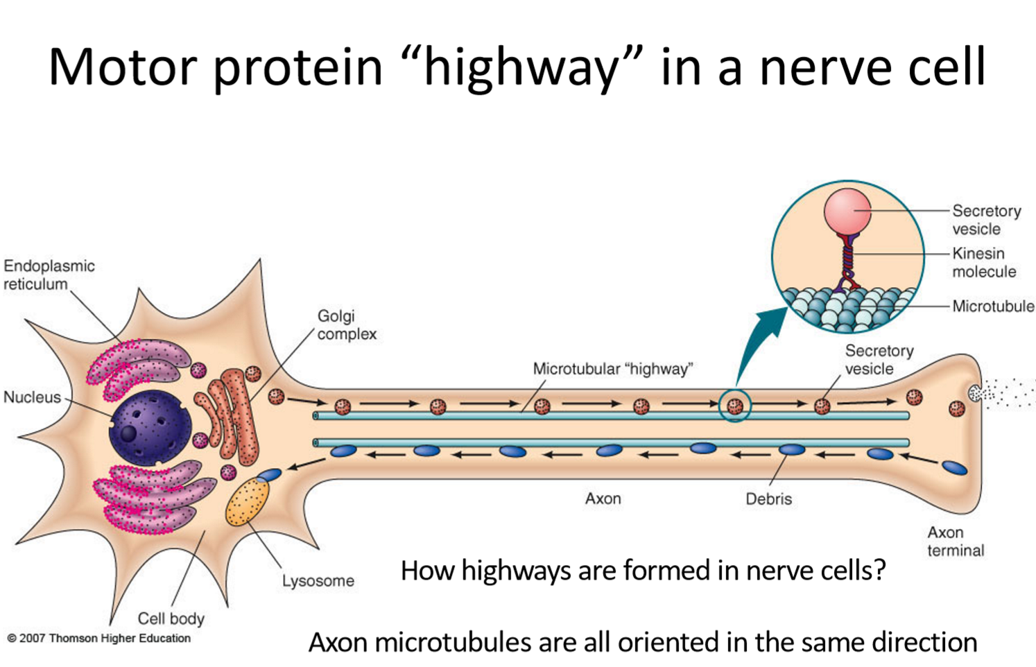
motor protein highway
motor protein highway
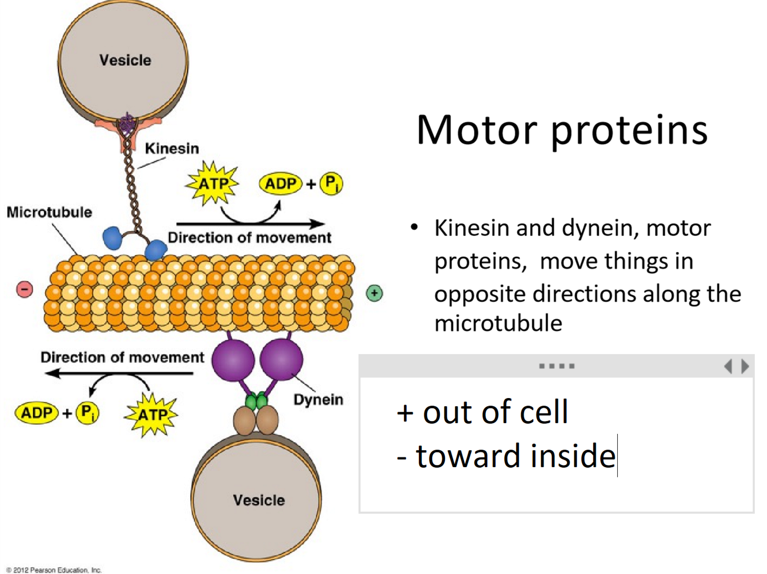
motor proteins
motor proteins
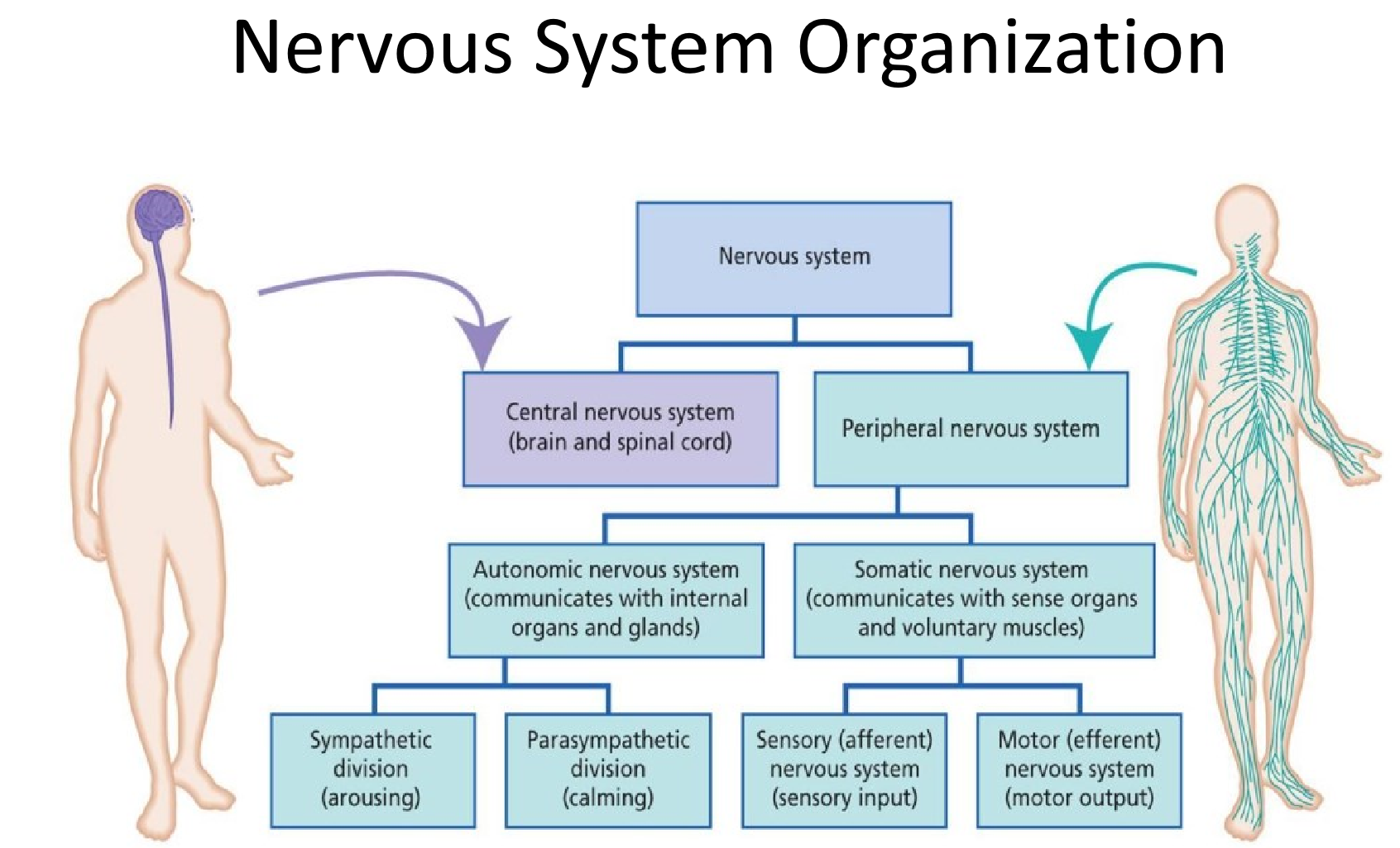
nervous system organization
nervous system organization
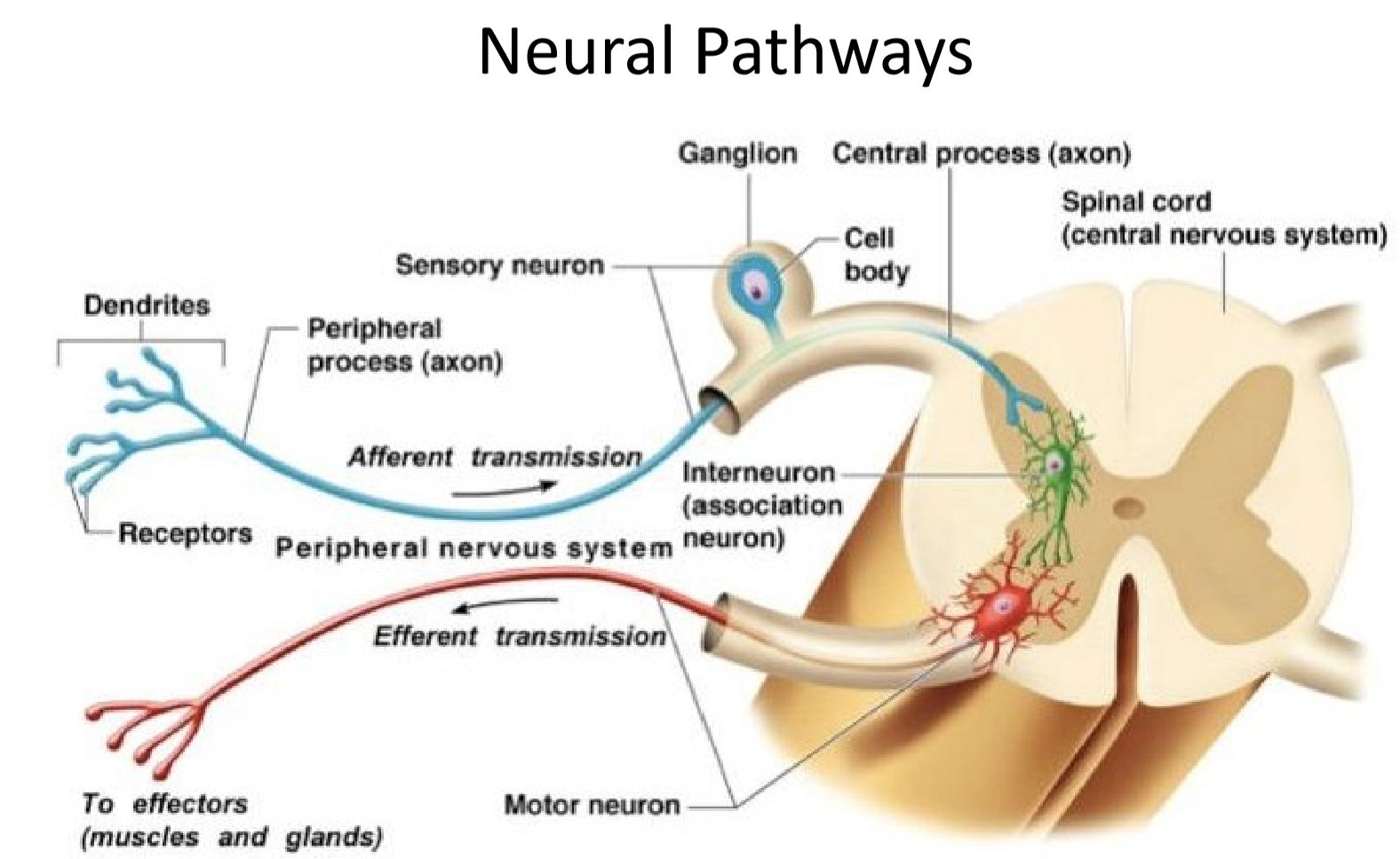
neural pathways
neural pathways

neurotransmitters
neurotransmitters
reproductive cycles
reproductive cycles
sex hormones and feedback loops
sex hormones and feedback loops
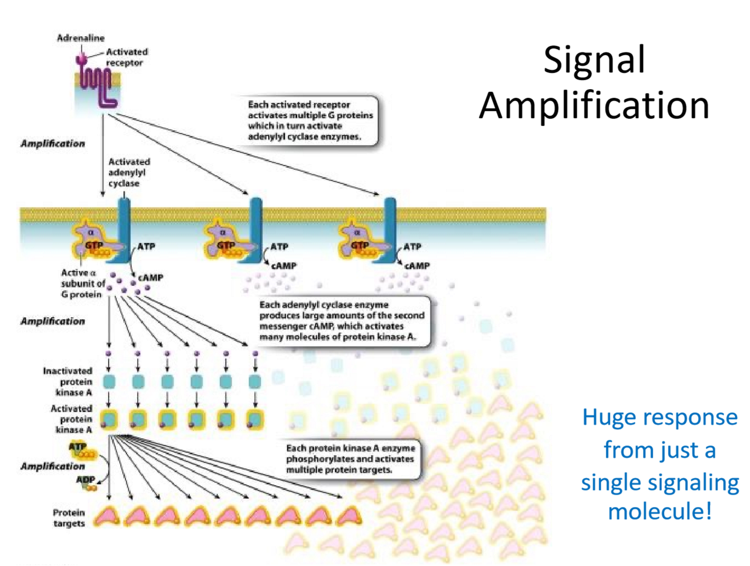
signal amplification
signal amplification
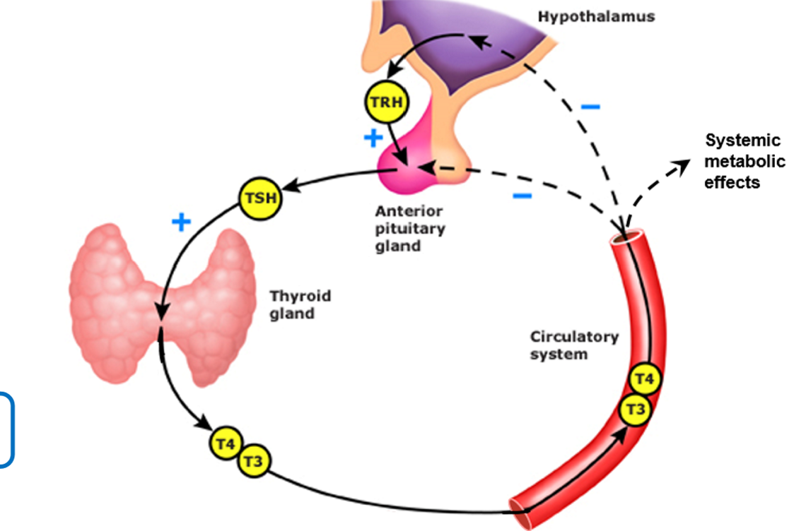
T3 and T4
T3 and T4
uterus
uterus
Define and differentiate between the terms microbiome, microbiota, metagenome, and holobiont
microbiome: organisms and their genetic material
microbiota: community of microbes living in a specific environment
metagenome: the sum of all genetic material of microorganisms
holobiont: combination of the host and all its microorganisms
Explain how and when the human microbiome is formed
formed at birth from environment of birth (skin microbiome of mother inherited in C-sections, vaginal microbiome of mother inherited in vaginal birth)