Musculoskeletal system and DNA
1/142
Earn XP
Description and Tags
For extended response term 2 wk 10
Name | Mastery | Learn | Test | Matching | Spaced |
|---|
No study sessions yet.
143 Terms
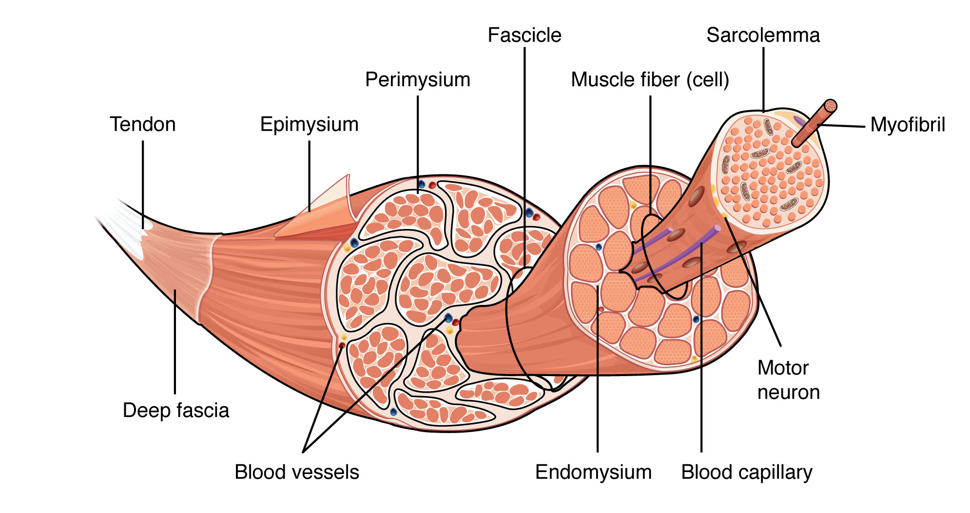
TEPFEMSMSM (structure of skeletal muscle hierarchy)
Tendon
Epimysium (holds bundles together)
Perimysium (surrounds each bundle so that it can function as an individual unit)
Fascicles (bundles of muscle fibres)
Endomysium
Muscle fibre (individual muscle fibres)
Sarcolemma
Myofibril (each muscle fibre consists of thin …)
Sarcomeres
Myofilaments (each myofibril consists of myofilaments that are either thick myosin or thin actin)
. other: blood vessels, blood capillaries and motor neurons
. muscle contains bundles of skeletal muscle fibres, covered in fascia (dense connective tissue) that becomes the tendon
Function of muscles
. decrease the distance between parts they’re connected to
. decrease the space they surround
. produce movement or generate tension
. principle function: contraction = shorten distance between bones
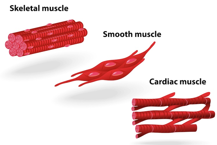
Skeletal muscle
. muscle attached to bone
. enable humans to move
. under voluntary control (contracts voluntarily)
. runs the entire length of the muscle
. has striated, tubular, multinucleated fibres
Fibres:
. occur in muscles attached to skeleton
. very elongated, striated, have many nuclei per cell, under voluntary control
. cells can be very large (2-3cm long + 100 micrometers in diameter in adult) and are referred to as muscle fibres because of their highly elongated shape
Smooth muscle
. located in the walls of hollow internal organs
. contract involuntarily (not under conscious control) to narrow the space they surround (intestines/arteries)
. visceral muscle
. has spindle shaped, non striated uninucleated fibres
Fibres:
. in the walls of visceral organs (stomach, liver, pancreas, intestines)
. appear spindle shaped, have a single nucleus per cell, under involuntary control
Cardiac muscle
. forms the heart wall to reduce the space in the chambers, forcing blood through to the vessels
. contracts involuntarily
. has striated, branched, uninucleated fibres
Fibres:
. make up walls of heart
. are branched, striated, have many mitochondria but a single nucleus, under involuntary control
What do the 3 types of muscles look like?
Functions of skeletal muscles (*clarify this is for skeletal MUSCLES not skeleton)
. support body
. movement of bones (most are attached to bone + move bones of the skeleton, but exception is facial muscles responsible for facial expression)
. maintenance of constant body temp
. assist movement in cardiovascular + lymphatic vessels
. protect internal organs
. stabilise joints
Characteristics of muscle tissue (EEEC)
. Excitability: can receive + respond to stimuli
. Extensibility: ability of muscle fibers to be stretched when pulled
. Elasticity: ability of muscle fibers to return to their original length after being stretched
. Contractability: cells can shorten (when stimulated) + thicken (when stimulation is removed cells relax and return to their original shape)
. muscle tissue consists of highly specialised, elongated cells that have elastic properties
Draw/label the skeletal muscle structure
What is the sarcolemma and where can it be found?
. a thin, transparent membrane surrounding a muscle cell
. the cell membrane of a muscle fiber (muscle cell)
. surrounds the entire muscle fiber
What is the sarcoplasm and where can it be found?
. the cytoplasm of striated muscle fibers
. found inside the sarcolemma
Which is bigger: myofibrils or myofilaments?
. myofibrils are bigger
—> are long, thread-like structures that run the length of the muscle fiber (found in the cytoplasm of muscle fibers)
. myofilaments (actin + myosin) are the thin + thick protein filaments that make up myofibrils of skeletal muscle fibres
Myofibril
. thread-like individual parallel muscle protein strands (containing actin + myosin) within sarcoplasm
What are myofilaments made of?
. protein
—> actin (thin filaments)
—> myosin (thick filaments)
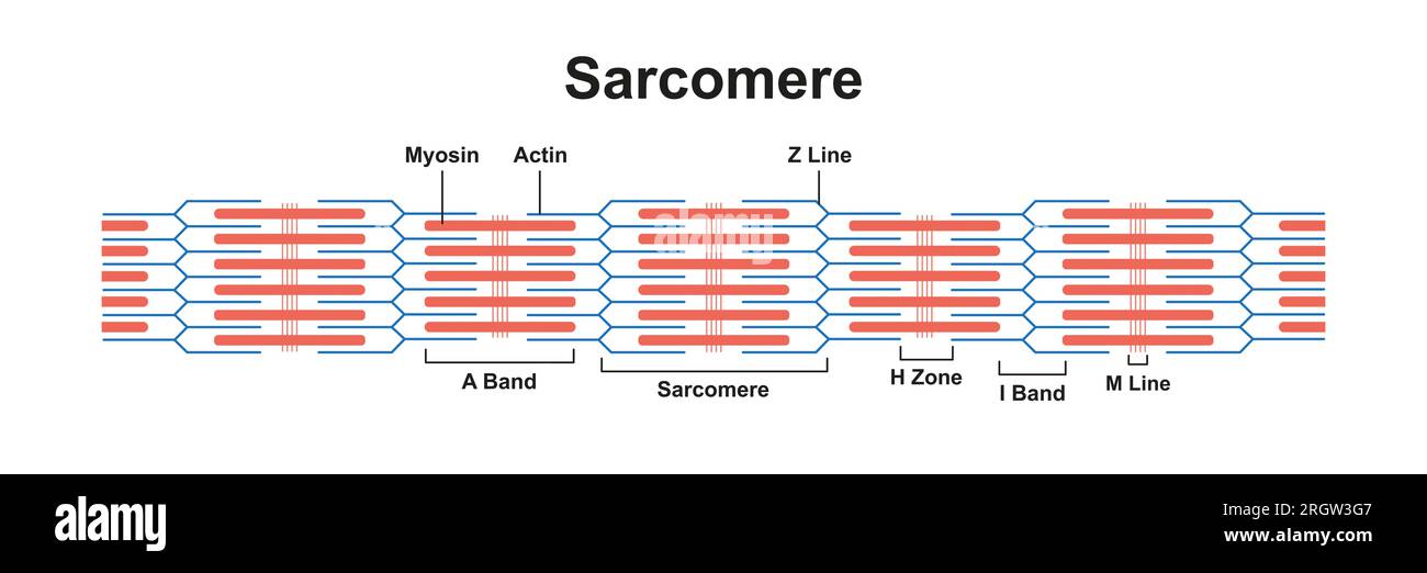
What are sarcomeres and where can they be found?
. the repeating contractile units (of striated muscle tissue) within a myofibril
. consists of actin + myosin filaments
. found between two Z lines in a myofibril + are responsible for the striated appearance of skeletal muscle and muscle contraction
Why does skeletal muscle appear striated
. because of the regular arrangement of actin + myosin filaments within sarcomeres
. the alternating light (I bands) and dark (A bands) give the muscle its striped appearance under a microscope
Fascicles
. muscle bundle surrounded by connective tissue
. a (skeletal) muscle contains bundles of skeletal muscle fibres (muscle cells)
. muscle fibre structure:
—> muscle cells occur in fascicles (bundles)
—> the fascicles are surrounded by a layer of tough connective tissue (perimysium) that allows adjacent bundles to slide over one another as they contract
. together bundles of fascicles make up a muscle
What are muscles covered with (describe fascia)
. muscles are covered with fascia (dense connective tissue) that becomes tendons
. Individual muscles are separated by fascia, that also forms tendons
. fascia is a thin casing of connective tissue that surrounds and holds every organ, blood vessel, bone, nerve fiber and muscle in place
. the tissue does more than provide internal structure: fascia has nerves that make it almost as sensitive as skin
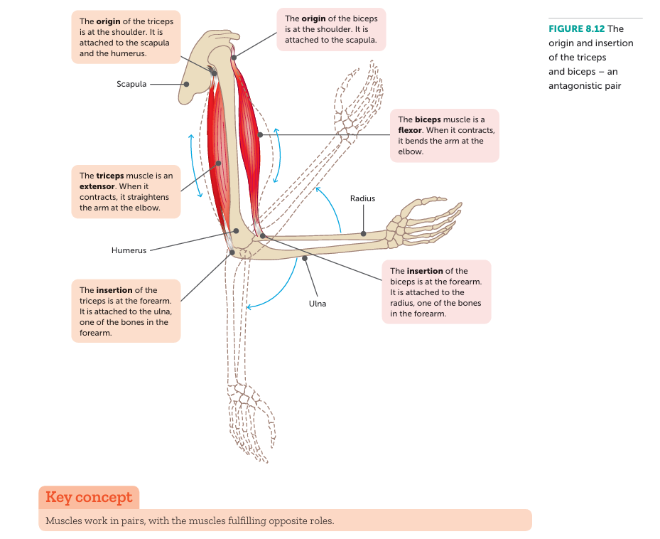
Where do muscles originate and insert
. muscles originate on the stationary bone (origin) and insert on the bone that moves (insertion)
What do cooperating muscles pairs have?
. prime movers, synergists and antagonists
In what way do muscles usually work in
. antagonistic pairs which work opposite one another to move in opposite directions
Agonist/prime mover
. muscle that causes the desired action (does the contracting)
Antagonist
. muscle that has an action opposite to that of the prime mover
Synergist/fixator
. assists agonist - groups work together (hamstring group contracts together to extend lower leg)
. synergist:
—> muscles that help the prime mover
—> a muscle that acts indirectly in steadying a joint during a particular movement
—> may produce same movement as prime mover OR steady a joint during a particular movement so that unwanted movement is prevented + the prime mover can function more efficiently
—> eg: wrist muscles during fist clenching
. Fixator: a muscle that contracts to immobilise a joint
—> acts as a stabiliser of 1 part of the body during movement of another part
—> EG: the many muscles that attach the scapula to the axial skeleton
Flexion
. a movement that decreases the angle between articulating bones
. to bend
. EG: when the elbow is flexed, the lower arm (with radius + ulna) moves closer to upper arm (with humerus)
. contraction causing upward movement
Extension
. lengthening of a muscle
. increases angle between articulating bones
. straightening of joint
. EG: when the knee is extended, the lower leg (with tibia + fibula) moves further away from upper leg (with femur)
Rotation
. movement of a bone around its long axis
. EG: moving head from left to right
Circumduction
. the circular movement of a limb/body part/joints, where the distal end moves in a circle, while the proximal end remains relatively fixed
Abduction
. movement of a limb away from the midline of the body
. EG: lifting arms up away from body = abduction at the shoulder joint
Adduction
. movement of a limb towards midline of the body
. EG: bringing arms inwards towards the body
Describe how skeletal muscles produce movement
. exert force on tendons
. attached to articulating bones, forming a joint
. when muscle contracts, 1 bone moves toward the other
Attachments of skeletal muscle (3)
. origin: end of a muscle that’s fixed to the stationary bone
. insertion: end of a muscle fixed to the movable bone
. belly: thicker + fleshy portion in the middle of a muscle (between tendons/origin + insertion)
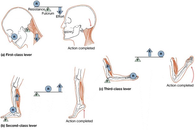
Levers
. rigid structures that move on a fixed point (fulcrum)
Describe lever systems in relation to bones and joints
. bones = levers
. joints = fulcrums
. levers are acted upon by resistance + effort
. leverage is responsible for a muscle’s strength + range of movement
Describe the 3 types of levers
1st class lever: fulcrum placed between effort + resistance
2nd class lever: fulcrum is at 1 end, effort is at opposite end, resistance is in between
3rd class lever: fulcrum is at 1 end, resistance is at opposite end, effort is in between
Describe the histology of skeletal muscle tissue (muscle fibres and stuff within)
. muscle fibres (the long cylindrical cells that make up skeletal muscles)
. sarcolemma
. sarcoplasm
. sarcoplasmic reticulum (a specialized type of smooth endoplasmic reticulum found in muscle cells, that regulates calcium ion concentration - calcium storage, release, reuptake organelle)
. T tubules
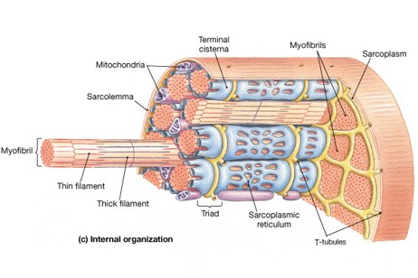
What do muscles fibres consist of
. myofibrils: run the entire length of a skeletal muscle fibre
What do myofibrils consist of?
. myofilaments (thin + thick filaments)
Myofilaments characteristics
. don’t extend entire length of fibre
. stacked in compartments = sarcomeres
. partitioned by Z lines
. A band (dark) contains thick + thin filaments
—> includes H zone, zone of overlap
. I band (light): thin filaments only
. H zone: thick filaments only
Describe thin filaments
. composed mostly of actin
. contain tropomyosin + troponin
. double stranded coil
Describe thick filaments
. composed mostly of myosin
. rod - shaped (tail) with head
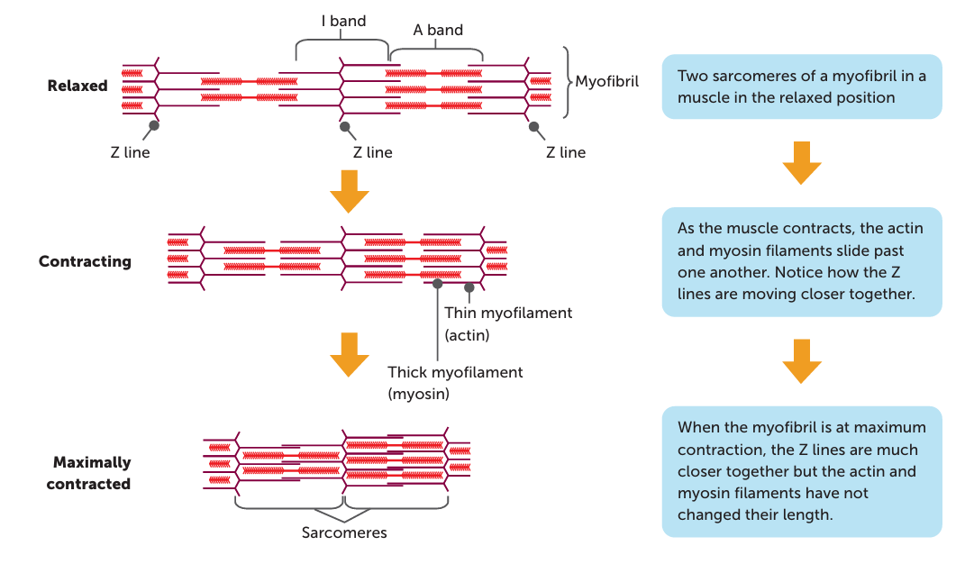
The sliding filament theory
. the theory used to explain what happens during muscle contraction, with actin and myosin filaments moving over one another and shortening the length of the muscle (sarcomeres within)
—> when muscle fibres are stimulated to contract, myofilaments (actin anchored on Z line and myosin anchored to M line) slide past one another, causing sarcomeres (muscle units) to shorten and the whole muscle fibre shortens
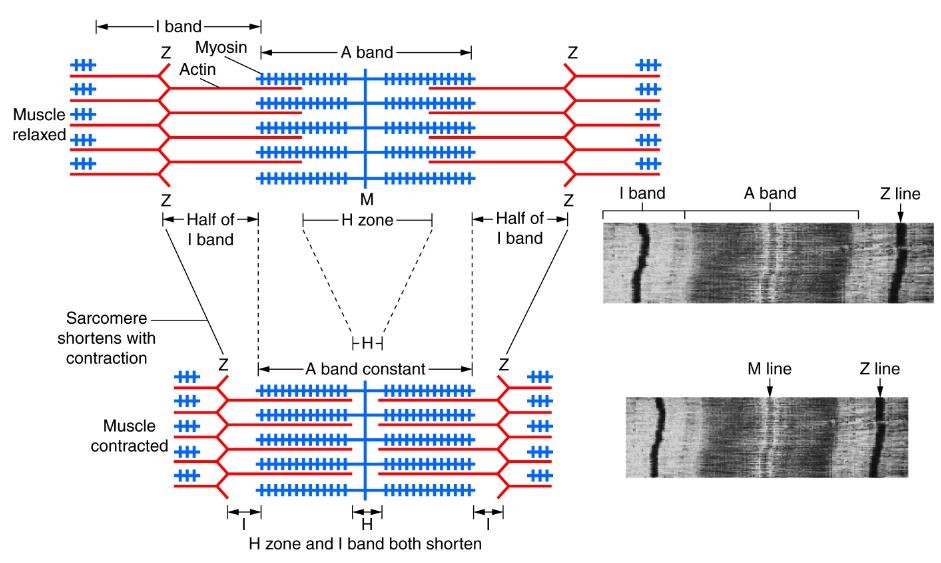
Describe the anatomy of a sarcomere
. composed of protein filaments, primarily actin (thin filaments) and myosin (thick filaments)
. Z lines:
—> protein discs at middle of thin filaments
—> boundaries of sarcomere
—> distance between successive Z lines = sarcomere
. A band:
—> length of thick filament (myosin)
—> darker band, containing actin + myosin and region where they overlap (at ends of A band)
—> middle is lighter as it contains thick filaments only
. H zone:
—> lighter area containing only thick myosin filaments (no overlap)
—> located within A band
. I band:
—> distance between successive thick filaments
—> contains only thin actin filaments
—> lighter band
. M line:
—> a dark line in the middle of the sarcomere (centre of H-zone) where myosin filaments are anchored together
. Actin (thin filaments):
—> filaments anchored to the Z-discs
—> extend towards the center of the sarcomere, interacting with myosin during contraction.
. Myosin (thick filaments):
—> filaments located in the A-band
—> have "heads" that can bind to actin and pull it during muscle contraction
Describe what happens at the sarcomere level during contraction (sliding filament theory)
. As the thin actin filaments slide inward over the thick myosin filaments toward H zone, Z lines drawn closer together toward the A band (distance between each Z line decreases) and sarcomere shortened
. Results in a shortening of the muscle fibres and hence a shortening of the whole muscle
. Myofilaments don't change size (meaning A band stays same length), just overlap more with gaps between filaments shortening (causing the fibril to shorten)
. I band reduces in length as the thick myosin filaments of each sarcomere draw closer together
. H zone is reduced in length as the actin filaments are brought closer together (occurs when the myosin heads bind to actin filament/cross bridges of thick filaments connect with actin + pull up and back, moving the actin filament backwards toward the M line, which stays the same size)
Draw and label what occurs during muscle contraction at the sarcomere level
What occurs during muscle relaxation, according to the sliding filament theory
. when muscle relaxes, actin + myosin filaments can be pulled past one another in the opposite direction and the muscle fibre returns to its original, uncontracted state
. muscle is lengthened at relaxation
Does muscle contraction need energy?
. YES: energy is required for shortening of the muscle fibres
. energy comes from the breakdown of ATP in the muscle cells
. energy is released when ATP breaks down to ADP and a phosphate group
. the breakdown of ATP deforms the heads of myosin molecules, with the simultaneous deformation of millions of myosin heads causing the myosin filament to crawl along the actin filament, resulting in the muscle cell getting shorter/contracting
Describe the 6 steps to the sliding filament theory
. don’t need?
Compare the structures of the 3 types of muscle
Skeletal muscle:
. striated (fibres contain alternating light and dark bands)
. fibres packed into regular parallel bundles
Cardiac muscle:
. striated
. bundles are branched but connected at both ends
. contraction not under conscious control (involuntary)
Smooth muscle:
. compared to skeletal, cells are small
. are spindle shaped + no striations
. have bundles of thin + thick filaments
Briefly describe the gross skeletal muscle structure
. muscles are composed of many fibres (cells) arranged in bundles (fascicles)
What are myofibrils made of
. contain the proteins actin + myosin:
. enable the cells to contract/shorten
. as the muscles are anchored to bones this contraction produces movement
. the arrangement of these proteins gives the muscles their striated look, creating dark + light bands
. the alternating dark + light bands of skeletal muscle fibres results from the overlapping bundles of actin + myosin filaments (thus myofibril = source of muscle fibre’s striations)
. A band = dark = myoSIN = thick filaments
. I band = light = actin = thin filaments
. actin = thin filaments
—> one of the contractile proteins of skeletal muscle, making up the thin myofilaments of a myofibril
. myosin = thick filaments
—> one of the contractile proteins of skeletal muscle, making up the thick myofilament of a myofibril
Describe skeletal myocyte microstructure
. muscle cells themselves are made up of fibres
—> contain numerous contractile cylindrical organelles = myofibrils
—> myofibrils consist of bundles of actin + myosin proteins (that give striations)
What are antagonistic muscle pairs
. groups of muscles that work in opposition to each other to produce movement at a joint
—> When one muscle contracts and shortens (agonist), the other relaxes and lengthens (antagonist), allowing for controlled movement
. most skeletal muscles work antagonistically in pairs/groups
. when the flexor/abductor muscle contracts, the antagonistic extensor/adductor muscle relaxes and vice versa
. smaller muscles that assist the primary antagonistic muscles = synergists
Explain how the triceps and biceps are antagonistic muscles
. they produce movements that are opposite
. triceps straightens arm, biceps bends arm
EG:
Flexion
—> the biceps (agonist) contracts to bend arm upwards
—> the triceps (antagonist) relaxes so arm can bend
Extension
—> triceps (agonist) contracts to straighten the arm
—> biceps (antagonist) relaxes so arm can straighten
Explain how the quadriceps and hamstring are antagonistic muscles
. they produce movements that are opposite
Flexion:
—> hamstring (agonist) contracts to bend lower leg
—> quadriceps (antagonist) relaxes and the leg bends
Extension:
—> quadriceps (agonist) contracts to straighten leg
—> hamstring (antagonist) relaxes to let lower leg straighten
Outline what muscle tone is and why it is useful/important
. maintaining partial contraction of skeletal muscles
. the amount of tension (resistance to movement) in muscles
—> helps us to hold our bodies upright when sitting + standing
—> changes in muscle tone are what enable us to move
—> contributes to the control, speed, and amount of movement we can achieve
Describe the features of the human skeleton in general
. humans have an internal skeleton called an endoskeleton
. consists of hard, mineralised structures located within the soft tissue of organisms
. adults have 206 bones
What is the skeletal system made up of (its parts)
. bones (skeleton) - and their associated structures:
. joints
. cartilages
. ligaments (bone to bone)
. tendons (bone to muscle)
5 functions of skeletal system
supports the weight of the body
protection of soft, vital internal organs
Movement due to attached skeletal muscles
Storage + release of minerals + fats
Blood cell formation (erythrocytes, leucocytes, thrombocytes)
Describe what the Axial skeleton is
. the part of the skeleton that forms and consists of the bones that lie around the central axis of the body
. provides the main support for erect posture + protection of the CNS and the organs contained within the thorax
. bones that make up are divided into 3 parts:
Bones that form the skull
Vertebral column
Ribs + sternum (together called bony thorax)
Describe what the Appendicular skeleton is
. the part of the skeleton made up of the bones of the upper and lower limbs, and the bones of the shoulders (pectoral girdle) and pelvis (pelvic girdle)
—> these 2 girdles allow for the articulation of the limbs with the axial skeleton
Describe the features of long bones
. longer than wide
. mostly compact bone
. have a thick outside layer of compact bone + an inner medullary cavity containing bone marrow
. have a shaft with heads at both ends
. ends contain spongy bone
. condyle = rounded bump
. trochanter = large bump like projection, larger than a tuberosity
. EG: femur, humerus
Describe the features of short bones
. cube-shape
. contain mostly spongy bone, surrounded by thin layer of compact bone
. EG: carpals, tarsals
Describe the features of flat bones
. thin + flattened
. thin layers of compact bone around a layer of spongy bone
. protect internal organs
. EG: skull, ribs, sternum, scapula
Describe the features of irregular bones
. irregular shape
. don’t fit into other bone classification categories
. primarily spongy bone that’s covered with a thin layer of compact bone
. EG: vertebrae, hip
Describe the features of sesamoid bones
. small bone commonly found embedded within a muscle or tendon near joint surfaces
. EG: patella
Describe bone as a connective tissue
. bone is a connective tissue
. connective tissues consist of cells separated from each other by large amounts of non-cellular material called matrix
. in bone tissue, inorganic salts of calcium + phosphate are deposited in the matrix
—> increase its rigidity + strength and make it the hardest of the tissues
Contrast the 2 categories of bone
Compact/cortical bone: a dense form of bone
Spongy/cancellous bone: bone that contains many large spaces, appearing spongy and consists of an irregular arrangement of bony plates (trabeculae)
Describe what a bone is made of
. bone is made of the matrix and cells
Bone matrix:
. non-cellular material between the cells of a tissue
. a combination of inorganic salts (eg: calcium + phosphate) and organic molecules (collagen)
—> inorganic:
. 70% of a bone is inorganic minerals
. they’re found in the matrix
. include hydroxyapatite (Ca10(PO4)6(OH)2) which contains calcium + phosphorus
. are other minerals too, but in smaller amounts
—> organic:
. 29% of a bone is made of collagen + other proteins
Bone cells:
. surrounded by the matrix
. 1% of a bone are cells such as osteoblasts, osteoclasts and osteocytes
Identify the structures of a long bone (what it consists of)
. diaphysis
. medullary cavity
. epiphysis
. periosteum
. articular cartilage
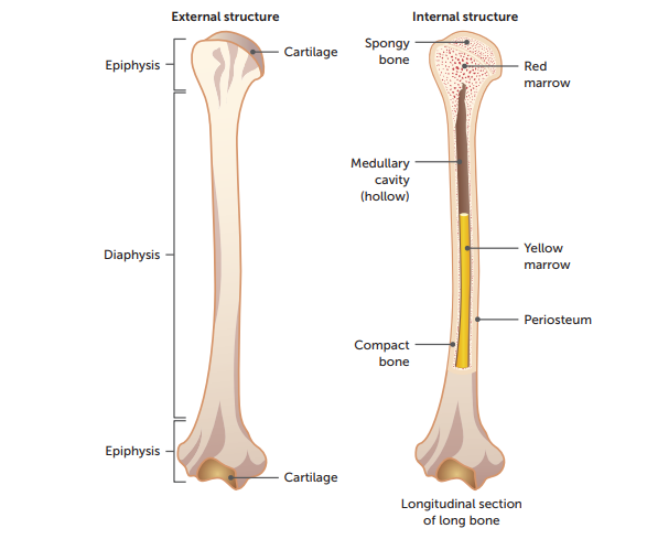
Structure of a long bone: Diaphysis
. shaft making up the main portion of the bone (middle)
. composed of a hollow cylinder of compact bone surrounding the medullary cavity
Structure of a long bone: Medullary Cavity
. cavity within the shaft/diaphysis
. contains yellow marrow which is used for fat storage (called yellow bone marrow cavity)
Structure of a long bone: Epiphysis/epiphyses
. enlarged ends of the bone
. covered by articular cartilage (thin layer of cartilage)
. composed mostly of spongy bone, surrounded by a thin layer of compact bone
. have compact bone on outside but their central regions contain spongy bone (is more porous than compact bone + contains many large spaces filled with marrow)
—> in certain bones, this may be red bone marrow, where blood cell production takes place
Structure of a long bone: Periosteum
. dense, white, fibrous outer covering of bone
. outside covering of diaphysis
. fibrous connective tissue membrane
. no periosteum at joints, where the bone is covered with an articular cartilage
. nutrient arteries branch off into networks supplying the bone with nutrients + oxygen
Structure of a long bone: Articular cartilage
. covers the external surface of the epiphyses
. made of hyaline cartilage
. decreases friction at joint surfaces
3 types of bone cells and their roles
Osteoblasts: bone-forming cells
Osteocytes: mature bone cells
Osteoclasts: bone-destroying cells, break down bone matrix for remodelling + release of calcium
Describe the general structure of compact bone
. consists of many similar units called osteons or haversian systems
—> run parallel to the long axis of the bone, giving the bone its max strength
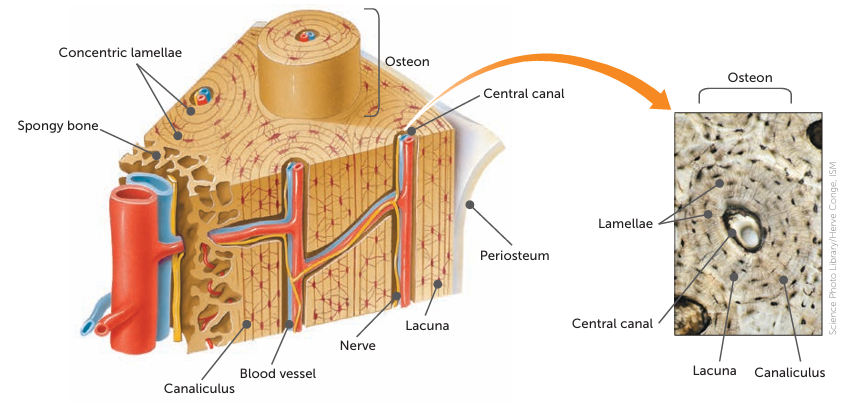
Osteon (the Haversian system)
. the basic unit of structure of compact bone
. consists of a central canal surrounded by concentric layers of hard matrix and bone cells
Describe the 6 main structures of an osteon
a central canal (haversian canal: a network of tubes in compact bone occupied by nerves + blood vessels) at its centre, containing at least 1 blood capillary and possibly nerves + lymph capillaries
Concentric layers of bony matrix called lamellae surrounding the central canal
Lacunae, which are small spaces in the matrix between the lamellae
an osteocyte occupying each lacuna
Tiny canals (canaliculi) running between the lacunae
Projections from the bone cells entering the canaliculi + making contact with adjacent bone cells, allowing materials to be passed from cell to cell
Describe the structure of spongy bone
. not organised into osteons with concentric layers of lamellae
. consists of an irregular arrangement of thin, bony plates (trabeculae)
. the bone cells occupy spaces in the trabeculae + nerves and blood vessels pass through irregular spaces in the matrix
. osteocytes are located in lacuna (like with compact bone)
. whereas compact bone tissue forms the outer layer of all bones, spongy bone forms the inner layer of all bones
. in compact bone, the lacunae + osteocytes are in concentric circles, whereas in spongy bone the lacunae + osteocytes are found in a lattice (like network of matrix spikes) = trabeculae —> between the trabeculae are spaces
. the spaces in some spongy bone contain red bone marrow, protected by the trabeculae where RBCs are made
Where does spongy bone occur and why is this useful?
. the trabeculae may appear to be a random network, but each trabecula forms along lines of stress to direct forces out to the more solid compact bone providing strength to the bone
. reduces the density of bone + allows the ends of long bones to compress as the result of stresses applied to the bone
. prominent in areas of bones that aren’t heavily stressed or where stresses arrive from many directions
Cartilage
. a type of flexible and firm connective tissue containing fibres of collagen
Describe cartilage as a connective tissue
. is a connective tissue
. connective tissues consist of cells separated from each other by large amounts of non-cellular material called matrix
. in cartilage, there’s a firm matrix made of a protein-carbohydrate called chondrin
Describe the main features of cartilage in general
. fibres of collagen protein are embedded in the firm matrix
. within the matrix are spaces that contain cartilage cells called chondroblasts which produce the matrix
. eventually these cells get trapped in small spaces (lacunae) where the cells become mature and called chondrocytes
. cartilage is avascular (doesn’t contain blood vessels) = has a low metabolic rate + heals slowly
. firm matrix enables cartilage to function as a structural support, while the presence of fibres gives cartilage a certain amount of flexibility
. found on the surface of bones at the joints, in the trachea + bronchi and forms the nose, larynx and outer ear
Why is cartilage classified into 3 types
. collagen fibres in matrix vary in thickness and arrangement, from extremely fine to quite coarse
. variation in the fibrous structure of cartilage is used to classify into 3 types:
Hyaline cartilage
Elastic cartilage
Fibrocartilage
Function of cartilage
. provides support and shape, cushions joints by absorbing shock, helps reduce friction between bones
Main differences between bone and cartilage
. unlike bone, cartilage is softer and doesn’t contain blood vessels (bones are vascular, having a rich blood supply)
What is cartilage made up of?
. made up of chondrocytes, collagen fibres and gel-like matrix called chondrin
Types of cartilage: Hyaline cartilage
. flexible supporting cartilage
—>contains many fine, closely packed collagenous fibres throughout matrix
—> fine fibres give the cartilage strength + flexibility (smooth as well)
EG:
—> C-shaped rings of trachea + bronchi made of
—> articular cartilage of joints is made of
. trachea, nose, end on bones
Types of cartilage: Elastic cartilage
. cartilage that contains elastic fibres
—> has conspicuous elastic fibres
—> contains collagenous fibres similar to those in hyaline but not so closely packed
—> provides flexible elastic support in places such as external ear
Types of cartilage: Fibrocartilage
. a type of cartilage that contains bundles of fibres
—> densest cartilage
—> coarse appearance from parallel bundles of thick collagenous fibres
—> fibres aren’t compacted as much as hyaline = can be compressed slightly
—> intervertebral discs of spinal column are made of, where it provides a cushion between the vertebrae, where there’s a need to withstand heavy pressure
Joints
. the connection between 2 bones
. site at which 2 or more bones come together
. articulation/movement of bones
. classified according to structure (functional + structural classification)
Functions of joints
. holds bones together
. allow for mobility
Fibrous/Fixed Joints
. a joint at which no movement occurs between the bones
. the bones are held in place by fibrous connective tissue
. EG: sutures of skull/between the bones of skull
. joint is fibrous, fixed, immovable
Cartilaginous joints
. joint at which only limited movement occurs between the bones
. bones are held in place by cartilage, allowing slight movement to occur
.EG: the junction of the 2 pelvic bones (pubic symphysis), joints between adjacent vertebrae, joints between ribs + sternum
Synovial joints
. freely movable joint
. the amount of movement possible is limited by ligaments, muscles, tendons and adjoining bones
. categorised by the type of movement that occurs between the articulating surfaces of the bones
Synovial joints: 1. Ball and socket joints
. form when the spherical head of 1 bone fits into a cup-like cavity of another
. EG: between hip + femur
Synovial joints: 2. Hinge joints
. allow movement in 1 plane only
. form when the convex surface of 1 bone fits into the concave surface of another
. movements include flexion + extension
. EG: between humerus + ulna
Synovial joints: 3. Pivot Joints
. formed when the rounded, pointed or conical end of 1 bone articulates with a ring, formed partly by bone and partly by a ligament
. EG: between vertebrae
Synovial joints: 4. Saddle joints
. where 2 bones forming the joint are both saddle-shaped (concave in 1 direction and convex in the other)
. movements include side to side + back and forth
. EG: between metacarpal and carpal