Histology of Salivary Gland

0.0(0)
studied byStudied by 0 people
learnLearn
examPractice Test
spaced repetitionSpaced Repetition
heart puzzleMatch
flashcardsFlashcards
Card Sorting

1/5

encourage image

There's no tags or description

Looks like no tags are added yet.

Study Analytics
Name
Mastery
Learn
Test
Matching
Spaced

No study sessions yet.

6 Terms

1
New cards
<p><span style="color: blue"><strong>Differentiate </strong></span><span style="color: purple"><strong>Serous</strong></span><strong> </strong>and <span style="color: green"><strong>Mucous</strong></span><span style="color: blue"><strong> Glands </strong></span></p><ul><li><p>Nucleus </p></li><li><p>Secretion <strong> </strong></p></li></ul><p></p>

Differentiate Serous and Mucous Glands

  • Nucleus

  • Secretion

Serous:

  • Nucleus: Round and circular

  • Secretion: Thinner

Mucous:

  • Nucleus: Flattened and basal

  • Secretion: Thicker

  • Cytoplasm: Foamy

<p><span style="color: purple"><strong><mark data-color="purple" style="background-color: purple; color: inherit"><u>Serous:</u></mark></strong></span></p><ul><li><p><strong><u>Nucleus:</u> </strong><span style="color: purple"><strong>Round</strong></span><strong> </strong>and <span style="color: purple"><strong>circular</strong></span></p></li><li><p><strong><u>Secretion:</u> </strong><span style="color: purple"><strong>Thinner</strong></span></p></li></ul><p><span style="color: green"><strong><mark data-color="green" style="background-color: green; color: inherit"><u>Mucous:</u></mark></strong></span></p><ul><li><p><strong><u>Nucleus:</u> </strong><span style="color: green"><strong>Flattened</strong></span><strong> </strong>and <span style="color: green"><strong>basal</strong></span></p></li><li><p><strong><u>Secretion:</u> </strong><span style="color: green"><strong>Thicker</strong></span></p></li><li><p><span style="color: #000000"><strong><u>Cytoplasm:</u> </strong></span><span style="color: green"><strong>Foamy </strong></span></p></li></ul><p></p>
2
New cards

Salivary Glands

  • Consist of

  • Originate from

  • 3 major salivary glands

    • Type of secretory cell

Consist of: Series of secretory units (glandular epithelium)

Originate from: Oral ectoderm, but grows in to underlying mesoderm

3 major salivary glands:

  • Parotid

    • Secretory cell: Serous

  • Sublingual

    • Secretory cell: Mucous

  • Submandibular

    • Secretory cell: Mixed

3
New cards

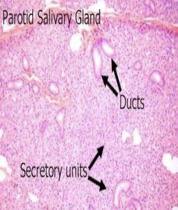
Histological Structure of Salivary Gland

  • What divides each lobules

  • What is lobule made up of

  • Types of ducts

What divides each lobules: Connective tissue capsule

What is lobule made up of: Secretory unit

Types of ducts: Intralobular and Interlobular

4
New cards

Intralobular vs Interlobular Duct

  • Location

  • Types

  • Function

  • Histology

Intralobular Duct:

  • Location: Inside lobule

  • Types: Intercalated and striated ducts

  • Function:

    • Intercalated: Collect secretions

    • Striated: Modify secretions

  • Histology: Simple cuboidal (one layer of cells)

Interlobular Duct:

  • Location: Between lobules

  • Function: Collects from multiple intralobular ducts and transports it towards main excretory duct

  • Histology: Stratified columnar (multiple layers of cell)

5
New cards

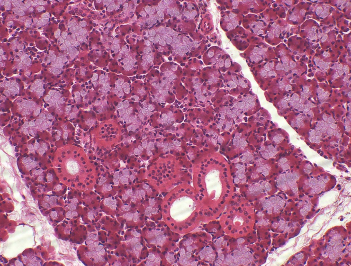
Salivary Gland: Parotid Gland #ff8e00

  • Compose of what secretory unit

  • Lobules separated by

    • Contains

  • Secrete into

    • Lined with

Compose of what secretory unit: Serous secretory unit

Lobules separated by: Loose connective septa

  • Contains:

    • Nerves

    • Blood vessels

    • Larger secretory ducts

Secrete into: Intercalated ducts

  • Lined with: Low cuboidal epithelium

<p><strong><u>Compose of what secretory unit:</u> </strong><span style="color: #ff8e00"><strong>Serous secretory unit</strong></span></p><p><strong><u>Lobules separated by:</u> </strong><span style="color: #ff8e00"><strong>Loose connective septa</strong></span></p><ul><li><p><strong><u>Contains:</u></strong></p><ul><li><p><span style="color: #ff8e00"><strong>Nerves </strong></span></p></li><li><p><span style="color: #ff8e00"><strong>Blood vessels </strong></span></p></li><li><p><span style="color: #ff8e00"><strong>Larger secretory ducts</strong></span></p></li></ul></li></ul><p><strong><u>Secrete into:</u> </strong><span style="color: #ff8e00"><strong>Intercalated ducts</strong></span></p><ul><li><p><strong><u>Lined with:</u></strong><span style="color: #ff8e00"><strong> Low cuboidal epithelium </strong></span></p></li></ul><p></p>
6
New cards
<p><span style="color: blue"><strong><mark data-color="blue" style="background-color: blue; color: inherit">Salivary Gland:</mark></strong></span><strong> </strong><span style="color: purple"><strong>Submandibular Gland</strong></span></p><ul><li><p>Type of secretion </p></li><li><p>Lobules separated by</p><ul><li><p>Contains</p></li></ul></li><li><p>Which cells form main lining </p></li><li><p>Which cells form periphery lining </p><ul><li><p>When grouped together, they are called </p></li></ul></li><li><p>Each secretory unit surrounded by </p><ul><li><p>Function</p></li></ul></li></ul><p></p>

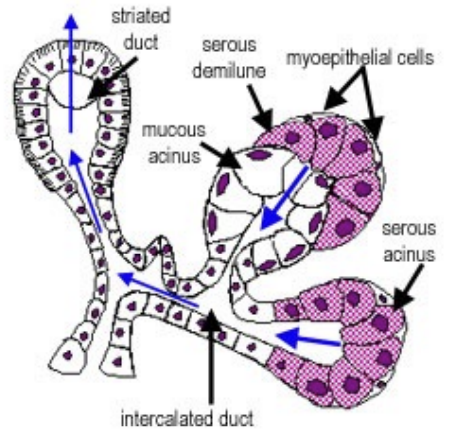
Salivary Gland: Submandibular Gland

  • Type of secretion

  • Lobules separated by

    • Contains

  • Which cells form main lining

  • Which cells form periphery lining

    • When grouped together, they are called

  • Each secretory unit surrounded by

    • Function

Type of secretion: Mixed, either serous or mucous

Lobules separated by: Loose connective septae

  • Contains:

    • Nerves

    • Blood vessels

    • Secretory ducts

Which cells form main lining: Mucous secreting cells

Which cells form periphery lining: Serous secreting cells

  • When grouped together, they are called: Serous demilune

Each secretory unit surrounded by: Contractile myoepithlial cells

  • Function: Contract and squeeze contents of lumen into duct

<p><strong><u>Type of secretion:</u> </strong><span style="color: purple"><strong>Mixed</strong></span><strong>, </strong>either <span style="color: purple"><strong>serous</strong></span><strong> </strong>or <span style="color: purple"><strong>mucous</strong></span></p><p><strong><u>Lobules separated by:</u> </strong><span style="color: purple"><strong>Loose connective septae</strong></span></p><ul><li><p><strong><u>Contains:</u></strong></p><ul><li><p><span style="color: purple"><strong>Nerves</strong></span></p></li><li><p><span style="color: purple"><strong>Blood vessels</strong></span></p></li><li><p><span style="color: purple"><strong>Secretory ducts</strong></span></p></li></ul></li></ul><p><strong><u>Which cells form </u></strong><span style="color: green"><strong><mark data-color="green" style="background-color: green; color: inherit"><u>main</u></mark></strong></span><strong><u> lining:</u> </strong><span style="color: green"><strong>Mucous secreting cells</strong></span></p><p><strong><u>Which cells form </u></strong><span style="color: blue"><strong><mark data-color="blue" style="background-color: blue; color: inherit"><u>periphery</u></mark></strong></span><strong><u> lining:</u> </strong><span style="color: blue"><strong>Serous secreting cells</strong></span></p><ul><li><p><strong><u>When grouped together, they are called:</u> </strong><span style="color: purple"><strong>Serous demilune</strong></span></p></li></ul><p><strong><u>Each secretory unit surrounded by:</u> </strong><span style="color: purple"><strong>Contractile myoepithlial cells</strong></span></p><ul><li><p><strong><u>Function:</u> </strong><span style="color: purple"><strong>Contract</strong></span><strong> </strong>and <span style="color: purple"><strong>squeeze</strong></span><strong> </strong>contents of <span style="color: purple"><strong>lumen</strong></span><strong> </strong>into <span style="color: purple"><strong>duct</strong></span></p></li></ul><p></p>