Neurobiology: Neuronal Signalling
1/14
There's no tags or description
Looks like no tags are added yet.
Name | Mastery | Learn | Test | Matching | Spaced |
|---|
No study sessions yet.
15 Terms
What is the structure (3) and function of a neuron
Neurons are specialized cells of the brain that transmit electrical and chemical signals throughout the nervous system.
Excitable cells → communicate via Action Potential following EPSP and IPSP
Neuron signaling → Chemical (NT release) and electrical (Gap Junctions) + synapse (junction between the terminal end of an axon and another cell)
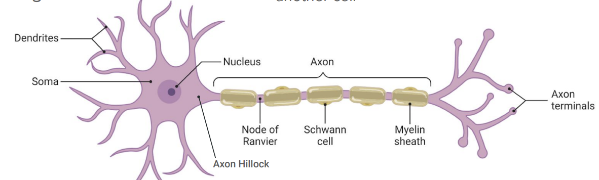
Their structure includes 3 major parts
Dendrites for receiving signals
branched extensions of soma
Soma (Cell body) for processing information
contains cells organelles
axon hillock
nissel bodies (RER + Ribosomes)
Axon for sending signals to other neurons or muscles
contains various ion channels
nodes of ranvier (unmyelinated segment with ion channels)
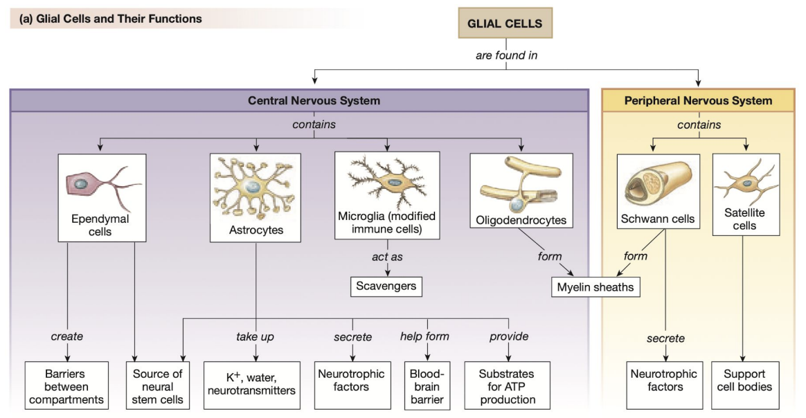
What does CNS and PNS mean
CNS stands for Central Nervous System, which includes the brain and spinal cord
PNS stands for Peripheral Nervous System, consisting of all the nerves outside the CNS that connect the body to the brain and spinal cord.
What are glial cells and how do they each contribute supporting the neural environment
Glial cells are non-neuronal cells in the nervous system that provide support, modifications, nourishment, and protection to neurons
non-excitable cell group
many types based on location and function
all originate form neural stem cell
They include astrocytes, oligodendrocytes, and microglia, each playing unique roles in maintaining homeostasis, myelinating axons, and responding to injury
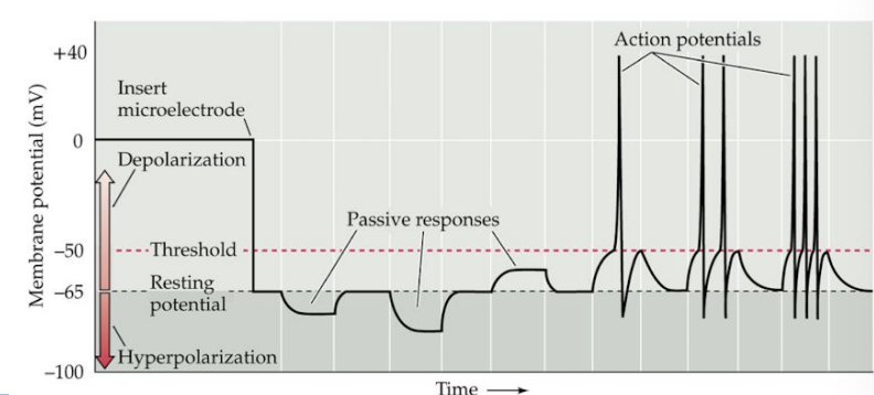
What is an Action Potential and what are its characteristics
An action potential is a rapid all-or-nothing electrical signal that travels the length of a neuron in order to communicate with another neuron, muscle, or gland
Its characteristics include:
All-or-nothing principle
Unidirectional
Communication from Soma to axon terminal
Lasts about 1-2 milliseconds
Constant strength (amplitude)
Frequency encodes strength of stimulus
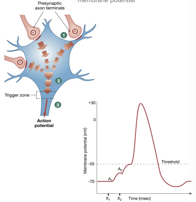
Hows does signal integration affect the generation of Action Potentials
Signal integration refers to the process by which a neuron combines excitatory and inhibitory signals from its dendrites to determine whether to generate an action potential
This involves summing the inputs and, if the membrane potential reaches a certain threshold, triggering the rapid depolarization and re-polarization that comprise the action potential
Summation: to add together
Graded potential: small localized change in membrane
spatial summation: graded potentials from multiple synapses converge to summate or cancel
temporal summation: graded potentials from one synapse occurring at slightly different times to summate or cancel
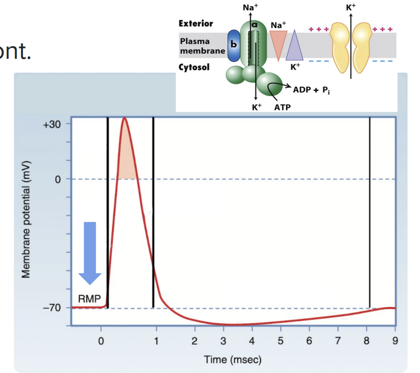
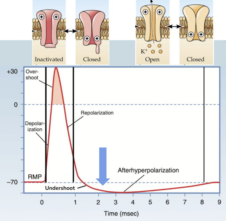
What are the phases of an action potential along with its ion activity and driving forces for ion movement
The phases of an action potential include resting membrane potential (RMP), depolarization, repolarization, hyperpolarization, and refractory periods
What is resting membrane potential (RMP)
The resting membrane potential (RMP) refers to the net charge difference across the neuronal membrane when the neuron is not actively transmitting signals
Net charge difference between intracellular and extracellular fluid at rest
-70 mV
Na+ /K+ ATPase: P-class pump & 3 Na+ out/ 2 K+ in
K+ leak channels (Ion channel)
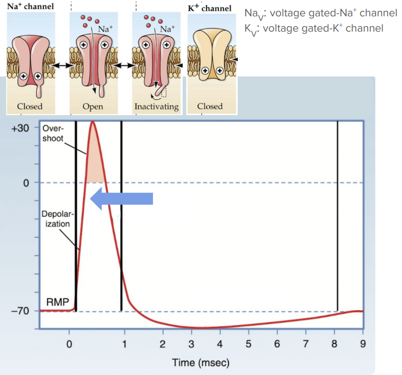
What is depolarization
The phase during an action potential where the membrane potential becomes more positive due to the influx of Na+ ions, reaching a threshold that triggers further action potential propagation
Only occurs if Threshold is reached (-55mV)
NaV open for Na+ influx
2 driving forces: Concentration gradient + Equilibrium potential
KV channels begin to open
Overshoot after 0mV → Na+ channels inactive
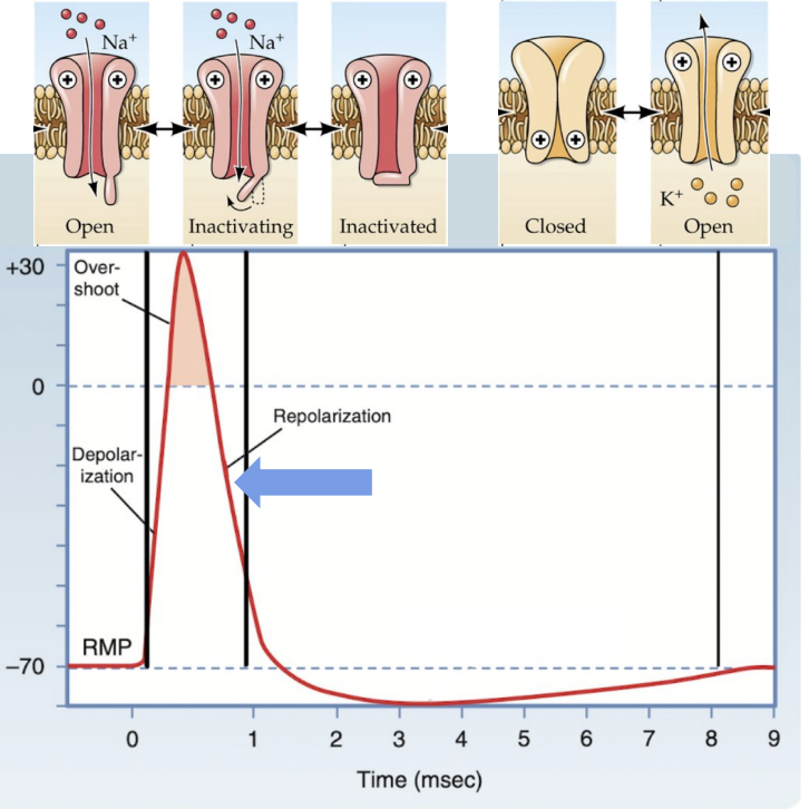
What is repolarization
The phase following depolarization where the membrane potential returns to a more negative value, primarily due to the efflux of K+ ions as K+ channels open
this process helps restore the resting membrane potential and prepares the neuron for the next action potential.
KV channels open for K+ efflux (2 driving forces)
Delayed rectifiers
NaV still inactivated
What is hyperpolarization
The phase during an action potential where the membrane potential becomes more negative than the resting potential, typically due to the continued efflux of K+ ions or influx of Cl- ions, making the neuron less likely to fire an action potential
Undershoot below -70mV
KV channels slowly close
NaV channels closed but not inactivated
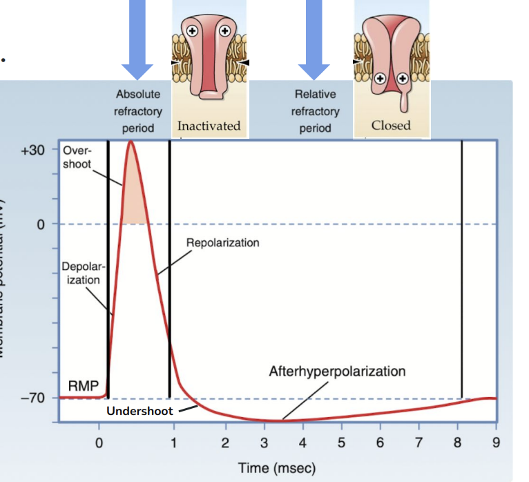
What are refractory periods
The periods following an action potential where a neuron is less responsive to stimulation
this includes the absolute refractory period, where no new action potential can be generated, and the relative refractory period, where a stronger stimulus is required to elicit an action potential
two types:
Absolute refractory period: No subsequent opening of NaV channels and promotes unidirectionality
Relative refractory period: NaV channels can open with sufficient stimulus
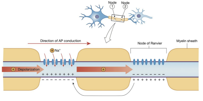
How can speed of conduct be affected
by factors such as axon diameter and myelination
larger diameters and myelination increase conduction velocity, allowing faster signal transmission.
Saltatory conduction: rapid propagation of action potentials along myelinated axons, where the electrical signal “jumps” from one Node of Ranvier to the next instead of traveling continuously down the axon
Combination of passive and active flow
Dramatically increases speed of neural signalling
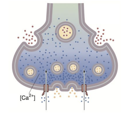
What are the steps in neurotransmitter release, what proteins are used, and what are the common results of neurotransmitter binding
Neurotransmitter release involves steps such as the action potential arriving at the axon terminal, calcium influx through voltage-gated calcium channels, vesicle fusion with the presynaptic membrane, and release of neurotransmitters into the synaptic cleft
Proteins like SNARE and synaptotagmin facilitate vesicle docking and fusion, while neurotransmitter binding commonly results in excitatory or inhibitory postsynaptic potentials.
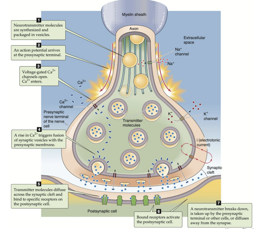
What are the 2 neurotransmitter and receptor types
Conventional NT (all different sizes)
Synthesized and stored within the presynaptic terminal
Released in response to depolarization and Ca2+ mediated
NT receptor exists on the postsynaptic membrane
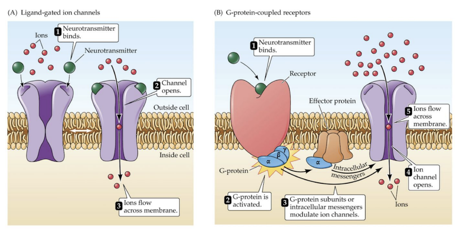
Types of receptors: Ionotropic and Metatropic
How do ionotropic and metabotropic receptors differ from each other
Ionotropic receptors are ligand-gated ion channels that mediate fast synaptic responses
Metabotropic receptors are G protein-coupled receptors that trigger slower, modulatory effects through second messenger systems.