Cell Structure
1/85
There's no tags or description
Looks like no tags are added yet.
Name | Mastery | Learn | Test | Matching | Spaced | Call with Kai |
|---|
No analytics yet
Send a link to your students to track their progress
86 Terms
How are cells organised?
Groups of cells are organised into tissues
Tissues are organised into organs
Organs may be organised into organ systems
Cell definition
Smallest, basic membrane bound unit of life
Responsible for all of life’s processes
4 aspects of cell theory
All living organisms are composed of one or more units called cells
Each cell is capable of maintaining its vitality independently
Cells can only arise from other cells
Viruses are not cells
What things does a cell require in order to continue living?
Metabolic machinery capable of obtaining energy from the environment e.g. light energy, degradation of foodstuffs
The ability to use this energy to support essential life processes e.g. movement of materials from one part of the cell to another, transport of molecules in and out of the cell
A set of genes to control the synthesis of all the cell components » these are passed onto the next gen through cell division
A physical boundary between itself and the environment » the cell membrane
Stem cell definition
A type of cell that can produce other cells which can develop into any kind of cell in the body
Types of cells
Prokaryotic » bacteria
Eukaryotic » animal, plant, fungi
How many cells are present in a human body?
From 1 to 1012
What is every living cell surrounded by?
A plasma membrane
Inside is a viscous fluid, the cytoplasm
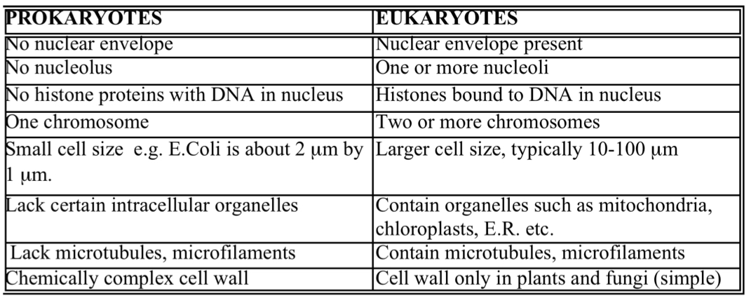
Differences between prokaryotes and eukaryotes
Bacteria (prokaryotes)
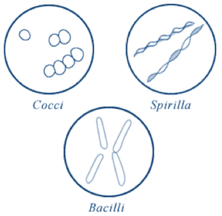
3 major shapes of bacteria cells
Coccus (round)
Bacillus (rods)
Spirilla (coils)
Bacteria (prokaryotes)
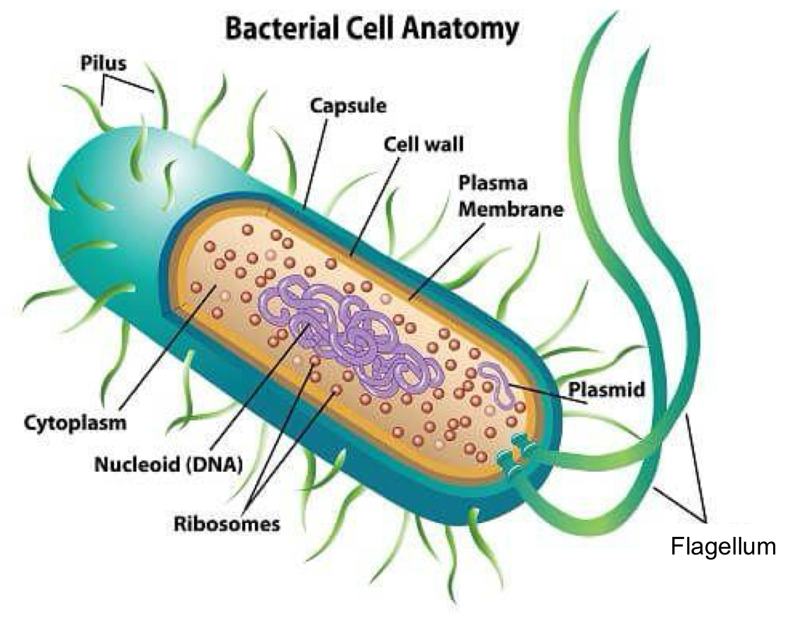
Label the structural features of a bacteria cell
Bacteria (prokaryotes)
State the function of each feature of a bacterial cell:
Pilus
Capsule
Cell wall
Plasma membrane
Plasmid
Flagellum
Ribosomes
Nucleoid (DNA)
Cytoplasm
Pilus: hair like appendage required for bacterial conjugation (transfer of genetic material)
Capsule: polysaccharide layer, contains water to prevent cell from drying out, protects cell from phagocytosis, helps cell adhere to surfaces
Cell wall: rigid structure responsible for shape of cell
Plasma membrane: consists of proteins and phospholipids, involved in transport, biosynthesis and energy transduction
Plasmid: circular DNA
Flagellum: enables movement and chemotaxis (movement away from harm and towards beneficial environments e.g. nutrients)
Ribosomes: translate genetic code from DNA to amino acids to proteins
Nucleoid (DNA): regulates growth, reproduction and function of the cell
Cytoplasm: fluid that fills the whole cell, maintains optimal environment for organelles
Bacteria (prokaryotes)
What 3 parts can the structure of a bacterial cell be divided into?
External » appendages and coverings
Cell envelope » cell wall and membranes
Internal organs
Bacteria (prokaryotes)
What are the appendages on bacteria for?
Movement (flagella)
Adhesion to surfaces (pilli, fimbriae, glycocalyx)
Bacteria (prokaryotes)
What does the cell envelope contain
Cell membrane
Cell wall (of varying thickness)
Bacteria (prokaryotes)
What does the cell wall contain?
Peptidoglycan
» made from long glycan chains cross-linked by peptides
Bacteria (prokaryotes)
What are the 2 types of glycan chains that make up peptidoglycan?
N-acetyl glucosamine (NAG)
N-acetyl muramic acid (NAM)
Bacteria (prokaryotes)
Why are cell walls strong and rigid?
Cell wall is made up of peptidoglycan
Cross-linking between amino acids in different glycan chains occurs with the help of DD-transpeptidase
This results in a 3 dimensional structure that is strong and rigid
Bacteria (prokaryotes)
What is an example of an antibiotic that interferes with bacterial cell wall synthesis?
Penicillin
Acts by binding to transpeptidases and inhibiting the cross-linking of peptidoglycan subunits
A bacterial cell with a damaged cell wall cannot undergo cell division and so will die
Bacteria (prokaryotes)
How are bacteria classified?
Bacteria are classified as Gram-positive or Gram-negative based on differences in their cell wall
» done through differences in the staining of their cell walls with crystal violet dye (Gram stain)
Bacteria (prokaryotes)
Gram-positive cell wall
Thick (20-80nm) layer of peptidoglycan
This layer is porous and contains teichoic acid
» teichoic acid maintains the cell wall and gives the cell surface an acidic (-) charge
The cell wall stains purple with Gram stain
Bacteria (prokaryotes)
Gram-negative cell wall
Thin (1-3nm) layer of peptidoglycan
Because cell is thin, gram-negative cells require an extra layer of protection called the outer membrane
» the outer membrane is a phospholipid membrane with tiny holes called porins
The cell wall stains pink with Gram stain
Bacteria (prokaryotes)
Why are Gram-negative bacteria more difficult to treat than Gram-positive bacteria?
Porins block the entrance of harmful chemicals and antibiotics
Bacteria (prokaryotes)
Why can Gram-negative bacteria induce fever and shock?
Attached to the outer membrane is a highly-branched fatty sugar called lipopolysaccharide (LPS)
LPS acts as an endotoxin causing shock and fever
Bacteria (prokaryotes)
Why do Gram-positive cell walls stain purple but Gram-negative cell walls stain pink?
Gram-positive cell walls have a thicker layer of peptidoglycan so can retain more dye
Bacteria (prokaryotes)
How can antibiotics affect bacterial cells?
Disrupt cell wall synthesis e.g. Penicillin
» bind to transpeptidases inhibiting the cross-linking of peptidoglycan subunits
» damaged cell walls mean cells cannot undergo cell division causing cell death
Inhibits DNA replication e.g. Quinolones such as Ciprofloxacin
Inhibits RNA synthesis by binding to RNA polymerase e.g. Rifampicin
Inhibits protein synthesis e.g. Macrolides such as Erythromycin
Eukaryotic cells
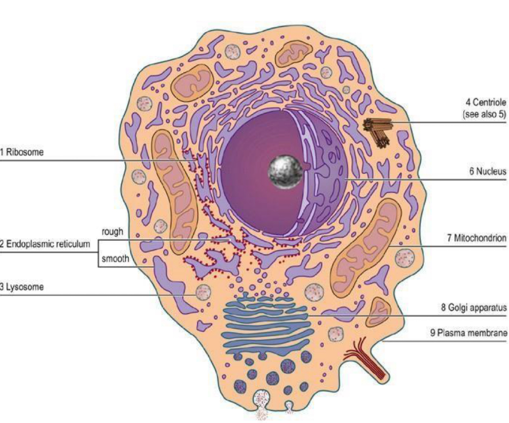
Label the structural features of eukaryotic cells
Eukaryotic cells
Ribosome
Contains RNA
Site of protein synthesis
Attached to the RER
Eukaryotic cells
Endoplasmic reticulum
3 dimensional network of membranes which extends throughout the cell
Large surface area
RER is covered with ribosomes and is involved in protein synthesis, proteins are folded inside the RER
SER is in involved in lipid metabolism
Eukaryotic cells
Lysosome
Vesicles containing hydrolytic enzymes
These enzymes break down proteins, lipids and nucleic acids
Eukaryotic cells
Peroxisome
Vesicles that produce hydrogen peroxide
They then destroy any excess hydrogen peroxide produced
Eukaryotic cells
Centriole
2 centrioles make up a centrosome
The cell’s cytoskeleton of microtubules is attached to the centrosome
Eukaryotic cells
Cytoskeleton
Makes up the framework of tubular proteins
Gives an animal cell its shape and provides basis for movement
Eukaryotic cells
Nucleus
Largest organelle in an animal cell
Contains genetic material in the form of DNA
DNA is held in chromatin fibres
During cell division the chromatin granules form chromosomes
Assembles ribosomes
Pore in the double layered nuclear envelope allow things to move in and out of the cytoplasm
Eukaryotic cells
Mitochondria
Has a double membrane
Inner membrane is folded forming cristae with a matrix on the inside
Matrix contains enzymes needed for respiration
Site of aerobic respiration » produces ATP
Eukaryotic cells
Golgi apparatus
Flattened sacs that receive vesicles from the Endoplasmic reticulum
Modify, sort and package the contents of the vesicles and secrete it to other organelles
In the Golgi carbohydrates are added to proteins coming from the ER to form glycoproteins
Eukaryotic cells
Plasma membrane
Double layer of phospholipids (bilayer)
Acts as a semi-permeable barrier
Embedded in the fluid mosaic are proteins, lipids and cholesterol
Acts as pumps and channels
Eukaryotic cells
Compare plant and animal cells
Similarities:
Ribosomes
Endoplasmic reticulum
Lysosome
Centriole
Cytoskeleton
Mitochondria
Golgi apparatus
Differences:
Plant cells have a cell wall, animal cells do not
Plant cells have a large fluid filled vacuole, animal cells do not
Plant cells contain chloroplasts, animal cells do not
Eukaryotic cells
Vacuole
Temporary food store containing sugars and amino acids
When vacuole is full, cell is turgid
Membrane surrounding vacuole is called the tonoplast
Eukaryotic cells
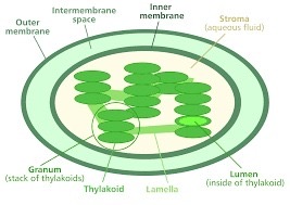
Chloroplast
Absorb light for photosynthesis to produce carbohydrates
Surrounded by double membrane
Thylakoid membranes inside which can form a stack called a granum
Grana linked by lamellae
What is the cell wall made of in plants, algae, fungi and prokaryotes?
Plants: cellulose
Algae: cellulose
Fungi: chitin
Prokaryotes: murein e.g. bacteria
Compare eukaryotes and prokaryotes
Similarities
Cell membrane
Cytoplasm
Ribosomes
DNA
Differences
P: no membrane bound organelles E: membrane bound organelles
P: smaller ribosomes E: bigger ribosomes
P: have a capsule E: no capsule
P: smaller E: bigger
P: circular DNA E: linear DNA
P: DNA not bound to histones E: DNA bound to histones
P: plasmids E: no plasmids
P: cell division by binary fission E: cell division by mitosis
What are viruses?
Tiny particles, not cells
Only visible in an electron microscope
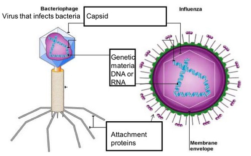
What do viral particles consist of?
Genetic material in the form of RNA or DNA
A protein coat (capsid) that surrounds RNA OR DNA
In some cases a lipid envelope that surrounded the protein coat when the virus is inside cells
What was the first virus to be discovered?
Tobacco mosaic virus
» Positive-sense single stranded RNA virus
» Infects a wide range of plants especially tobacco
Why can viruses only replicate by infecting a host?
Viruses do not contain their own biochemical machinery
So they bind to host cells, enter the cell and incorporate their genomes into the host cell’s DNA to replicate the virus particles
What do viruses target on cell surfaces for cell attachment and entry?
Glycolipid molecules (glycans) on the cell surface
Summarise the process of viral replication
Virus attaches to the host cell by targeting glycans on the cell surface
Virus injects its genetic material into host cell
Host cell transcribes and translates viral genes
These proteins form new virus particles
Virus particles burst out of host cell, so host cell is destroyed
Adenoviruses
Cause a wide range of illnesses e.g. fever, sore throat etc
Contagious » transmitted via droplets
Influenza virus
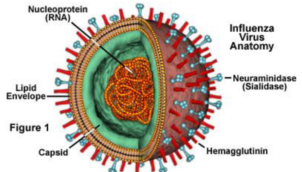
Structure of the virus particles
Spherical shape
2 antigens on the surface (glycoproteins) » HA and NA
These proteins determine the subtype of the influenza virus
Influenza virus
Why is there a new flu vaccine each year
Due to antigenic drift
» Mutations in the HA and NA antigens
Influenza virus
How does the influenza virus infect a host cell and replicate?
The virus’ hemagglutinin (HA) glycoprotein binds to sialic acid receptors on the cell’s surface.
The cell engulfs the virus by endocytosis
This vesicle then fuses with a lysosome » contains digestive enzymes and is acidic
The acidity inside the lysosome breaks down the virus’ coat.
The hemagglutinin protein changes shape and inserts itself into the membrane of the vesicle
The viral membrane fuses with the vesicle membrane, allowing the virus to release its RNA into the cell.
Host cell transcribes and translates viral RNA. These proteins form new virus particles
A enzyme neuraminidase helps cuts the sialic acid from the cell membrane so the new virus particles can escape
How does HIV infect a host cell and replicate?
1. The HIV's attachment proteins binds to the CD4 receptor proteins on the surface of the t helper cell
2. The virus's lipid envelope fuses with the cell membrane of the Th cell.
3. The protein capsid breaks down
4. RNA and enzymes (e.g. reverse transcriptase) of the virus are now released into the cytoplasm of the host cell.
5. Reverse transcriptase converts the viral RNA to DNA.
6. The viral DNA is incorporated into the cell's DNA.
7. The viral DNA can now be transcribed into mRNA
8. Viral mRNA passes through the nuclear pore and attaches to a ribosome
9. Viral mRNA is translated into viral proteins that can be assembled into new HIV particles.
10. HIV particles bud off the Th cell (so that the Th cell's membrane forms the lipid envelope of the virus)
How does Herpes Simplex Virus (HSV) infect a host cell and replicate?
HSV uses the glycoproteins on its surface (gB and gC) to bind to the heparan sulfate proteoglycans on the host cell
The virus fuses with the cell membrane and releases its capsid into the cytoplasm
Capsid travels to nucleus and injects its DNA into nucleus
Transcription and translation occur producing new virus particles
Can cause lytic infection » virus bursts out of host cell, destroying it
Can cause latent infection » viral DNA can remain dormant in the cell until activated
Treating viruses
Vaccines » stimulates immune system, producing antibodies and memory cells
Antiviral drugs » inhibit viral replication
What to antiviral drugs target?
Viral glycoproteins used to enter cells
Reverse transcriptase used to incorporate viral DNA into host DNA
Proteases used to assemble new virus particles
Antiviral drug targets should be…
Unlike any other proteins in humans as possible to reduce likelihood of side effects
Mitosis
Cell division which allows organisms to grow and replace cells
Produces 2 identical daughter cells
Meiosis
Division that results in 4 gametes with half the chromosome number
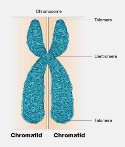
Chromosome
Threadlike structure of nucleic acids and proteins found in the nucleus
Consists of 2 sister chromatids joined by a centromere
Chromatid
2 threadlike strands that make up the chromosome
Centromere
Region of a chromosome where the microtubules of the chromosome join
Telomere
Cap at the end of each chromosome arm which maintains stability
Cell cycle
Series of events that occur when a cell divides and grows:
Interphase
Mitosis: prophase, metaphase, anaphase, telophase
Cytokinesis
Label the structure of a chromosome
Explain the levels of DNA organisation
DNA is associated with histone proteins forming chromatin
Chromatin condenses to form chromosomes
So chromatin is a lower order of DNA organisation whereas chromosomes are a higher order
Cell Cycle
Interphase
G1:
Cell grows
Protein synthesis
Synthesis of RNA
Replication of organelles
S:
DNA decondenses (not visible)
DNA is replicated (results in 2 sister chromatids attached by a centromere)
G2:
Synthesis of organelles such as mitochondria
Cell grows more and proteins for cell division made
ATP increases
Cell Cycle
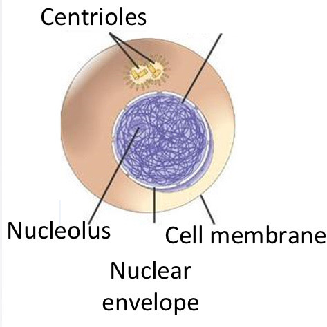
Mitosis: Prophase
DNA condenses and becomes shorter and thicker » more visible
Nuclear envelope breaks down
Centrioles migrate to opposite ends of the cell
Centrioles produce spindle fibres, made from microtubules
Cell Cycle
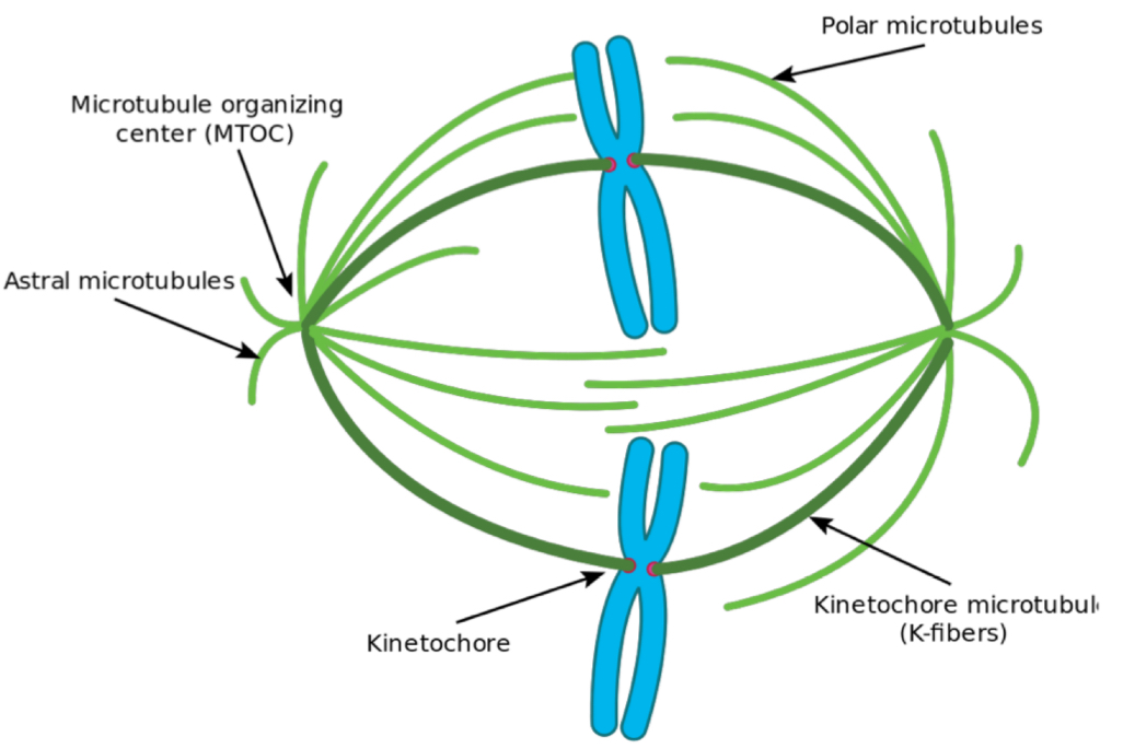
Mitosis: Pro-metaphase
Phosphorylation of nuclear lamins (proteins that support the nuclear envelope) by M-CDK causes the nuclear envelope to break down completely
Spindle fibres attach to each chromosome at its kinetochore (proteins located at the centromere)
Cell Cycle
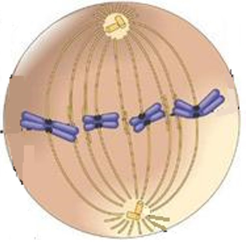
Mitosis: Metaphase
The spindle fibres move the chromosomes so that they line up along the equator of the cell
Cell Cycle
Mitosis: Anaphase
Cohesin which joins the sister chromatids is broken down by enzymes, causing the centromeres to split
Spindle fibres contract and shorten
This separates the sister chromatids, pulling them to opposite poles of the cell
Cell Cycle
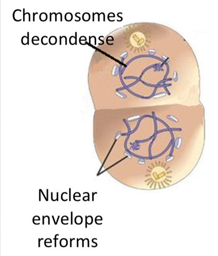
Mitosis: Telophase
2 new nuclear envelopes form around the 2 new sets of chromosomes
Chromosomes decondense
Nucleoli reform
Nucleus takes on a granular appearance
Cell Cycle
Cytokinesis
The cell membrane between the 2 nuclei separates
Cytoplasm separates
Forms 2 identical daughter cells
How many chromosomes do human cells contain
46 chromosomes
23 homologous pairs » one of the pair from mother, the other from father
What are homologous pairs of chromosomes?
Chromosomes that have the same genes at the same loci
What is different about a female’s chromosomes?
Females have 2 X chromosomes as the 23rd homologous pair
Why is it so important that gametes are haploid?
To maintain chromosome number at fertilisationd
Key parts of meiosis
2 divisions:
Separates homologous pairs
Separates sister chromatids
Describe the process of meiosis I
Interphase: DNA unravels + replicates so there is 2 copies of each chromosome (2 x 2n)
Prophase I:
Chromosomes condense and become visible >> each chromosome has a pair of sister chromatids joined by centromere
Homologous chromosomes pair up forming bivalents (2 x 2n) » this is called synapsis
Crossing over occurs
Metaphase I:
Spindle fibres join to the centromeres causing homologous pairs of chromosomes to line up next to each other along equator of cell (2 x 2n)
Independent segregation occurs
Anaphase I: spindle fibres contract pulling one of each pair of homologous chromosomes to opposite poles (2 x 2n)
Telophase I:
2 new nuclear envelopes form
Chromosomes uncoil, nucleoli reform, nuclei take on granular appearance
Cytokinesis: cytoplasm divides to form 2 daughter cells (2n)
What is synapsis?
When homologous chromosomes pair up to form bivalents
What are the 2 factors which cause gametes to vary (genetic variation)
Independent segregation
Crossing over
How does crossing over occur during Prophase I?
Homologous pairs of chromosomes pair up forming bivalents
Chromatids twist around each other forming crosslinks » called chiasmata
This leads to genetic material being exchanged
So chromatids now contain a different combination of alleles
Each of the 4 daughter cells will contain chromatids with a different combination of alleles
» results in shuffling of genes by RECOMBINATION
(E.g. if initially one chromatid in the pair was Ee and the other was Ff, due to crossing over one will now be Ef and the other eF)

How does independent segregation occur during Metaphase I?
The homologous chromosomes in each bivalent are arranged along the equator of the cell
The alignment of these homologous chromosomes is random (e.g. chromosome from mother could be on left and chromosome from father could be on right or vice versa and this can happen for all the homologous pairs)
When the homologous chromosomes are separated into two daughter cells during anaphase I, the combination of chromosomes in each daughter cell is a random mix of chromosomes from both parents
» results in shuffling of genes by REASSORTMENT
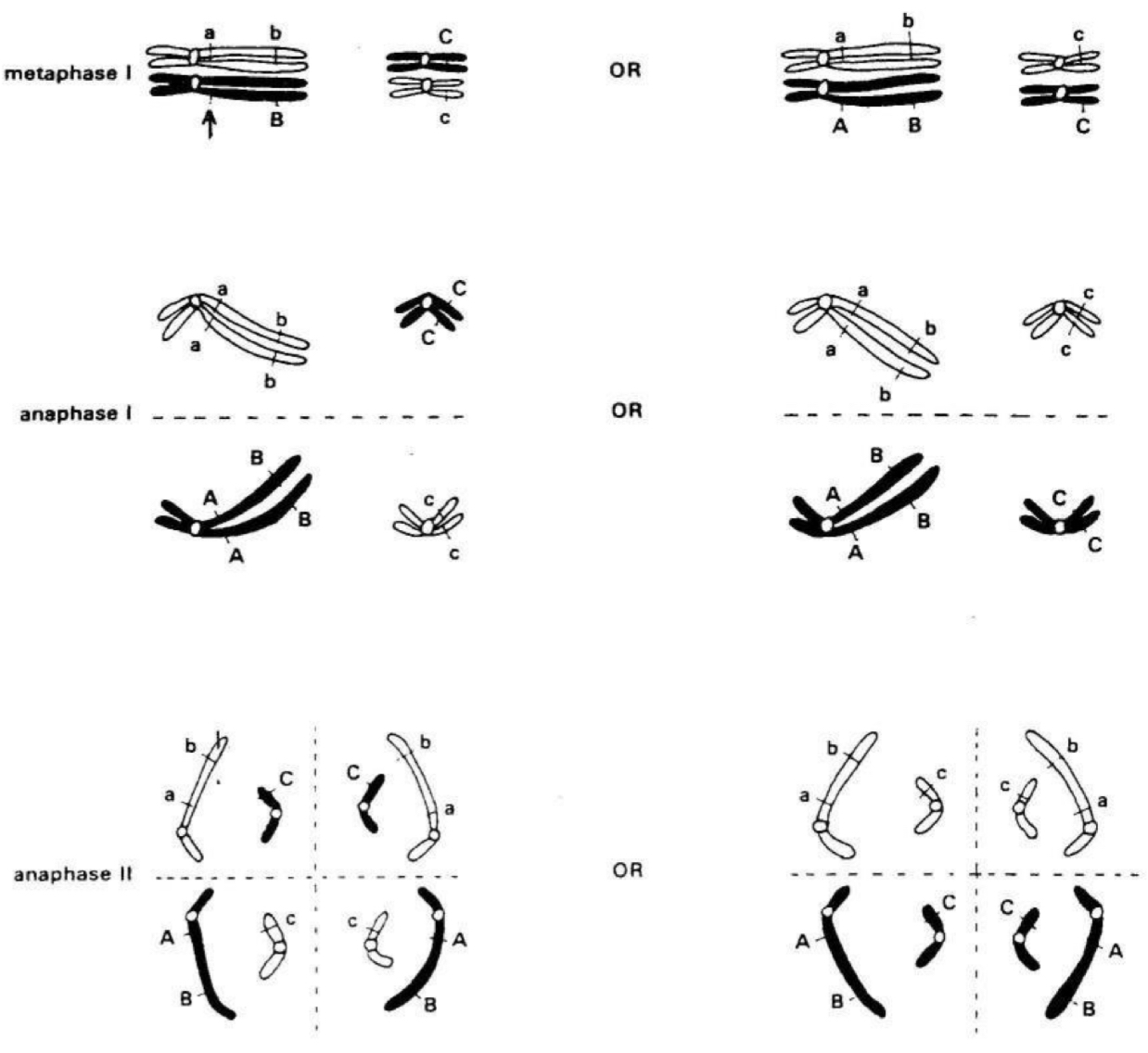
ALL GENETIC VARIATION OF GAMETES ONLY OCCURS DURING
MEIOSIS I
Describe the process of meiosis II
Prophase II: centrioles move to opposite ends of the cell and produce spindle fibres
Metaphase II: spindle fibres attach to centromeres of the chromosomes
Anaphase II: spindle contracts and sister chromatids pulled to opposite poles
Telophase II: chromatids reach opposite poles and decondense, nuclear envelope forms around each chromatid
Cytokinesis: cytoplasm divides
4 haploid daughter cells produced
Why does sexual reproduction not result in an organism that is exactly the same as the parents?
Because of recombination and reassortment, gametes have a varied combination of genes