BIOLOGY 315 WK 8 - Circulatory System II
1/97
There's no tags or description
Looks like no tags are added yet.
Name | Mastery | Learn | Test | Matching | Spaced | Call with Kai |
|---|
No analytics yet
Send a link to your students to track their progress
98 Terms
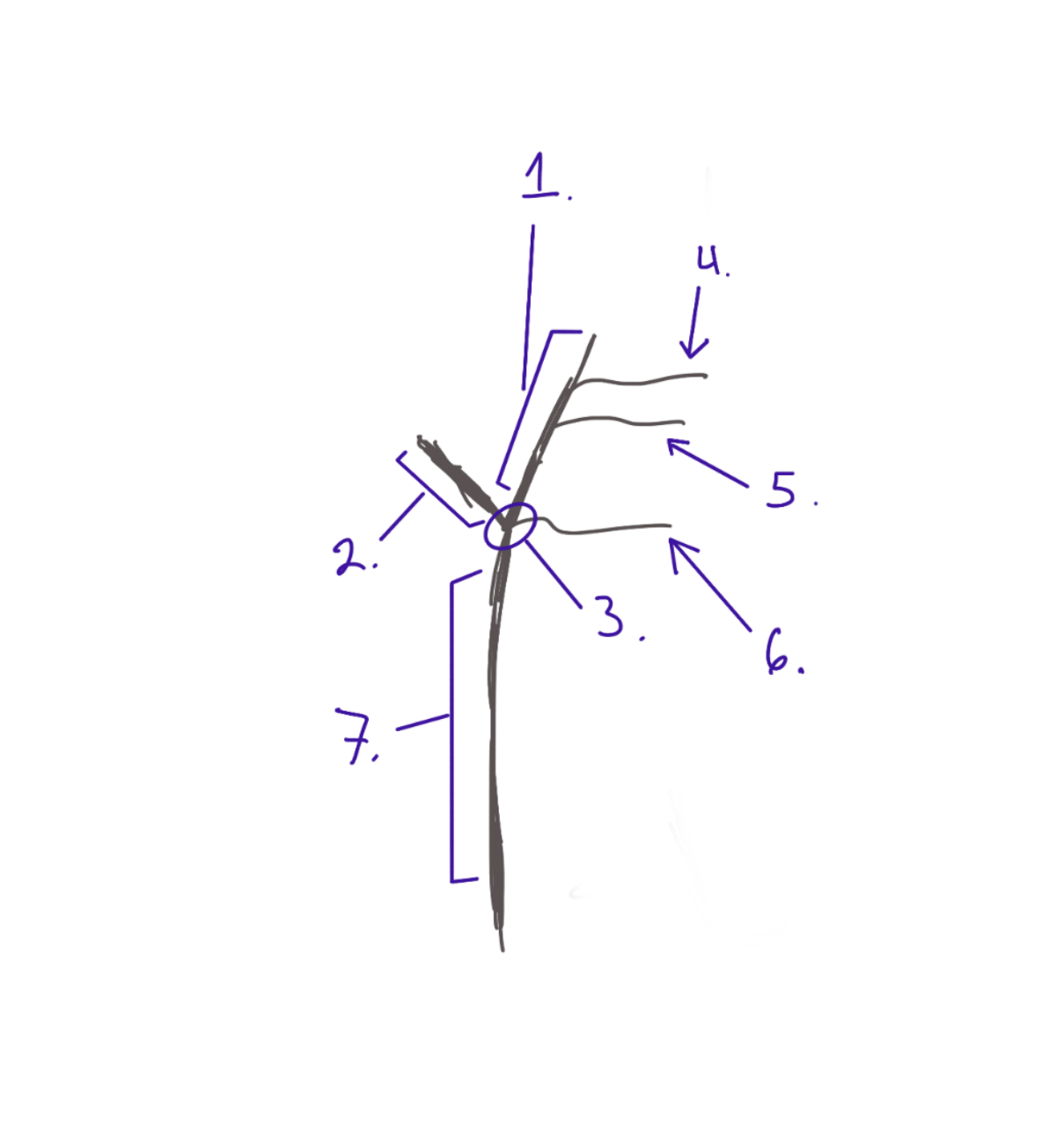
R common carotid artery
Structure. The bottom of the "Y". #7

R Carotid Body
Internal Structure. The middle of the "Y". #3

R external carotid artery
structure. The right of "Y", Has Branches. #1

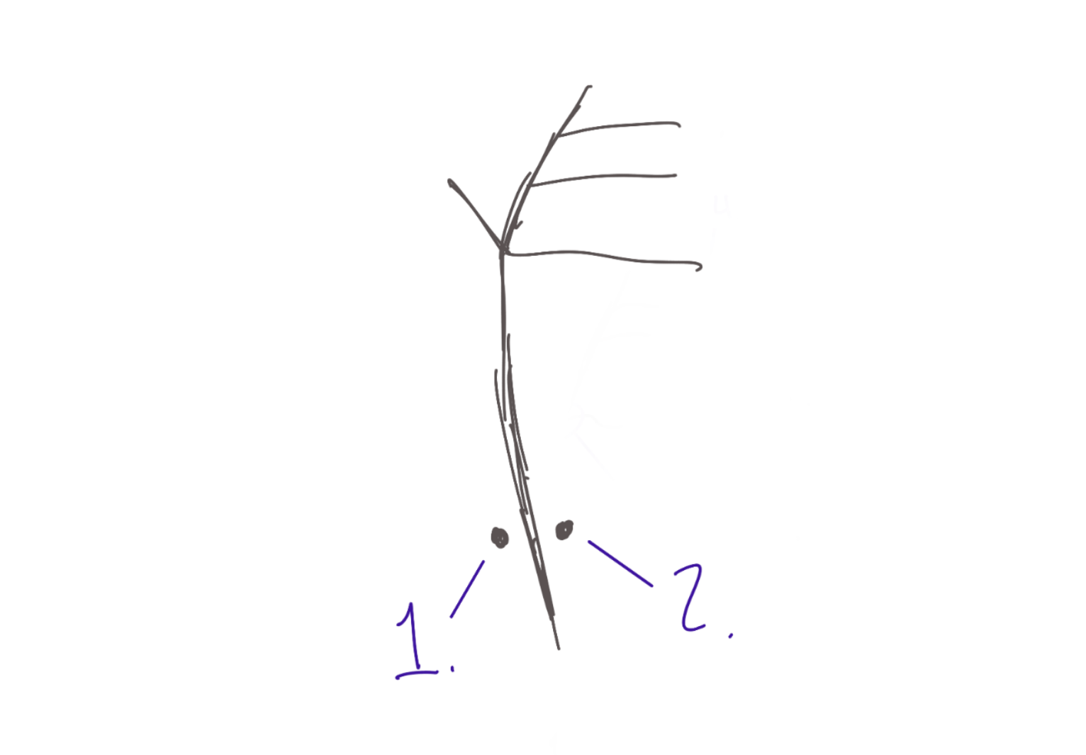
R superior thyroid artery
Structure. The most inferior branch on the Y. #6

R lingual artery
Structure. Most medial branch on the Y. #5

R Facial artery
Structure. The most superior branch on the Y. #4
R occipital artery
Structure. Most lateral/superior pin on the cheek. #1
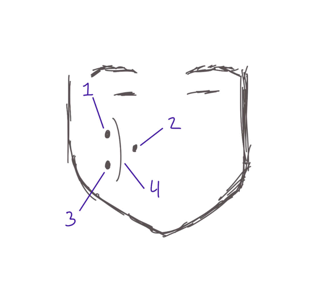
R Maxillary artery
Structure. Most deepest/medial pin on the cheek. #2

R middle meningeal artery
Structure. Most inferior pin on the cheek. #3

R Superficial temporal artery
Structure. Makes the arch shape in the cheek, between the 3 pins!. #4

R Internal carotid artery
Structure. The most lateral in the "Y" shape. #2

R subclavian artery
Structure. Above TA's probe.
R vertebral artery
Structure. Below the two pins.

R thyrocervical trunk
Structure. Blue pin, but most lateral pin. (2pins only) #1

R inferior thyroid artery
Structure. Pink pin, most medial pin. (2. pins only) #2
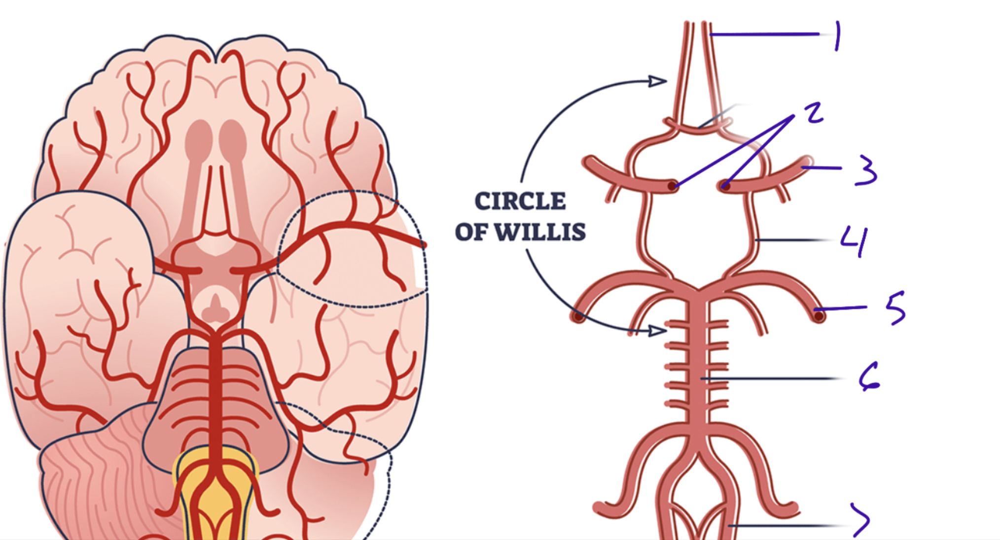
Anterior cerebral artery
Above the optic chiasm when looking inferior. #1

Internal carotid artery
TA puts probe inside the hole. #2

Posterior communicating artery
The cut one.#4

Middle cerebral artery
#3

Posterior cerebral artery
#5

Basilar artery
#6

Vertebral artery
Actually shown to the left side in our POV since some are missing #7
Superior sagittal sinus
#1. Longitudinal fissure.

Inferior sagittal sinus
#2. on the corpus calosum

Straight sinus
#3. Between the pons and brain

Transverse Sinus
#4. Creepy face purple color

Sigmoid sinus
#5. Light blue color.

Confluence of sinuses
#6. collective space.
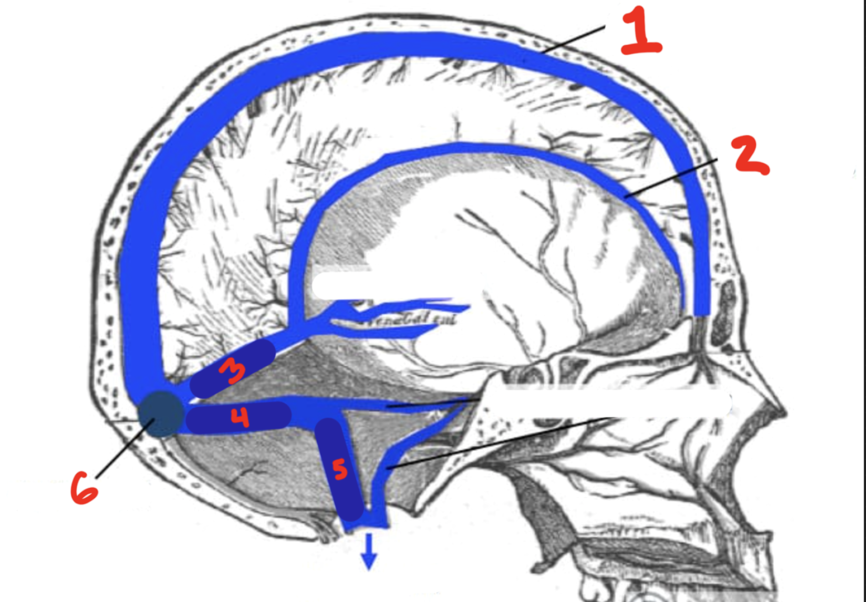
Cavernous sinuses
Space. Where the oculomotor nerve sit.
R subclavian vein
R internal jugular vein
R external jugular vein
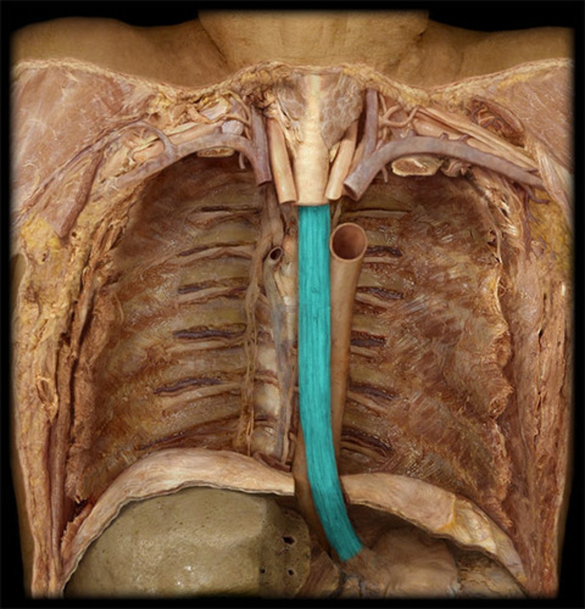
Thoracic esophagus
Greater curvatures of the stomach
Borders
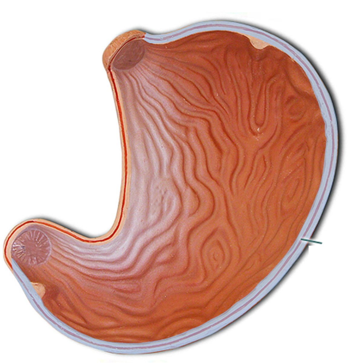
Lesser curvatures of the stomach
Borders
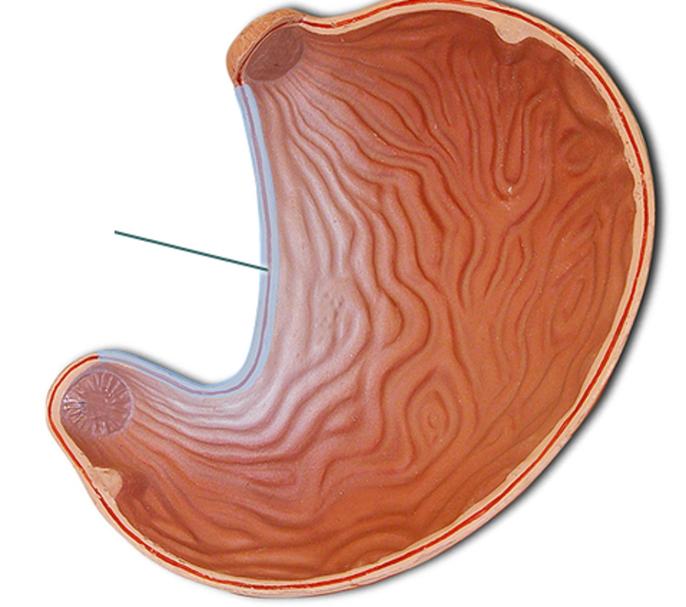
Cardia of the stomach
Feature. Above the stomach.
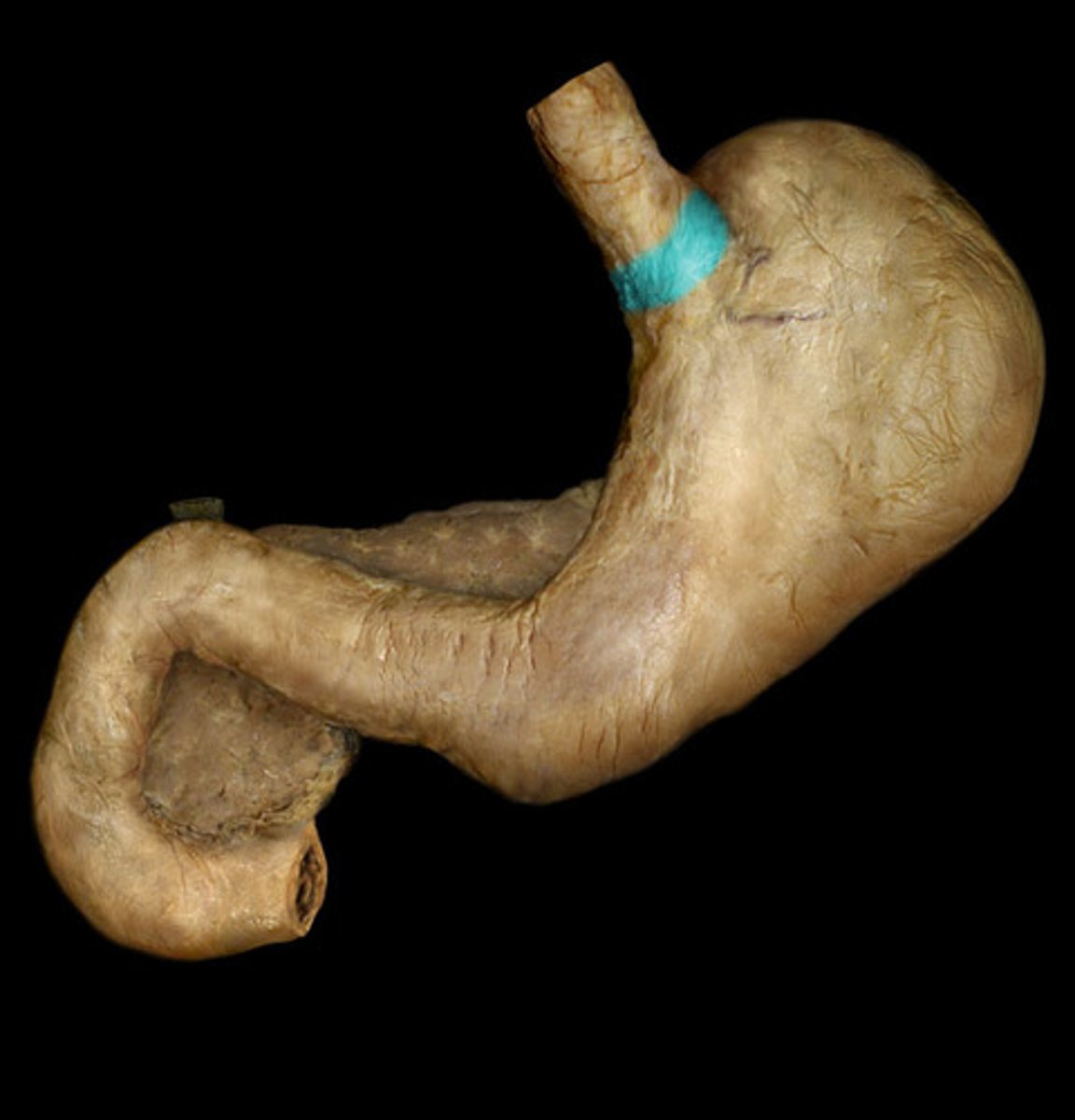
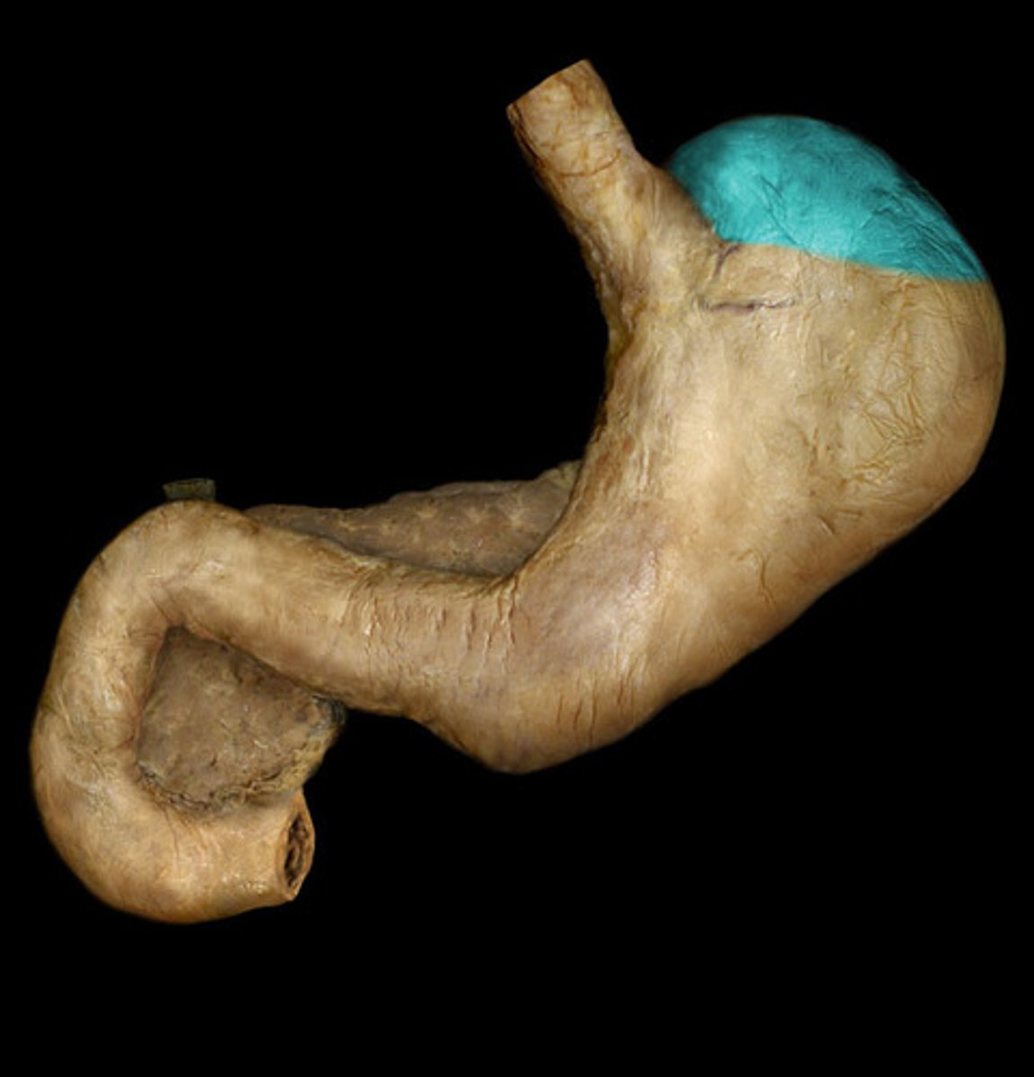
Fundus of stomach
region. Follow the great curvature then its the hump.
body of stomach
region
Pyloric antrum
region
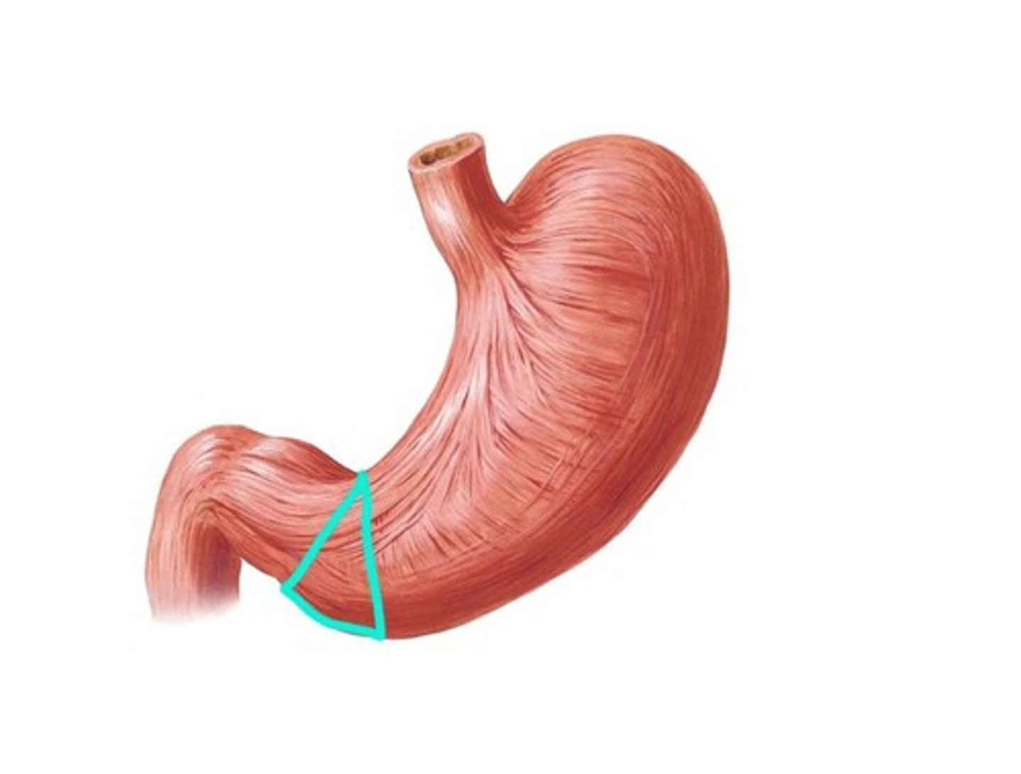
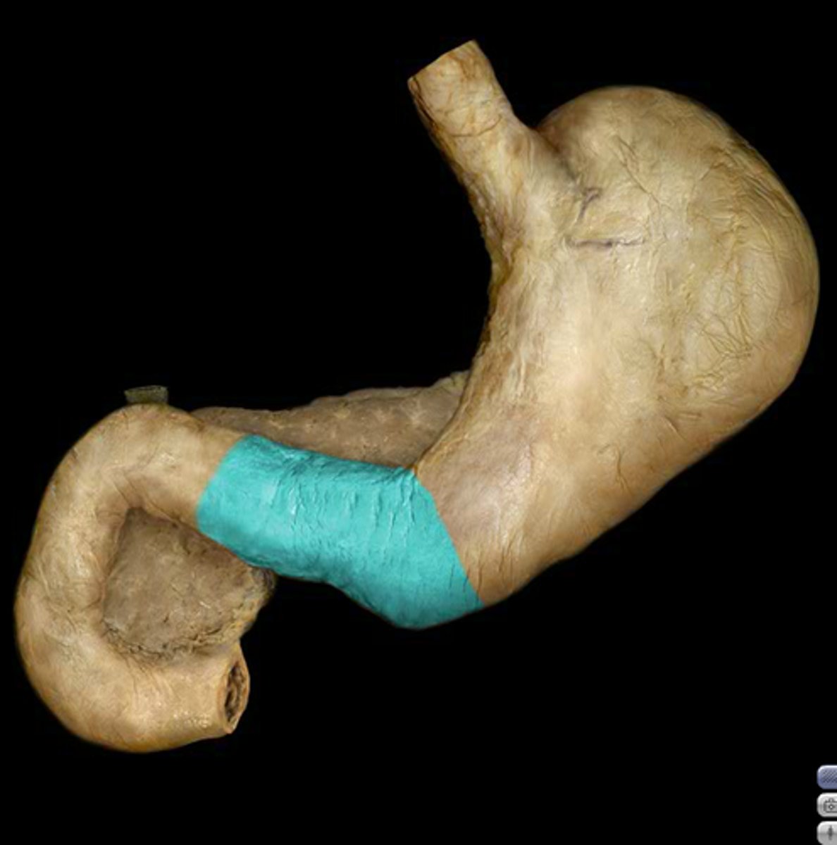
Pylorus
Feature
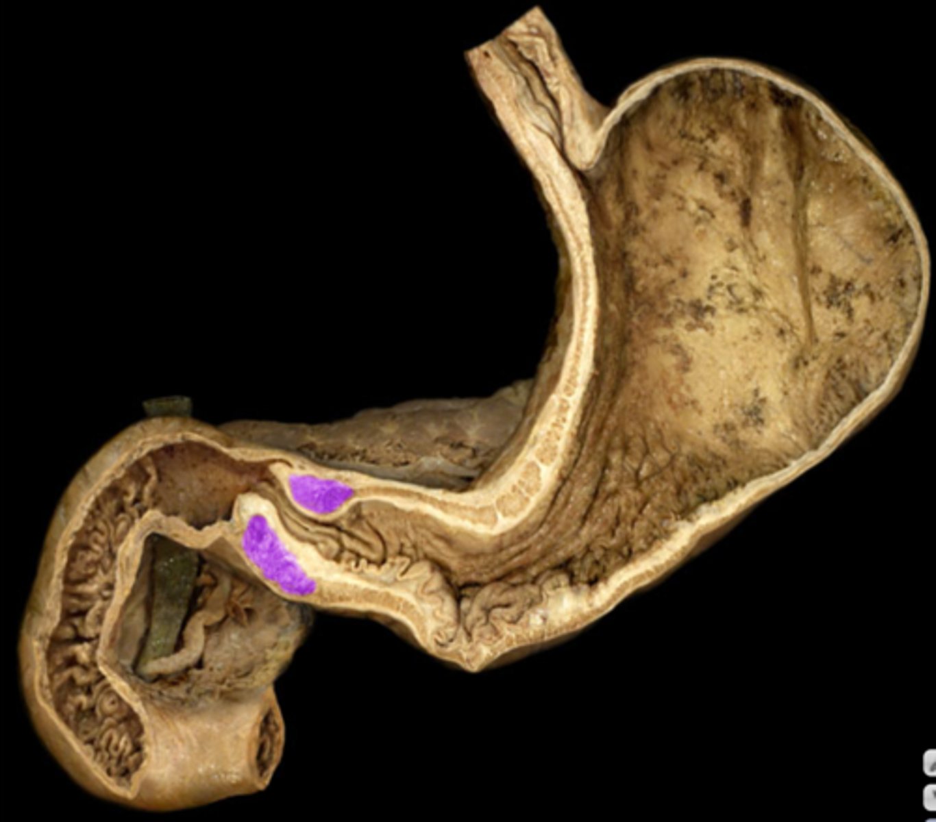
Pyloric sphincter Muscle
Internal Structure. Inside pylorus
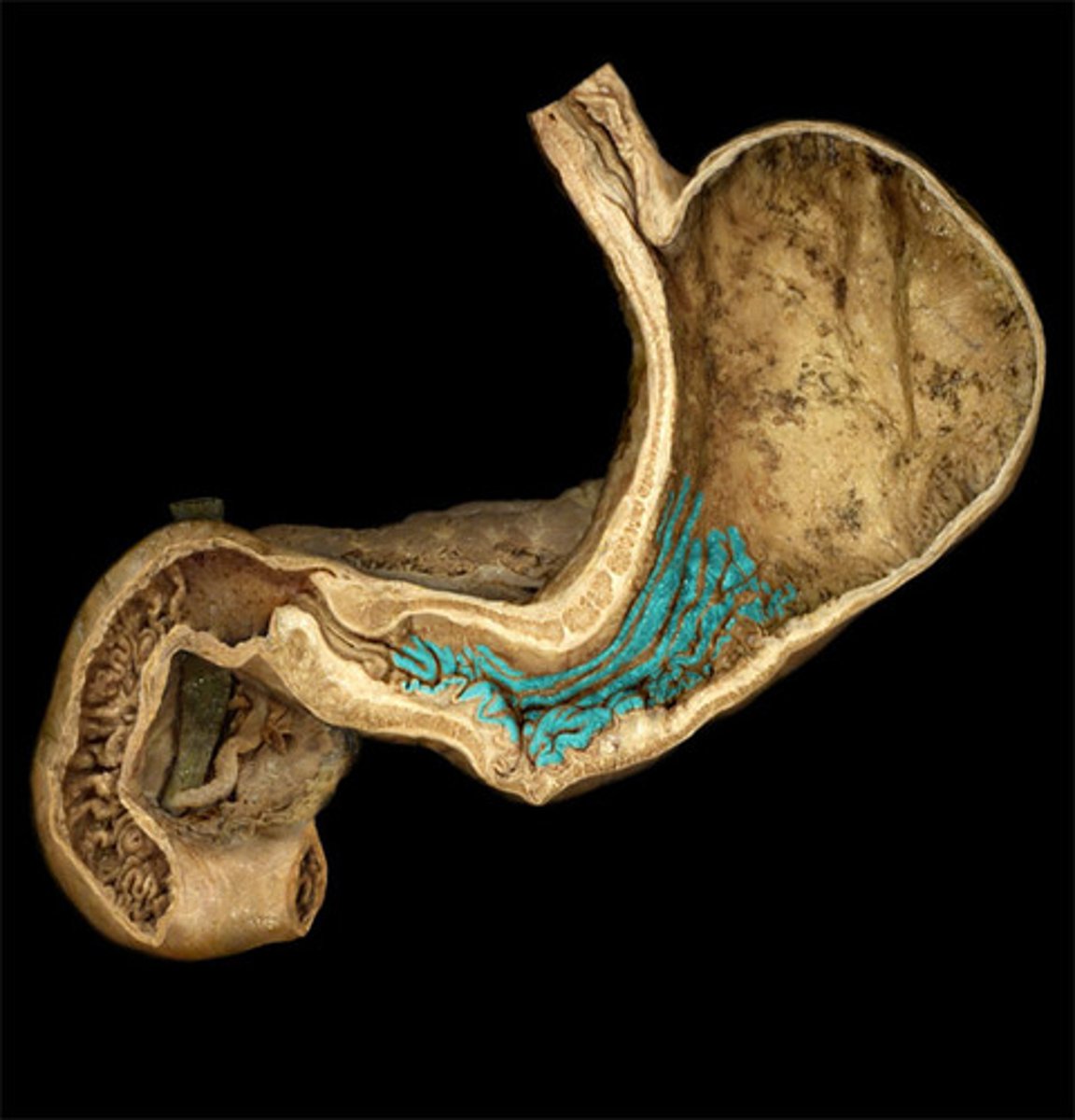
Gastric folds
internal structure. Inside the stomach.
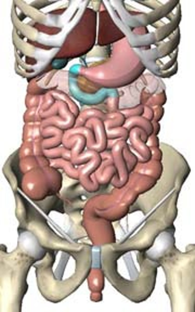
Duodenum
Portion
Superior Part of the duodenum
descending part of the duodenum
horizontal part of the duodenum
ascending part of the duodenum
Jejunum
Ileum
Cecum
It looks like a C
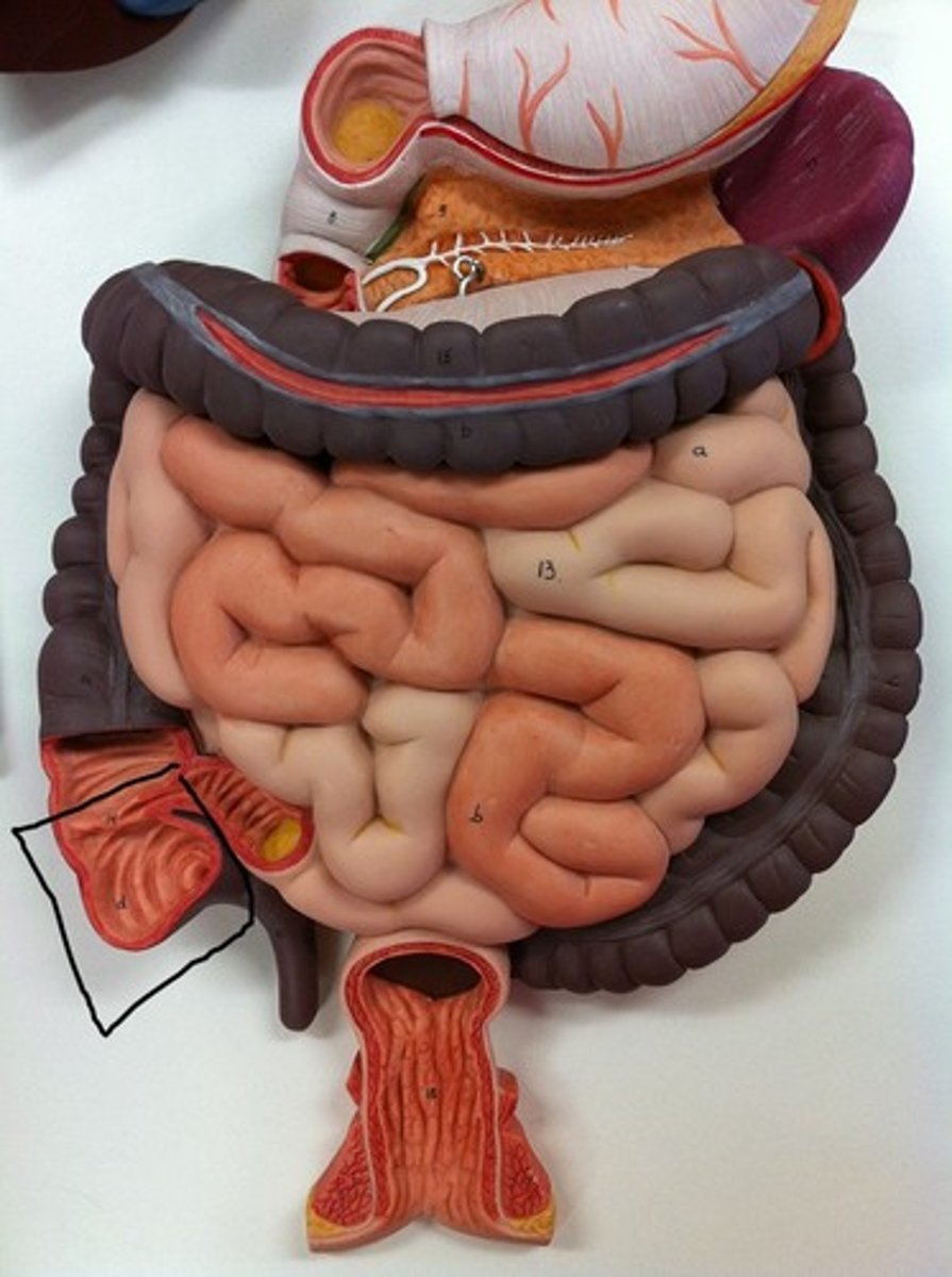
Ileal orifice
Junction
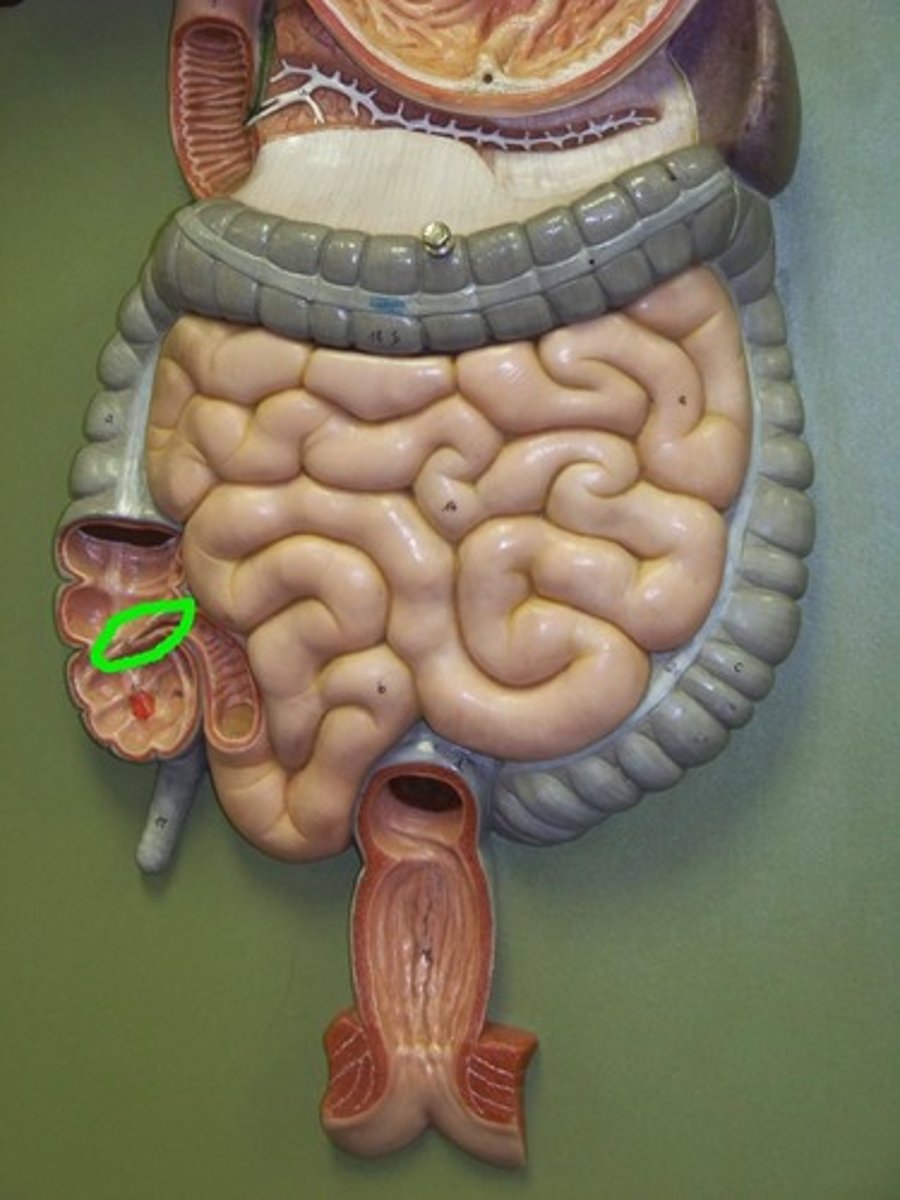
Appendix
Ascending colon
Structure
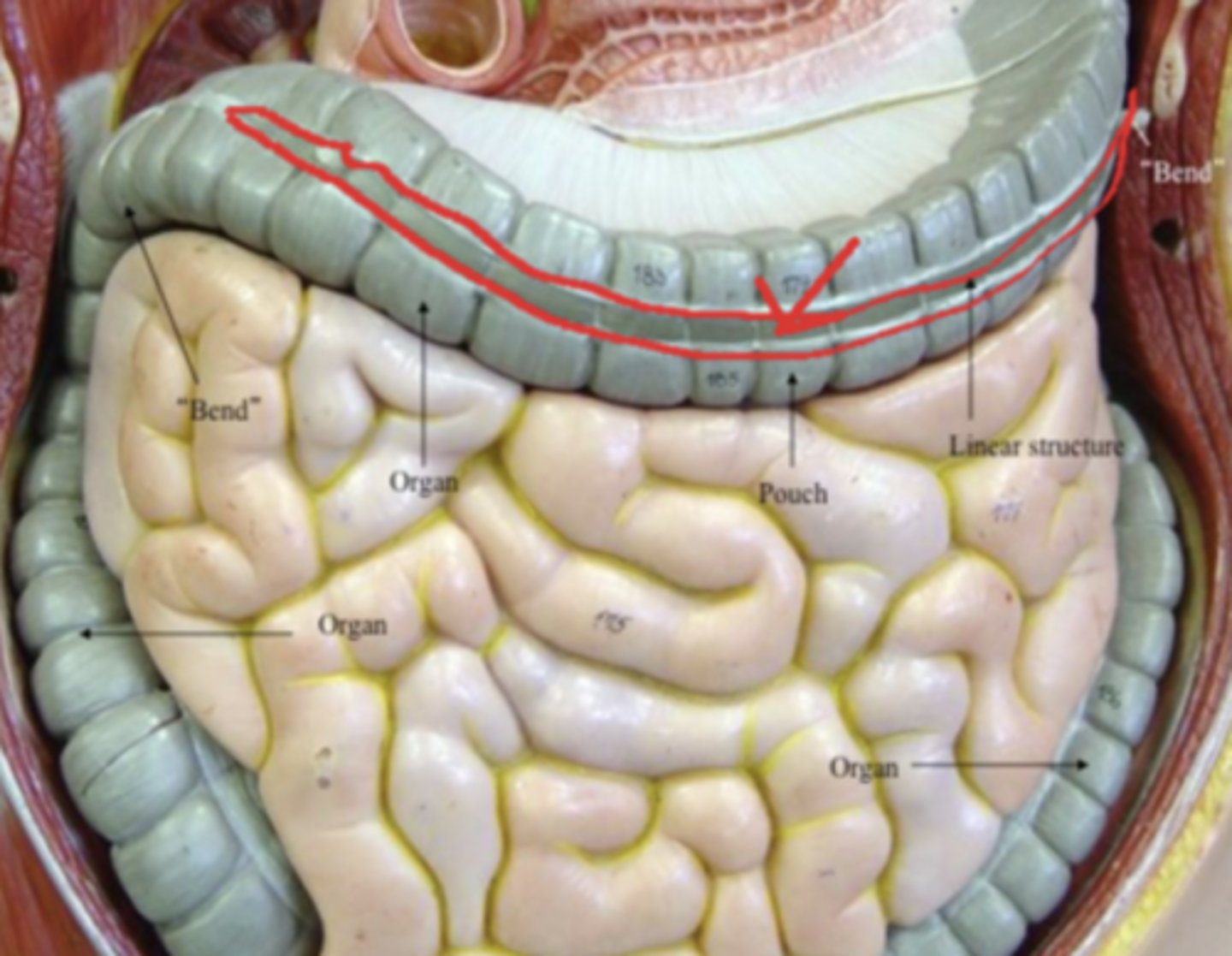
Right colic flexure
feature
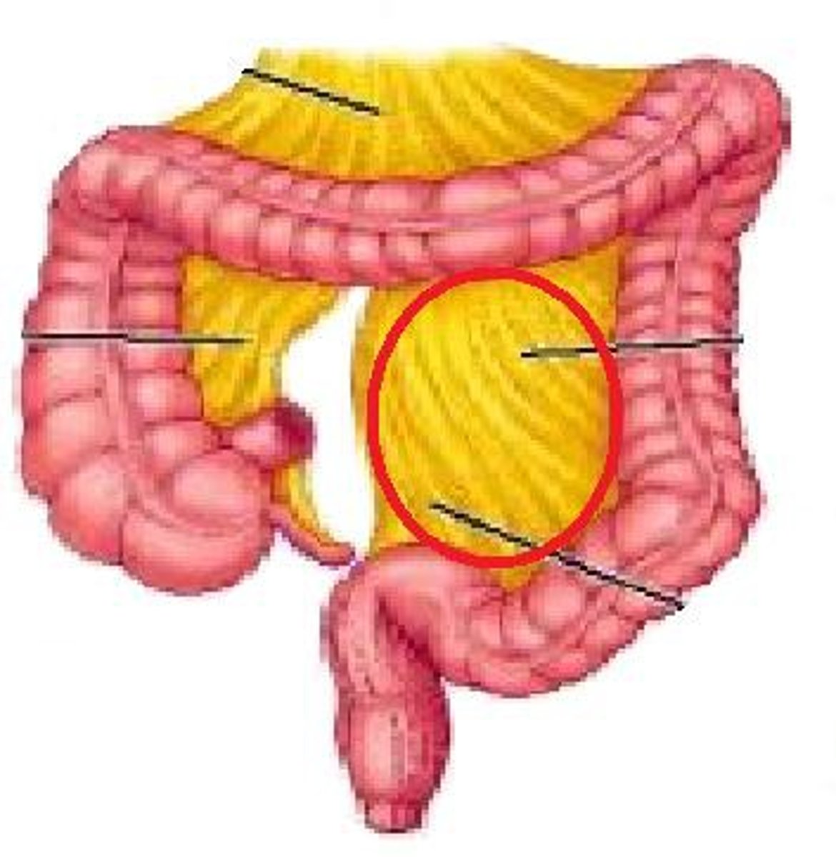
Transverse colon
Structure
Left colic flexure
feature
Descending colon
Structure
Sigmoid colon
Structure
taeniae coli muscle
structure.
Haustra of colon
Segments
Omental appendices
Feature
Rectum
Transverse folds
Anal canal
opening
Internal anal sphincter muscle
External anal sphincter muscle
Anal columns
Ridges
Anal sinuses
Depressions
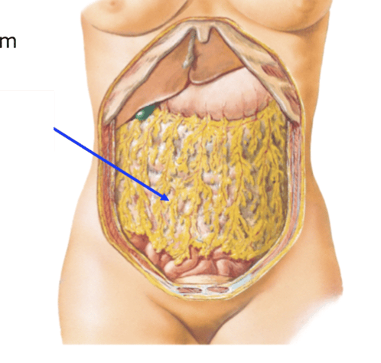
Greater omentum
Covering
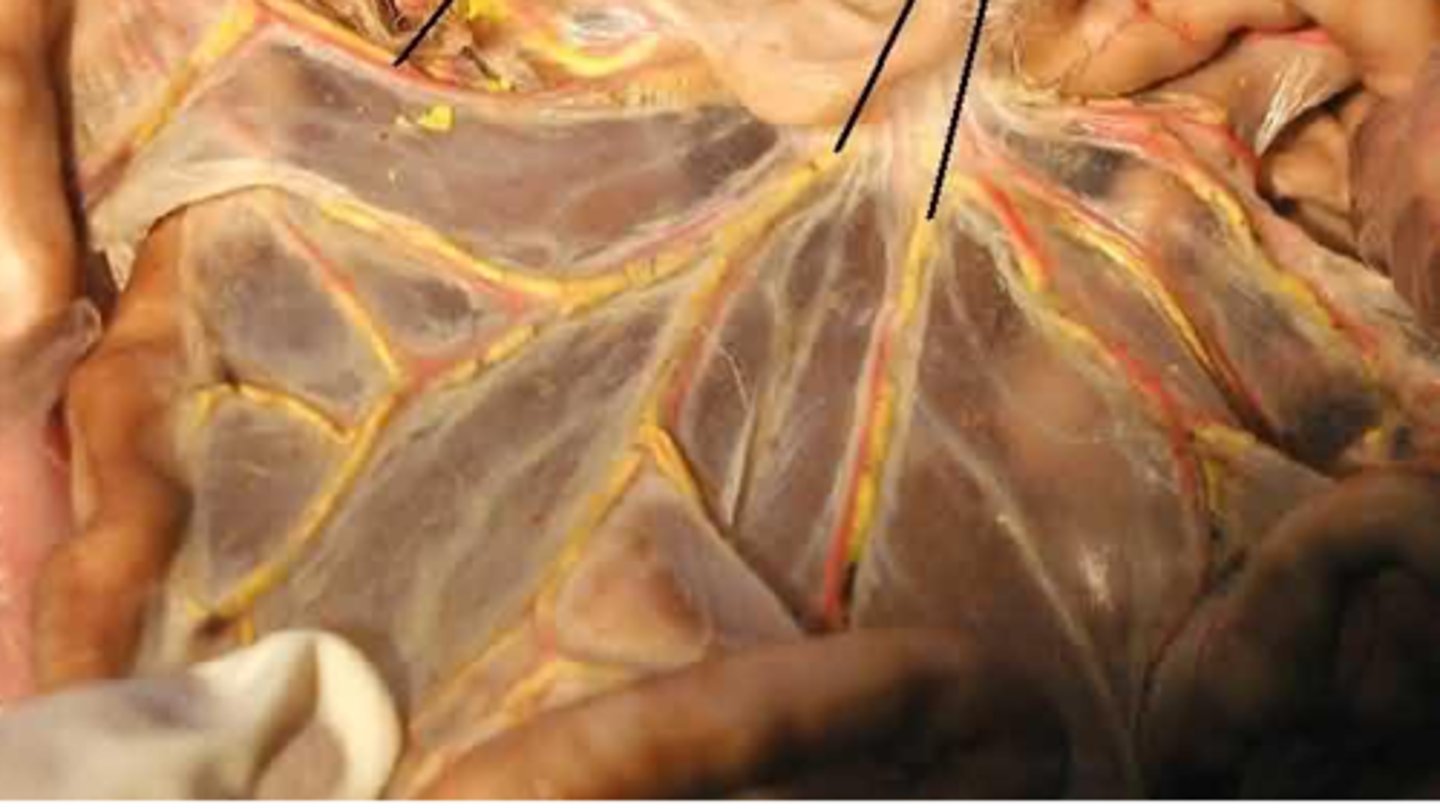
The mesentery
Covering. Like A fish flaring.
Sigmoid mesocolon
Covering. Below the sigmoid.
R subclavian artery
R axillary artery
R thoracodorsal artery
R lateral thoracic artery
R Subscapular arter
R Brachial artery
R deep artery of the arm
R radial artery
R ulnar artery
R radial veins
R Brachial veins
R axillary vein
R subclavian vein
R Cephalic vein
R Basilic vein
R Medial cubutal vein
R External iliac artery
R Femoral artery
R popliteal artery
R anterior tibial artery
R dorsalis pedis artery
R posterior tibial artery
R fibular artery
R Medial plantar arteries
R lateral plantar arteries
R posterior tibial veins
R poplitean veins
R Femoral veins
R great saphenous vein
R small saphenous vein