Uterine Pathology
1/9
There's no tags or description
Looks like no tags are added yet.
Name | Mastery | Learn | Test | Matching | Spaced | Call with Kai |
|---|
No analytics yet
Send a link to your students to track their progress
10 Terms
Posterior thickening of the myometrium is most often related to what?
a. adenomyosis
b. congenital malformations
c. cervical cancer
d. fibroids
Adenomyosis
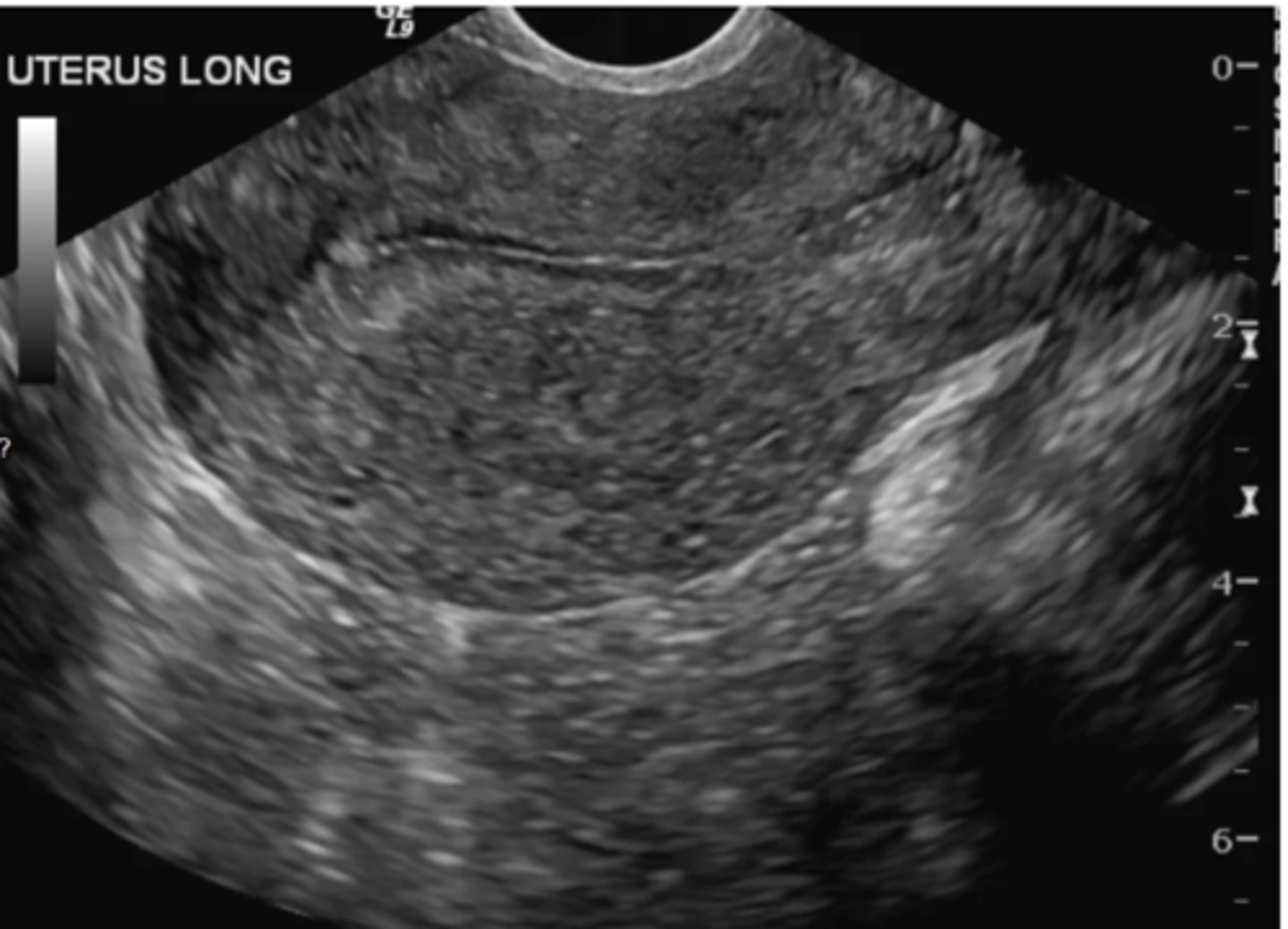
Where is the fibroid located in the image?
a. subserosal
b. intramural
c. submucosal
d. intracavitary
Submucosal
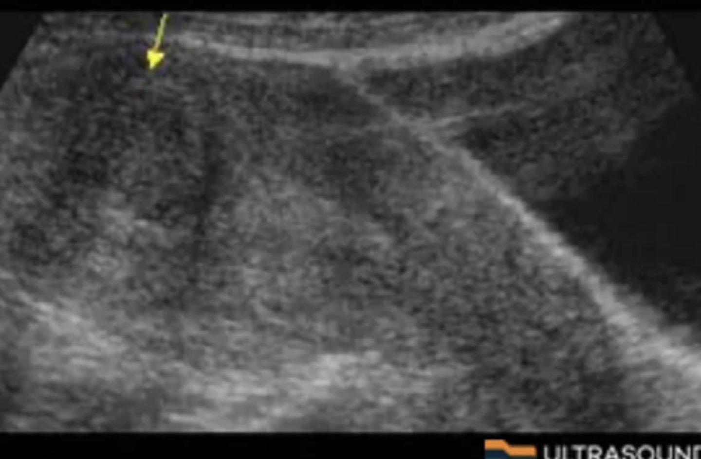
The diagnostic findings in the image are consistent with what pathologic process?
a. adenomyosis
b. myometrial hyperplasia
c. cervical cancer
d. leiomyosarcoma
Adenomyosis
-The ultrasound findings are subtle in this case but some features are present. They myometrium is mildly heterogeneous with small cystic areas and there is thickening specifically in the posterior uterus. These are all features of adenomyosis
Which artifact is most likely seen with an IUD?
a. reverberation
b. edge shadowing
c. posterior enhancement
d. mirror image
Reverberation
What's the most common gynecological malignancy under the age of 50?
a. cervical carcinoma
b. ovarian cystadenocarcinoma
c. leiomyosarcoma
d. endometrial carcinoma
Cervical carcinoma
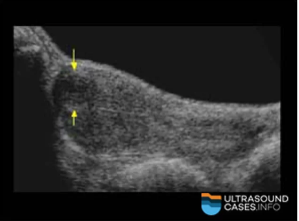
What is documented in the image below?
a. intrauterine device
b. endometrial calcifications
c. adenomyosis
d. endometrial scarring
Intrauterine device
Small myometrial cystic areas, thickening of the myometrium, abnormal bleeding, and dysmenorrhea all describe what condition?
a. adenomyosis
b. endometrial carcinoma
c. endometriosis
d. endometrial hyperplasia
Adenomyosis
What clinical history best corresponds to a diagnosis of adenomyosis?
a. dysmenorrhea
b. acute pelvic pain
c. infertility
d. secondary amenorrhea
Dysmenorrhea
What is the most common benign mass of the cervix?
a. nabothian cyst
b. garter duct cyst
c. cervical adenoma
d. leiomyoma
Nabothian cyst
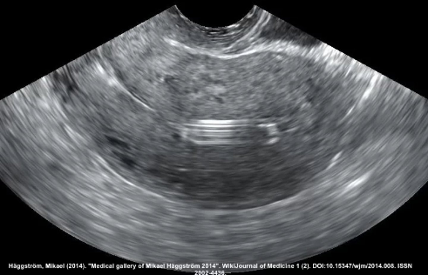
Where is the mass located in the image below?
a. intramural
b. subserosal
c. submucosal
d. pedunculated
Intramural