05 - Hearts and Circulatory Systems
1/58
There's no tags or description
Looks like no tags are added yet.
Name | Mastery | Learn | Test | Matching | Spaced |
|---|
No study sessions yet.
59 Terms
closed circulatory - blood vessel
serious of vessels are all connected
closed circulatory system - circulatory fluid
blood
Blood is separate from the extracellular fluid (ECF)
Blood Flow Pathway - closed circulatory system
Heart → Arteries → Capillaries → Veins → Back to the Heart
closed circulatory system - single circuit
fish
closed circulatory system - multiple circuit
advanatage of closed circulatory system
being high pressure systems - blood pressure can be kept high leading to high levels of blood flow to tissues and therefore high levels of oxygen and nutrient delivery.
allow for blood to be directed to specific organs - it is possible to regulate
blood flow to individual organs; increasing or decreasing flow as required
open circulatory system - circulatory fluid
haemolymph - can be considered to be a mixture of blood and extracellular fluid. I
open circulatory system - blood vessels
blood vessels (i.e., arteries, capillaries and veins) do not form a complete enclosed circuit starting and ending at the heart.
the haemolymph is pumped from the heart into arteries. It is then “dumped” into the body cavity or sinuses in the tissues before being picked up by veins and returned to the heart.
open circulatory system - pressure system
low pressure systems
that blood flow to organs is not as fast or efficient as seen in closed circulatory systems.
parts of the heart
two ventricles (left and right) and two atria (left and right).
purpose of left side of heart
high pressure pump that pumps oxygenated blood to the systemic tissues.
purpose of right side of heart
low pressure pump that pumps deoxygenated blood to the lungs
left atria
Oxygenated blood returning to the heart from the lungs enters the left atria
how does oxygynated blood enter the left atria
pulomnary veins
what happens to the blood after left atria
It then moves through an atrioventricular (AV) valve into the left ventricle. (Blood in)
right atria
Deoxygenated blood returning to the heart from the systemic circulation (all organs not including the lungs) enters the right atria
how does deoxygynated blood enter the right atria
superior and inferior vena cava.
what happens to the blood after right atria
moves through an atrioventricular valve into the right ventricle.
left ventricle
Oxygenated blood leaves the left ventricle
right ventricle
Deoxygenated blood leaves the right ventricle t
how does Oxygenated blood leave the left ventricle
through a semilunar valve and enters the aorta (Blood out)
how does Deoxygenated blood leave the right ventricle
hrough a semilunar valve and enters the pulmonary arteries (Blood out)
atrioventricular valves
valves that separate the atria and the ventricles
semilunar valves
separate the left ventricle from the aorta and the right ventricle from
the pulmonary artery.
pathway The Human/Mammalian Circulation
Deoxygenated blood → Superior and Inferior Vena Cava → Right Atria → Right
Ventricle → Pulmonary Arteries → Lung → Oxygenated Blood → Pulmonary Veins →
Left Atria → Left Ventricles → Aorta → Systemic Tissues → Deoxygenated Blood
how does mammalian fetus receive blood
from the placenta
blood pathway for fetal
From the placenta, blood flows, via the umbilical vein, into the right atria of the fetus.
what do fetals breath
amniotic fluid
why is blood shunted away from lungs
fetal lungs breathe amniotic fluid and not air
thus lungs not involved in oxygen exchange
thus no point in sending blood to lungs
how is blood shunted away from lungs
2 ways
foramen ovale and ductus arteriosus
shunted away from lungs - foramen ovale
blood flowing into the right atria (from the umbilical vein) is not sent to the right ventricle
it is shunted through a “hole-in-the-heart” called the foramen ovale into the
left atria.
shunted away from lungs - ductus arteriosus
if blood isnt shunted through foramen ovale
(moved from right atria to right ventricle to pulmonary artery)
it gets shunted through the ductus arteriosus into the aorta.
The ductus arteriosus is a hole between the pulmonary artery and the aorta.
what happens to oramen ovale and the ductus arteriosus at birth
close immediately at birth due to a complex series of changes in blood pressure and blood flow resistance that are triggered once the lungs start to breath air rather than amniotic fluid.
cephalopods - circulatory pathway
3 hearts
deoxygenated blood is pumped via two branchial hearts across the gills where it is oxygenated
Oxygenated blood then flows to the systemic heart which pumps it to the systemic circulation.
the branchial hearts are the equivalent to what of the mammalian heart
right side
pumping deoxygenated blood to the gas exchange organ – the gills for cephalopods; lungs for mammals
the systematic hearts are the equivalent to what of the mammalian heart
left side
pumping oxygenated blood to the systemic tissues
Why are both the oxygenated and deoxygenated blood blue of cephalopod
Unlike mammals (which use iron-based hemoglobin, making blood red), cephalopods use copper-based hemocyanin to transport oxygen.
Deoxygenated hemocyanin = Darker blue
Oxygenated hemocyanin = Lighter blue
afferent
inflow
efferent
outflow
examples of Cephalopods
squid and octopi
structure of fish heart
4 chambers in series (i.e., one following the other)
sinus venosus
atrium
ventricle
bulbus arteriosus
fish circulatory system path way
Venous (deoxygenated) blood enters the heart through the sinus venosus.
Blood then flows through the sinoatrial valve into the atrium (a very thin-walled chamber).
Blood then passes through the atrioventricular valve into a muscular ventricle.
Finally blood moves through the bulbal valve into the bulbus arteriosus
contractile force of fish circulatroy system
The atrium and the ventricle are both contractile but most of the contractile force arises from the ventricle.
Windkessel vessel
This means that it expands (fills with blood and bulges outward) when the heart is contracting and then collapses back into its initial “shape” when the heart is relaxing. This helps force blood through the arteries when the heart is relaxing
bulbous arteriosus functions as windkessel vessel
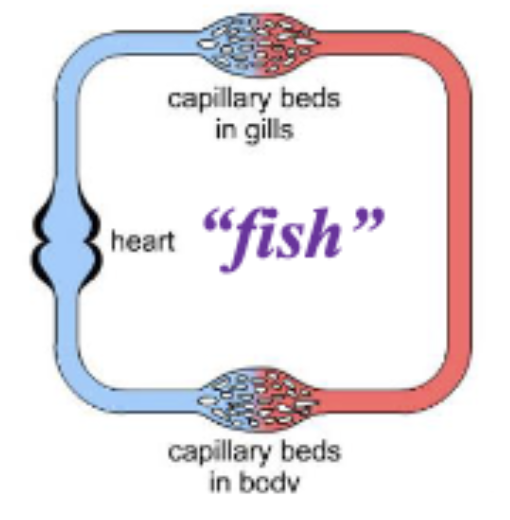
Circulation in Gill pathway
Deoxygenated blood from the tissues (systemic circulation) returns to the heart through the venous system.
It is then pumped through the ventral aorta (VA) to the gills where it is oxygenated.
Oxygenated blood leaves the gills via the dorsal aorta (DA) and flows into the systemic circulation.
The circulation is therefore a single loop – heart to VA to gills to DA to systemic circulation (arteries then veins) and then back to the heart.
heart → ventral aorta → gills → dorsal aorta → systemic circulation → heart
in fish circulatory system blood must be pumped in how many capillary beds
Blood must be pumped across two capillary beds – capillaries in the gills and capillaries in the systemic circulation.
Therefore, the heart must generate sufficient pressure to drive the blood through the entire circuit while simultaneously having a low enough pressure to prevent pressure-induced damage to the delicate gill tissue.
which circulatory system does the lung fish resemble to
more of a mammel than a fish even though its a fish
Circulation in the Lungfish
Deoxygenated blood returns to the heart from the tissues (i.e., the systemic circulation).
It is then pumped through the gills to a pulmonary artery (PA) and then into the lungs where it is oxygenated.
It then returns to the left side of the heart.
It is then pumped through the gills to the dorsal aorta (DA)
then to the systemic circulation (tissues) where it supplies oxygen to the tissues and becomes deoxygenated
It then returns to the right side of the heart.
right artria → right ventricle → gills → pulomnary artery → lungs → left atria → left ventricle → gills → dorsal aorta → systematic circulation →
simmilarty and differences between lung fish and mamalian circulatory system
differences:heart is not completely divided like a mammalian
simmilarity: The right side of the heart receives deoxygenated blood from the systemic circulation and the left side of the heart receives oxygenated blood from the lungs.
role of gills in lung fish circulatory system
the gills play very little role in gas exchange (i.e., O2 uptake from the water or CO2 excretion into the water) since they are much reduced in size
Amphibian Heart
3 chambers
left atrium, right atrium, single ventricle
Amphibian circulatory system
Deoxygenated blood from the tissues enters the sinus venous (part of the venous system)
Then flows into the right atria.
It enters the ventricle and then the conus arteriosus.
This deoxygenated blood then flows through the pulmocutaneous arteries to the lungs and the skin.
Within the lungs and skin, blood picks up oxygen (becomes oxygenated) and also loses CO2
Oxygenated blood from the lungs returns to the left atria via the pulmonary vein.
Then enters the ventricle and the conus arteriosus before moving through the systemic arteries to the tissues (systemic circulation)
where it gives up oxygen to the tissues and picks up CO2 (a waste product of metabolism).
tissue → sinus venous → right atria → ventricle → conus arteriosus → pulmocutaneous arteries → lungs/ skin → pulmonary vein → left atria → ventricle → conus arteriosus → systemic arteries → systemic circulation
how doesnt oxygenated and deoxygenated blood mix within the ventricle
very little mixing occurs. There are anatomical features of the ventricle and the conus arteriosus that prevent this. In addition, slight differences in the timing of contraction of the right and left atria also help prevent this
oxygynated blood = higher con vol %
deoxygynated = lower
Reptilian Hearts - non crocodiles
two atria and two ventricles
the ventricles are not completely divided (i.e., there is a connection between
the left and right ventricle such that blood can move between the two ventricles
three blood vessels that arise directly from the ventricles
pulmonary artery → transports blood to the lungs
two aorta → transport blood to the systemic circulation.
reptilian - circulation non crodile
Deoxygenated blood from the tissues enters the right atrium.
It will then move into the ventricles and ultimately be pumped to the lungs.
Oxygenated blood from the lungs enters the left atrium.
It will then move into the ventricles
Then be pumped through the aorta to the systemic circulation.
In the turtle, the left aorta actually receives deoxygenated blood rather than oxygenated blood.
Reptilian Hearts - crocodiles, parts, vessels, valves
two atria and two ventricles
The ventricles are completely divided into a left ventricle and a right ventricle
Blood cannot move from one ventricle into the other ventricle
pulmonary artery that leaves the right ventricle → deoxygenated blood to the lungs.
left aorta (systemic arch) that leaves the right ventricle → when open it carries
deoxygenated blood to the systemic tissues
right aorta (systemic arch) leaves the left ventricle
valve that separates the right ventricle from the left aorta.
valve that separates the left ventricle from the right aorta
connection between the right aorta and the left aorta → Foramen of Panizza.
Blood Flow from the Crocodilian Heart during Normal Breathing
Low resistance in the pulmonary artery (PA) → Blood easily flows to the lungs.
Deoxygenated blood from the right ventricle (RV) → into the pulmonary artery (PA) → reaches the lungs for oxygenation.
Pressure in the right ventricle (RV) is lower than in the left aorta (LA) → Keeps the RV-to-LA valve closed, preventing mixing.
Oxygenated blood from the left ventricle (LV) → flows into the right aorta (RA) and (to a lesser extent) into the left aorta (LA) through the Foramen of Panizza.
Crocodilian Heart during Normal Breathing simmilarity to humans
Right side pumps deoxygenated blood to the lungs.
Left side pumps oxygenated blood to systemic circulation.
Main difference: Crocodiles have two aortas (RA & LA), while humans have one.
Blood Flow from the Crocodilian Heart during Diving
crocodile doesnt breathe = Lung and pulmonary artery (PA) blood vessels constrict, increasing resistance to blood flow.
Less or no blood reaches the lungs, leading to a pressure build-up in the right ventricle (RV).
Pressure in the right ventricle (RV) exceeds that in the left aorta (LA) → Opens the RV-to-LA valve.
Blood that should have gone to the lungs is redirected into the systemic circulation via the left aorta (LA).
Deoxygenated blood is recycled back into the body instead of being reoxygenated.