TRIPLE: the urinary system
1/37
There's no tags or description
Looks like no tags are added yet.
Name | Mastery | Learn | Test | Matching | Spaced | Call with Kai |
|---|
No analytics yet
Send a link to your students to track their progress
38 Terms
Where are the kidneys located?
At the back of the abdomen
Key point
What is osmoregulation?
It is the process fo maintaining water and salt concentrations (osmotic balance) across membranes within the body
It is an example of homeostasis in the body
What is the importance of osmoregulation?
The cytoplasm of cells and the blood plasma is largely composed of water
Water moves by osmosis
If cells lose or gain too much water by osmosis they do not function properly:
too much water = cells swell and burst + excess water in faeces cannot be absorbed
too little water + too much salt = cells shrink
Water can be lost through ___, ___ and ___.
Exhalation (via the lungs), sweating (from the skin) and in urine (via kidneys)
Water lost through the lungs or skin cannot be controlled, but the volume of water lost in the production of urine can be controlled by the kidneys
What is something neither the skin nor the lungs can do but the kidneys can in relation to water?
Water lost through the lungs or skin cannot be controlled, but the volume of water lost in the production of urine can be controlled by the kidneys
The kidneys are part of which system? What other parts in the body compose this system?
The urinary system
Kidneys, bladder, ureter, urethra, sphincter muscles + renal arteries and renal veins
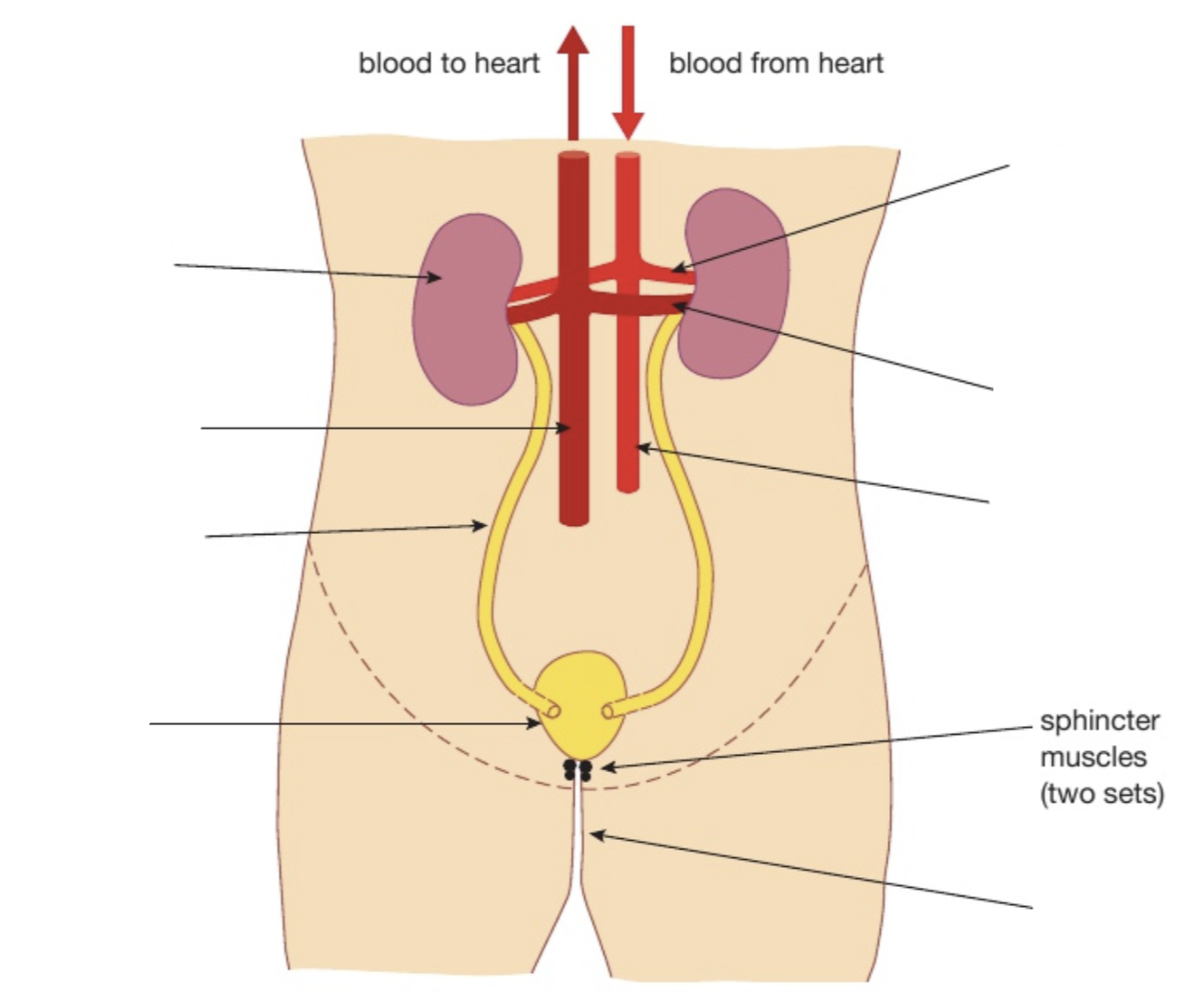
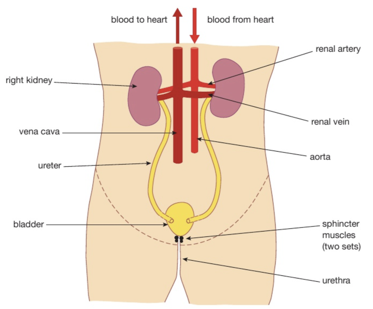
Kidneys: What is their function in the body + Explain?
Functions
Osmoregulation: regulating water content of the blood (vital for maintaining blood pressure and osmoregulation) + salt concentrations
Excrete urea (toxic waste product of metabolism)
Ureter
Tube that transports urine from the kidney to the bladder
Bladder
Organ that stores urine until it is excreted by the body
Urethra
A tube that carries urine from the bladder to the outside of the body
Renal artery
It supplies high pressure blood straight from the body’s main artery (the aorta) containing oxygen, urea, water, salt, glucose, etc
Renal vein
After the blood has been filtered, the ‘cleaned’ blood passes out through each renal vein to the main vena cava
What is the function of the nephron?
To filter the blood and form urine
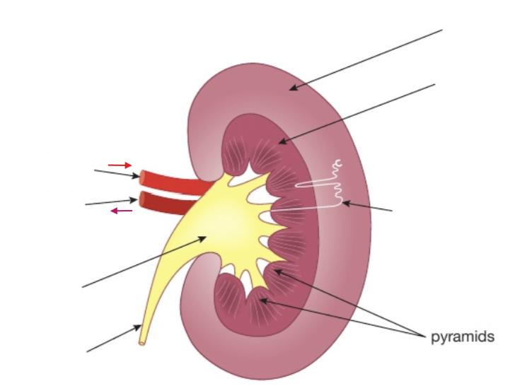
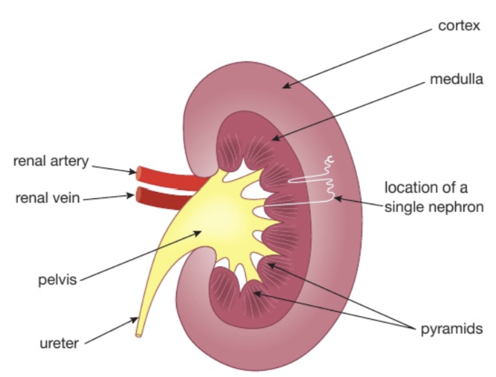
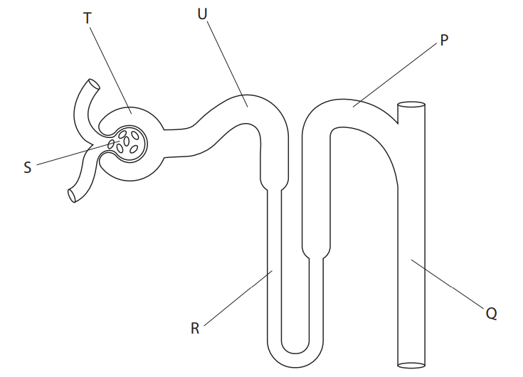
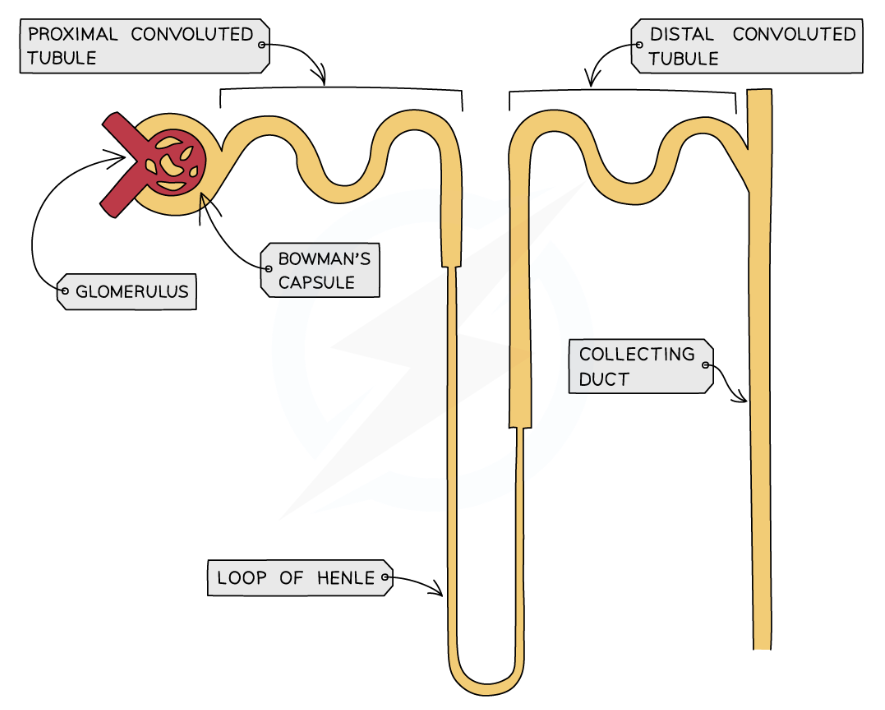
Different parts of the nephron are located in different areas of the kidney. Tell me which are located where.
Bowman’s capsule, glomerulus, first coiled tubule/proximal convoluted tubule and second coiled tubule/distal convoluted tubule are found in the cortex, which is the outermost layer of the kidney
The loop of Henle and collecting duct are found mainly in the medulla, the inner region of the kidney
What are the main processes that occur in the nephron to form urine?
1. Ultrafiltration
2. Selective reabsorption of glucose
3. Reabsorption of water
Describe what happens at the start of the formation of urine
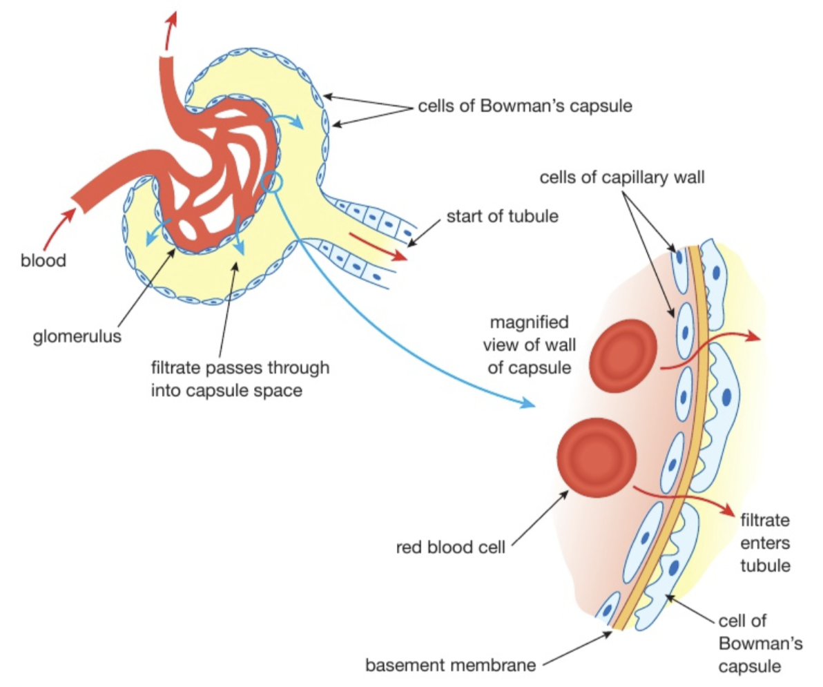
Ultrafiltration occurs in the glomerulus (surrounded by the Bowman’s capsule)
Blood enters the kidney through the renal artery and divides into smaller capillaries. The smallest arteries supply the capillaries of the glomerulus
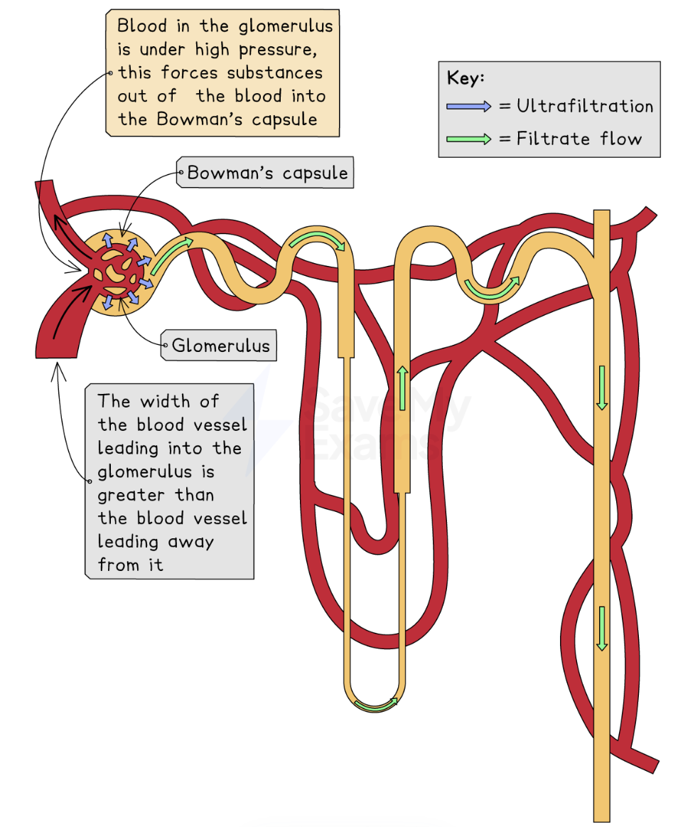
Blood in the arteries supplying blood to the glomerulus are at a high pressure, forcing the fluid from the blood through the walls of the capillaries of the glomerulus and the Bowman’s capsule into the capsule
Blood in the glomerulus and the space in the capsule are separated by two layers of cells, the capillary wall and the wall of the capsule. Between these two layers there is a third one called the basement membrane which is not made of cells.
These 3 layers act like a filter, allowing small molecules to pass through but holding back larger ones)
What is the name given to the smallest arteries that supply the capillaries of the glomerulus?
Arterioles
What causes the high pressure? Why do we need high pressure
The width of the lumen of the arteriole (blood vessel leading into the glomerulus) is greater than the width of the lumen leading away from the glomerulus
The difference in width increases the pressure of the blood in the glomerulus.
We need it for ultrafiltration, so that small molecules are forced out into the Bowman’s capsule
What is the fluid that collects in the Bowman’s capsule? What are the substances that are present there?
Glomerular filtrate
Water
Glucose
Urea
Ions (salts)
What happens after ultrafiltration?
The glomerular filtrate passes through the first coiled tubule and selective reabsorption occurs
All the glucose is selectively reabsorbed from the filtrate in the first coiled tubule back into the capillaries by active transport because glucose is a useful substance
All the glucose along with most of the sodium and chloride ions are reabsorbed
What do the cells of the first coiled tubule contain? Why?
They contain many mitochondria to provide energy (ATP) needed for active transport of glucose
What is selective reabsorption?
Process taking place in a kidney tubule whereby different amounts of substances (e.g glucose) are absorbed from the filtrate back into the blood
What happens after selective reabsorption?
The filtrate passes through the rest of the nephron: loop of Henle, second coiled tubule where reabsorption of water occurs
Some of this process occurs in the loop of Henle but most of it occurs from the collecting duct
Water is reabsorbed into the blood by osmosis
What is the reabsorption of water in the nephron an example of?
Osmoregulation (maintaining a constant water and salt content in the body)
How is the amount of water being reabsorbed from the filtrate controlled?
It is dependent of the concentration of the hormone ADH (antidiuretic hormone). REMEMBER this phrase ‘ADH increases the permeability of the collecting duct to water’
More ADH → it causes the collecting ducts to become more permeable so more water is reabsorbed back into the blood → urine is more concentrated, lower volume of water in urine
Less ADH → it causes the collecting ducts to become less permeable so less water is reabsorbed → urine is less concentrated (dilute), larger volume of water in ruine
What releases ADH?
Pituitary gland
Changes in blood water levels are detected by osmoreceptors in the hypothalamus in the brain
The hypothalamus sends signals to the pituitary gland, which releases ADH
Osmoregulation is an example of ___
Definition of this?
Negative feedback
Process where a change in the body is detected and brings about events that return conditions to normal
What is another substance present in urine? How does this solute end up inside the urine? What is the difference in the concentration of this substance in the urine vs in the blood?
Ammonium ions
These are secreted into the fluid as it passes along the tubule
The concentration of the ammonium ions in the urine is about 150 times what it is in the blood
The kidney ___ the concentration of urine and so ___ the water content of the blood
The kidney controls the concentration of urine and so regulate the water content of the blood
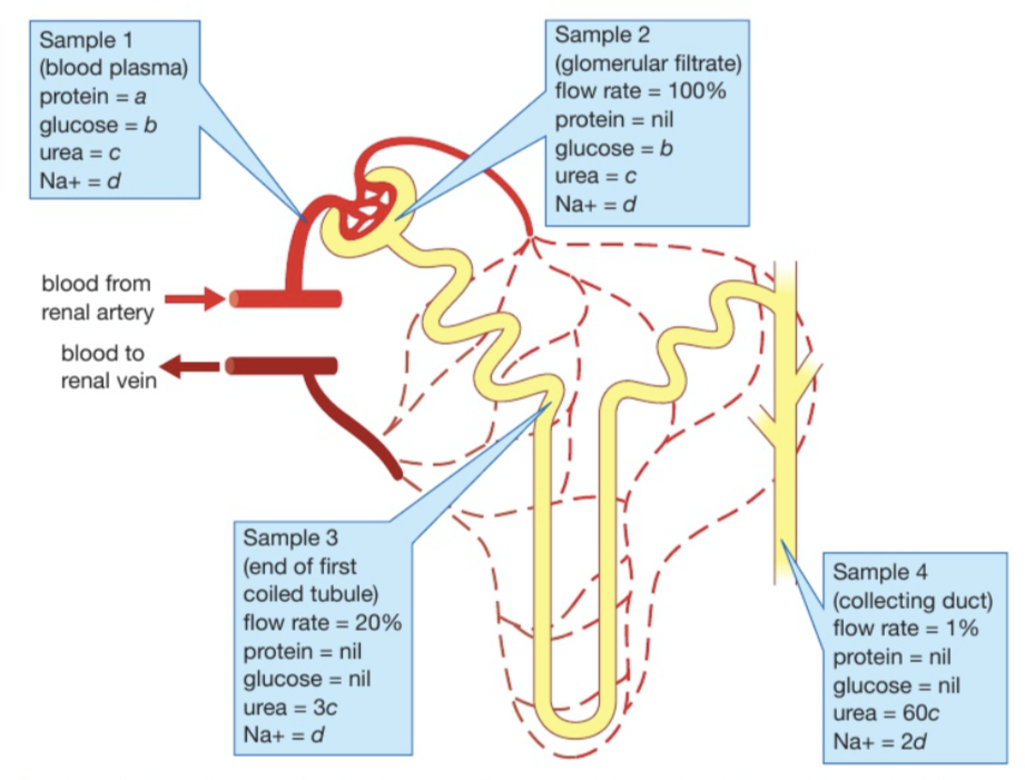
Describe and explain what happened to the concentration of urea during the formation of urine
The flow rate is a measure of how much water is in the tubule
At sample conc is 1c
At sample 2 conc is 1c (same conc as before because volume of solution has not changed. The flow rate is still 100%)
At sample 3, conc is 3c because flow rate has dropped to 20% = 80% of the water has been reabsorbed but some urea has also been diffused out into the medulla
At sample 4, no urea has been diffused out from point 3 to 4. This is because the flow rate has fallen from 20% → 1% so it has fallen 20x and 3c x 20 = 60c

How do we know that some of the urea has been diffused out from sample 2 to sample 3?
How do we know no urea has been diffused out form sample 3 to sample 4?
Because flow rate has decreased from 100% to 20% meaning that it is 1/5 of what it was
If we hadn’t lost any urea then concentration would be 5c, not 3c because its concentration at sample 3 should be 5 times what it was in sample 2.
We calculate this by dividing 100/20 = 5 so everything should be 5 times more conc
Because flow rate has fallen from 20% → 1% and 20/1 = 20. We should multiply conc at sample 3 (3c) by 20 which gives us 60c.
TIPS AND TRICKS:
Remember to correctly describe where substances are moving from and to in the kidneys and HOW they are moving
e.g small substances such as urea are forced out of the blood during ultrafiltration, they don’t diffuse out of the blood
Some exam questions might present a table that gives you details about the relative conc of substances in filtrate at diff parts of the nephron.
What would the conc of glucose, protein, urea in a healthy individual in the nephron?
What problem might an individual have if there is glucose present after the first tubule? What about if there is protein in the urine?
Healthy concs
No protein in the tubules at all (only in glomerulus)
No glucose after the first coiled tubule
Greatest conc of urea in the filtrate in the collecting duct
Problems
Glucose = blood glucose levels are higher than normal (e.g person is diabetic)
Protein in urine = blood pressure in arterioles are too high so causes damage to the glomerulus and protein molecules may be squeezed/forced out of the glomerulus into the Bowman’s capsule. The protein cannot be reabsorbed back into the bloodstream from the nephron, so it will end up in the urine.
What should be present int he urine?
Urea, excess ions, excess water
What are things that affect the conc of urine?
Water intake
Temperature — high temp = more conc urine as some water is lost in sweat so less will be lost in urine
Exercise — more exercise = more water lost as sweat (same as temp)