Looks like no one added any tags here yet for you.
cyanosis
light skin: bluish
dark skin: ashen gray
pallor paleness
light skin: loss of rosy glow
dark skin: ashen gray, yellowish brown
erythema redness
light skin: visible redness
dark skin: rely on palpation or warmth, edema
petechiae small pinpoint
light skin: purplish pinpoints
dark skin: usually invisible, check oral mucosa, conjunctiva, eyelids
malignant skin lesions
basal cell carcinoma
squamous cell carcinoma
malignant melanomas
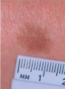
macules
solely a color change, flat & circumscribes, less than 1cm
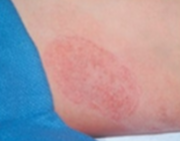
patches
macules larger than 1cm
papule
felt & caused by superficial thickening of the epidermis
plaques
papules coalescing by superficial thickening of the epidermis
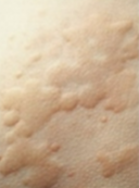
wheal
superficial, raised, transient, and erythematous; irregular in shape due to edema
urticaria (hives)
wheals coalesce to form extensive pruritic reaction
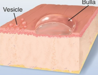
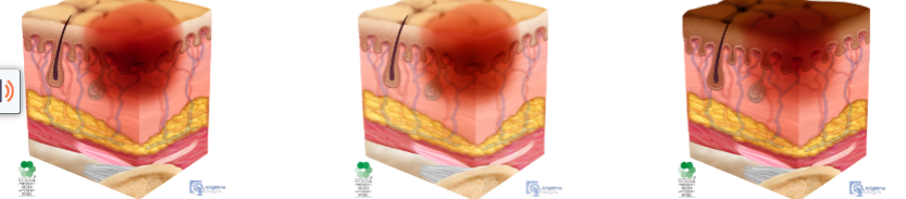
vesicles
elevated cavity containing fluid up to 1cm (blister)
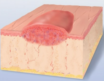
bulla
larger than 1cm, usually single chamber, superficial in dermis & ruptures easily
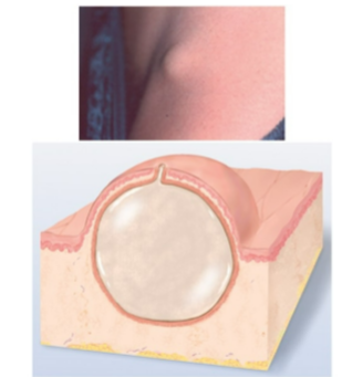
cysts
encapsulated fluid dilled cavity
pustules
pus in cavity that is circumscribed & elevated
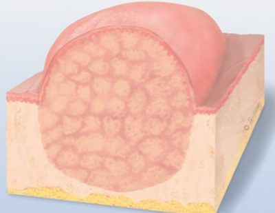
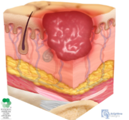
nodule
solid elevated, hard or soft, greater than 1cm, may extend deeper into dermis than papule
tumor
larger in diameter, firm or soft, deeper into dermis, benign or malignant
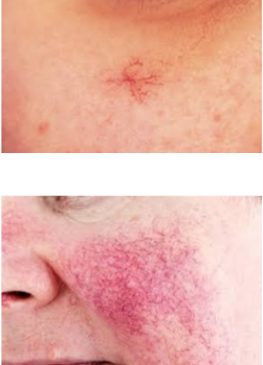
telangiectases
spider star or spider angioma, rosacea
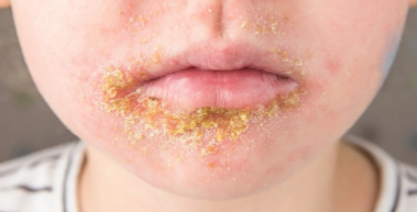
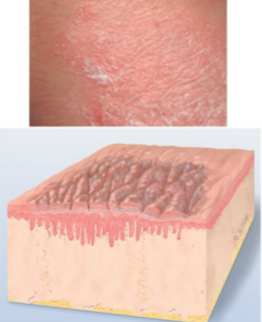
crust
thickened dried out exudate
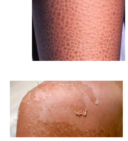
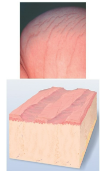
scale
compact flakes of desiccated skin from shedding dead excess keratin cells
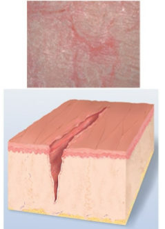
fissures
linear crack w/ abrupt edges extending into dermis
erosions
scooped out but shallow depression
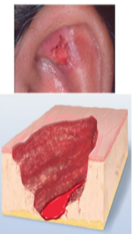
ulcers
deeper depression extending into dermis w/ irregular shape, may bleed, leaves scar
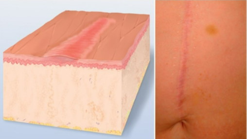
scar
permanent fibrotic change after healing
atrophic scares
resulting skin level is depressed w/ loss of tissue & thinning
lichenification
prolonged intense scratching leading to thickened skin producing tightly packed set of papules
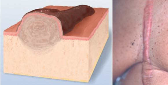
keloids
benign excess of scar tissue beyond original injury
excoriations
self-inflicted abrasion that is superficial
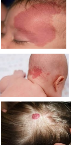
hemangiomas
port win stain (nevus flammeus)
strawberry mark (immature)
cavernous hemangioma (mature)
purpuric legions
petichae, ecchymosis, purpura
annular or circular
begins in center & spreads to periphery
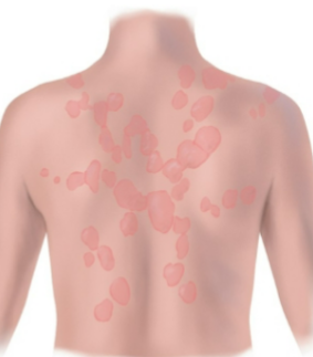
confluent
lesions run together
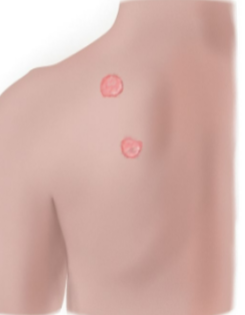
discrete
distinct & separate
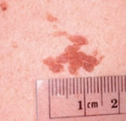
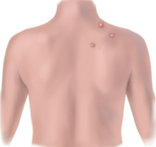
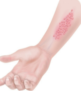
grouped
cluster of lesions
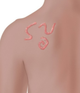
gyrate
twisted, coiled, or snakelike
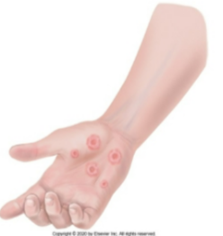
target or iris
resembles iris of eyes, concentric rings
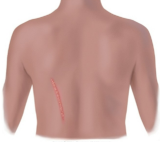
linear
scratch, streak, line, or stripe
polycyclic
annular lesions grow together
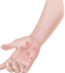
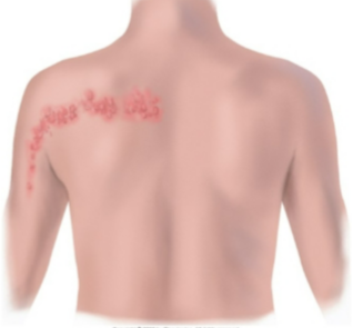
zosteriform
linear arrangement following a unilateral nerve route
stage 1 pressure ulcer
intact skin w/ localized are of non-blanchable erythema
stage 2 pressure ulcer
partial thickness loss of skin w/ exposed dermis
adipose not visible
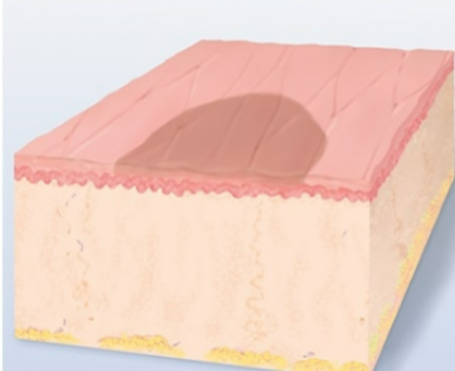
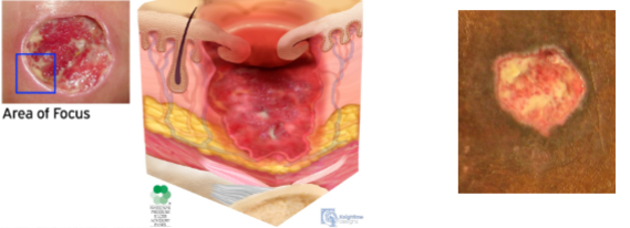
stage 3 pressure ulcer
full thickness loss of skin
adipose is visible
granulation tissue and epibole (rolled wound edges) often present
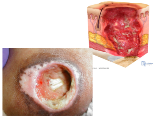
stage 4 pressure ulcer
full thicken skin & tissue loss w/ exposed fascia, muscle, tendon, ligament, cartilage, or bone in the ulcer
epibole, slough, and eschar often visible-
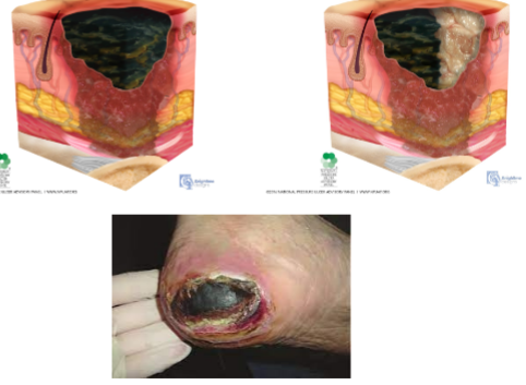
unstageable pressure ulcer
full thickness skin & tissue loss to extent that damage cannot be confirmed due to slough or eschar obscuring it
Braden scale
predicts pressure sore risks from
sensory perception
moisture
activity
mobility
nutrition
friction & shear