Bio 315 Circulatory System I
1/136
There's no tags or description
Looks like no tags are added yet.
Name | Mastery | Learn | Test | Matching | Spaced | Call with Kai |
|---|
No analytics yet
Send a link to your students to track their progress
137 Terms
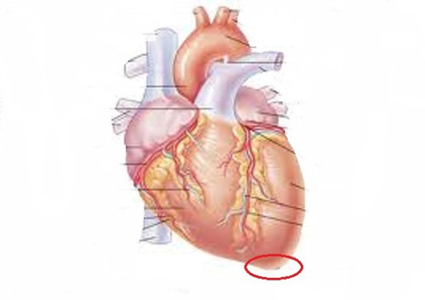
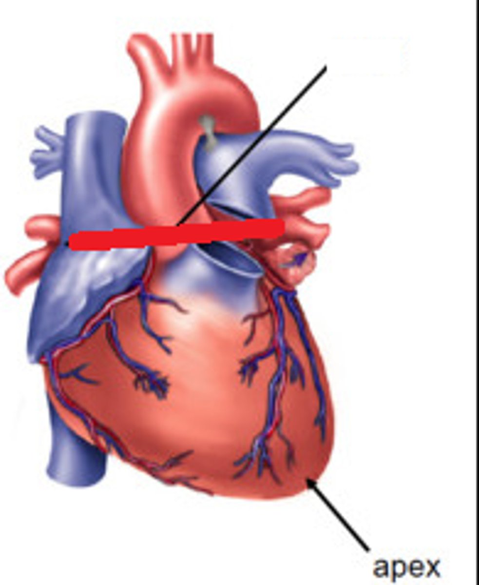
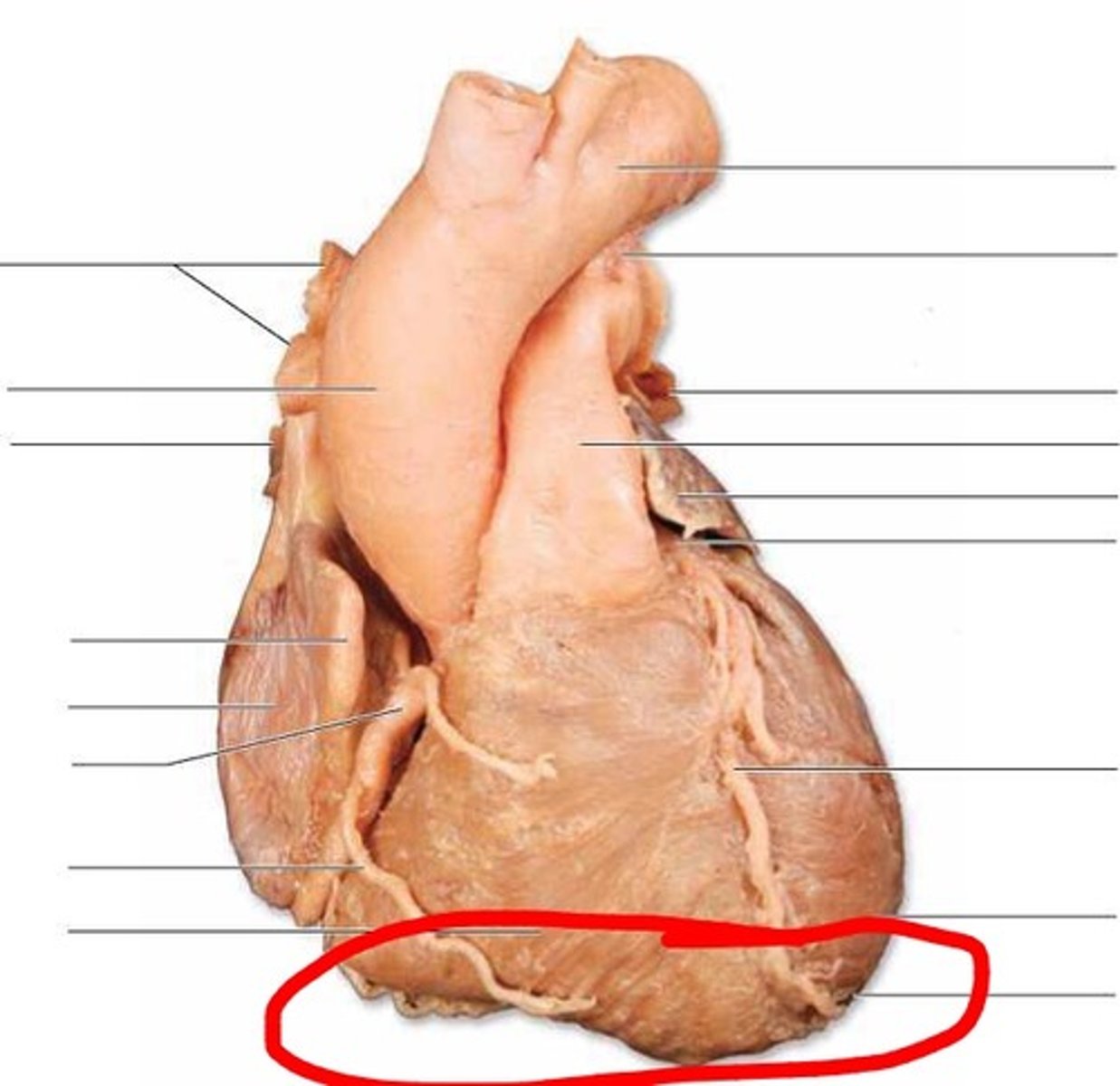
apex of the heart
Feature; bottom tip of heart, goes towards Left side of body
Base of the heart
Feature;
chock and everything below
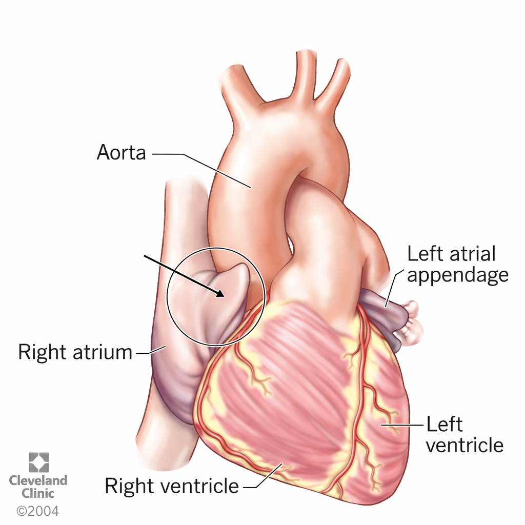
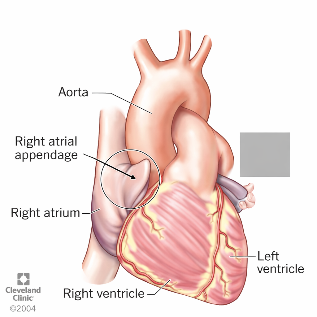
R atrium of the heart
Chamber; pinch top pt right side
H1
1
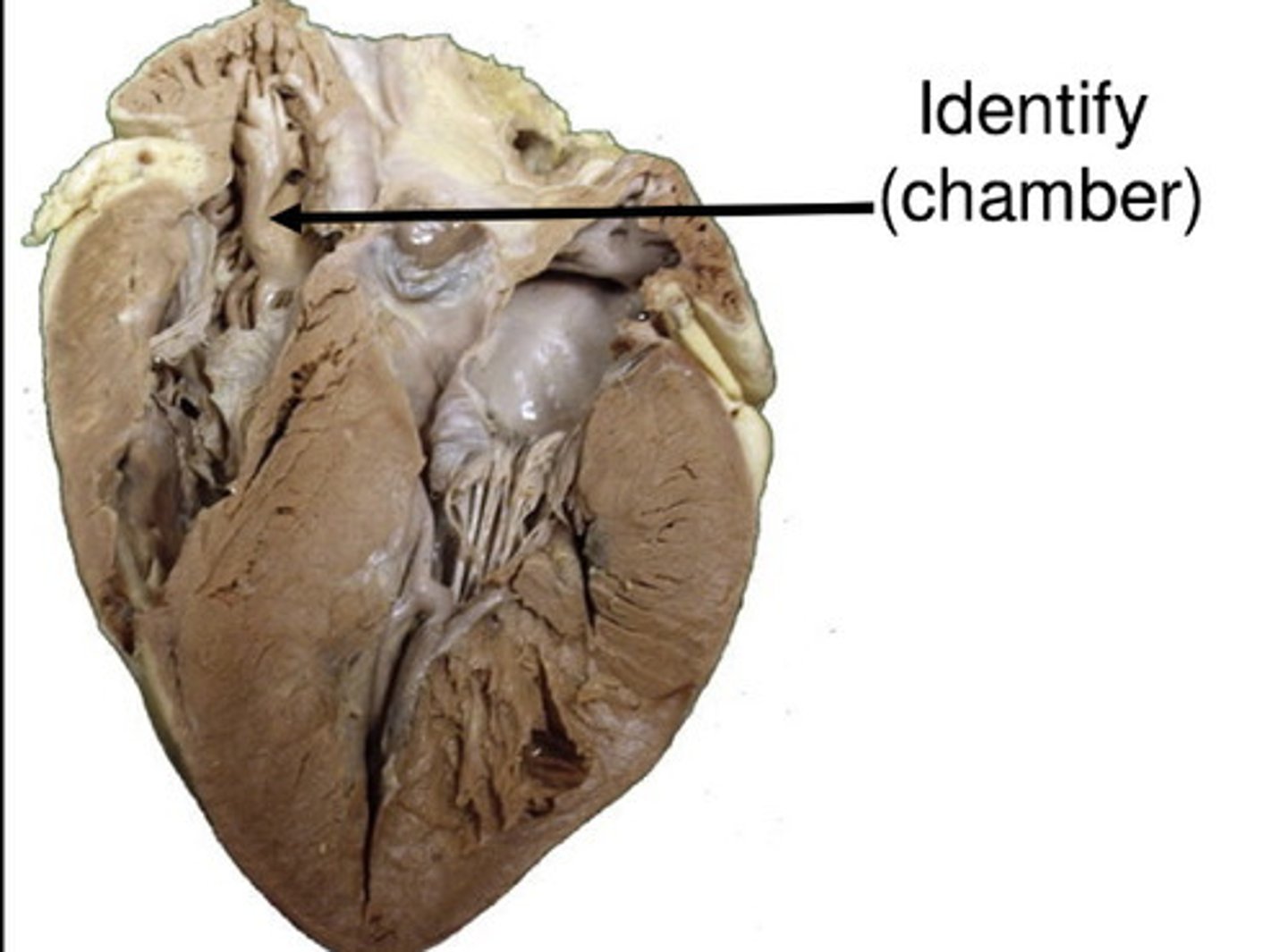
R auricle of the heart
Feature; above right atrium, black nub
smaller more charred
H5
2
L atrium of the heart
Chamber; left side top pt LL
LUQ
H1
28

left auricle of the heart
Feature; elephant ear, chunky nub on top of Left atrium
on pt L side more thick
H5
14
Right ventricle
Chamber; pinch pt right bottom
LRQ
5
L ventricle
Chamber; pinch pt bottom left, bigger
LLQ
15

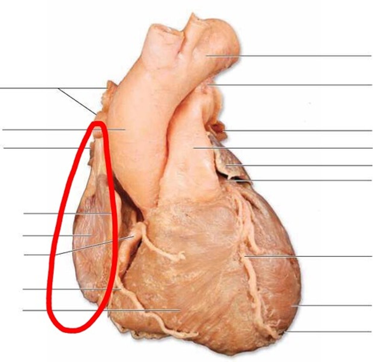
anterior surface of the heart
Region; hold heart where it sits in chest, wave hand in front
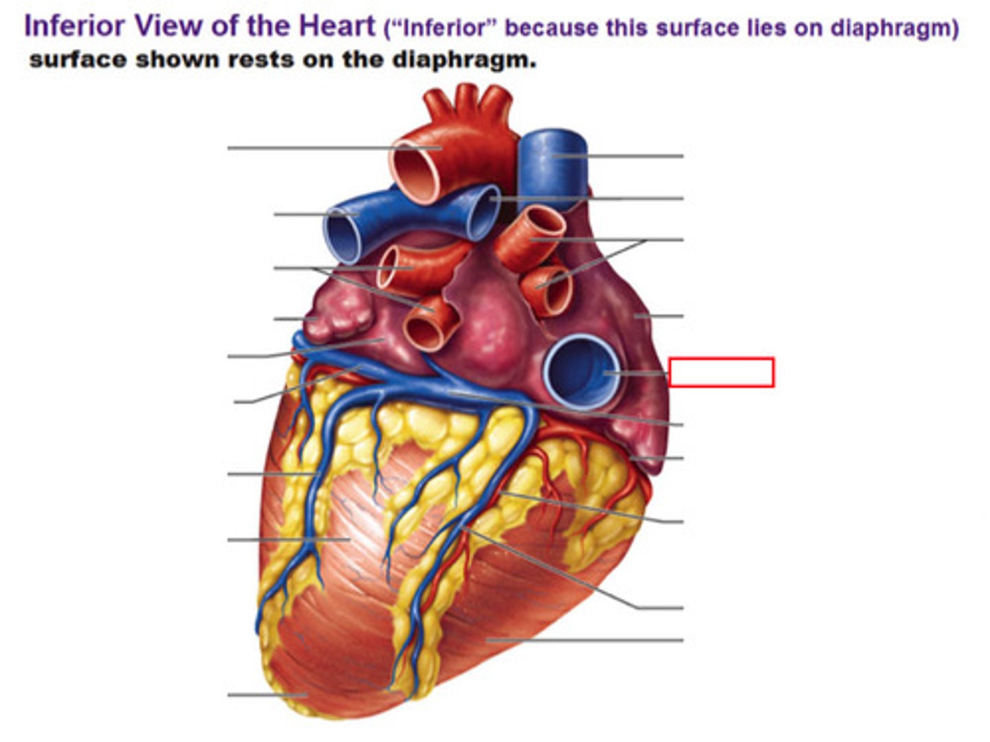
Diaphragmatic surface of the heart
Region, hold heart where it sits in chest, wave hand below it
R. pulmonary surface of the heart
Region; hold heart where it sits in chest, wave hand on medial side
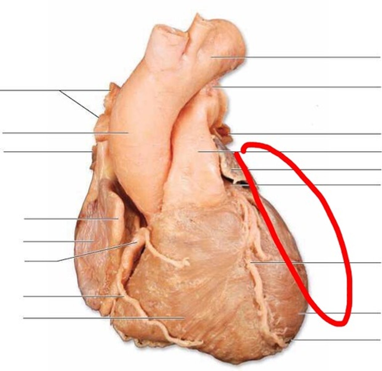
L pulmonary surface of the heart
Region; hold heart where it sits in chest, wave hand on lateral side
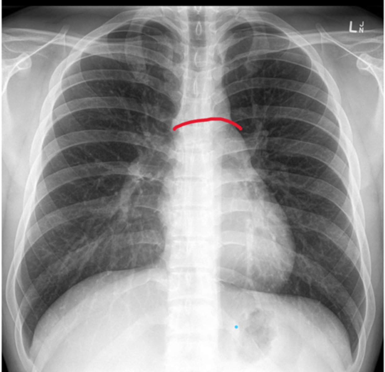
Right border of the heart
Boundary; X-ray, medial edge
L border of the heart
Boundary, X-ray, lateral edge, apex points this way

Inferior border of the heart
Boundary; X-ray, bottom edge
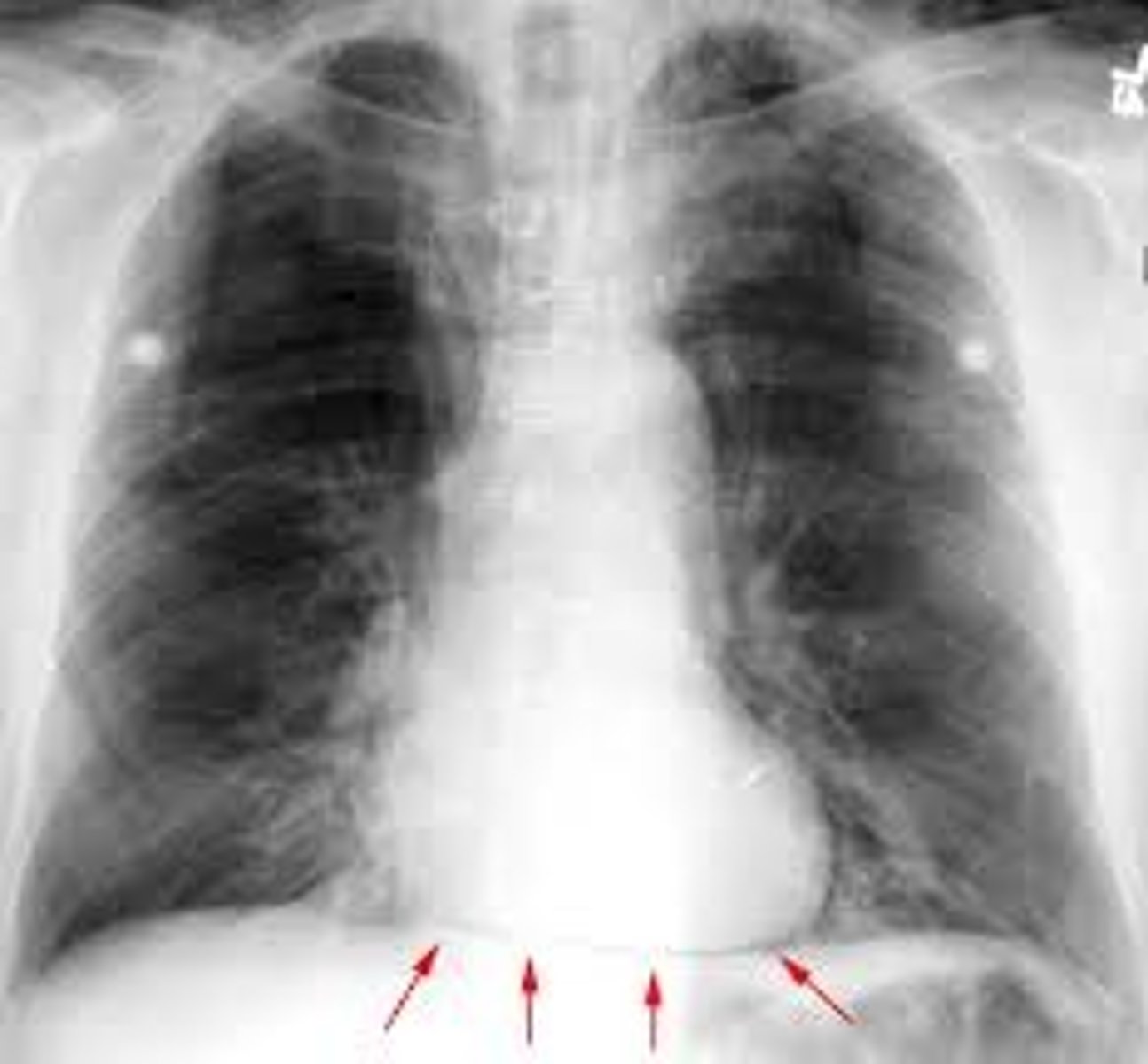
Superior border of the heart
Boundary; X-ray, top edge
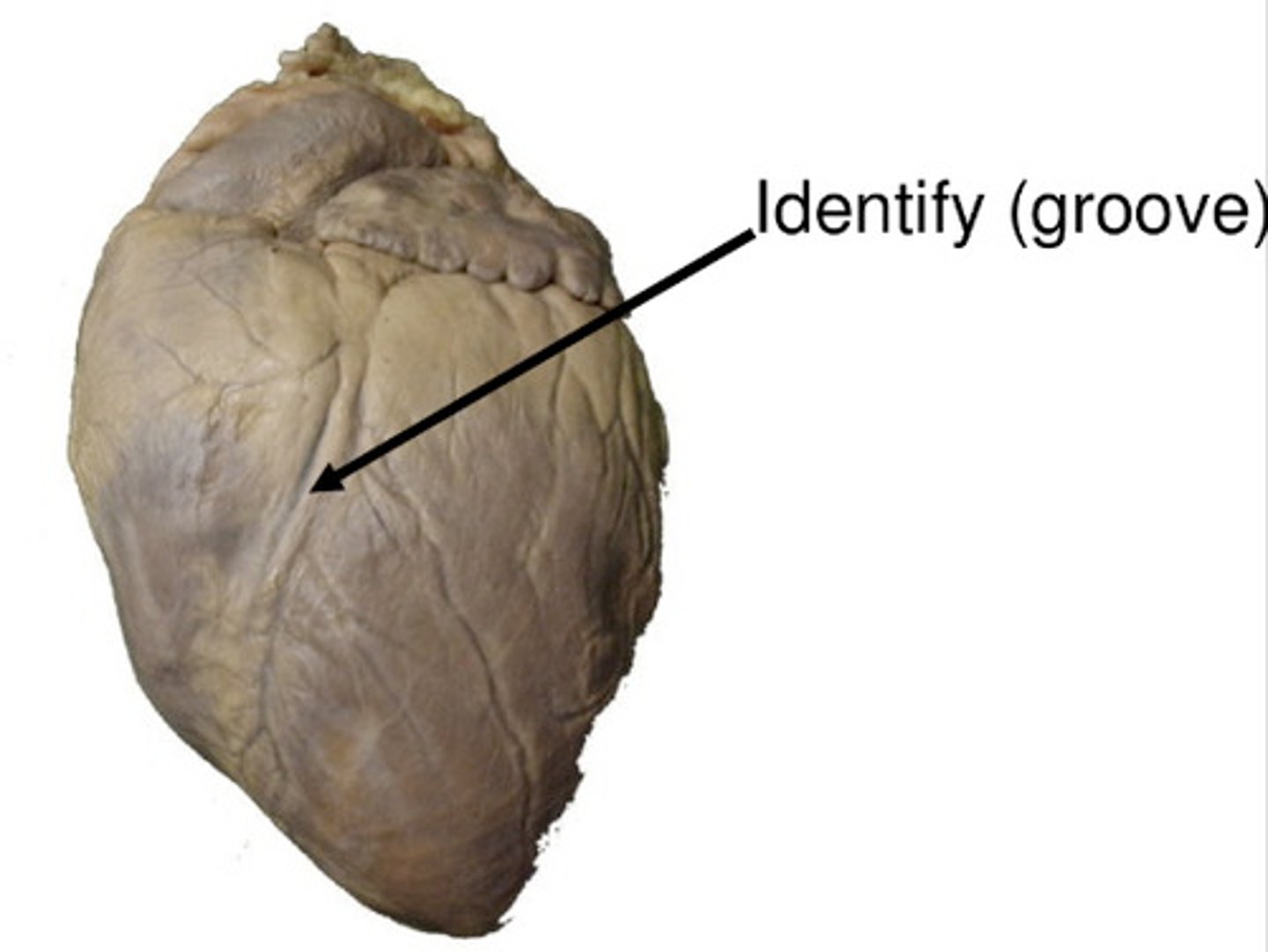
Coronary sulcus
Depression, goes from Right auricle to Left auricle, probe lays in
H1
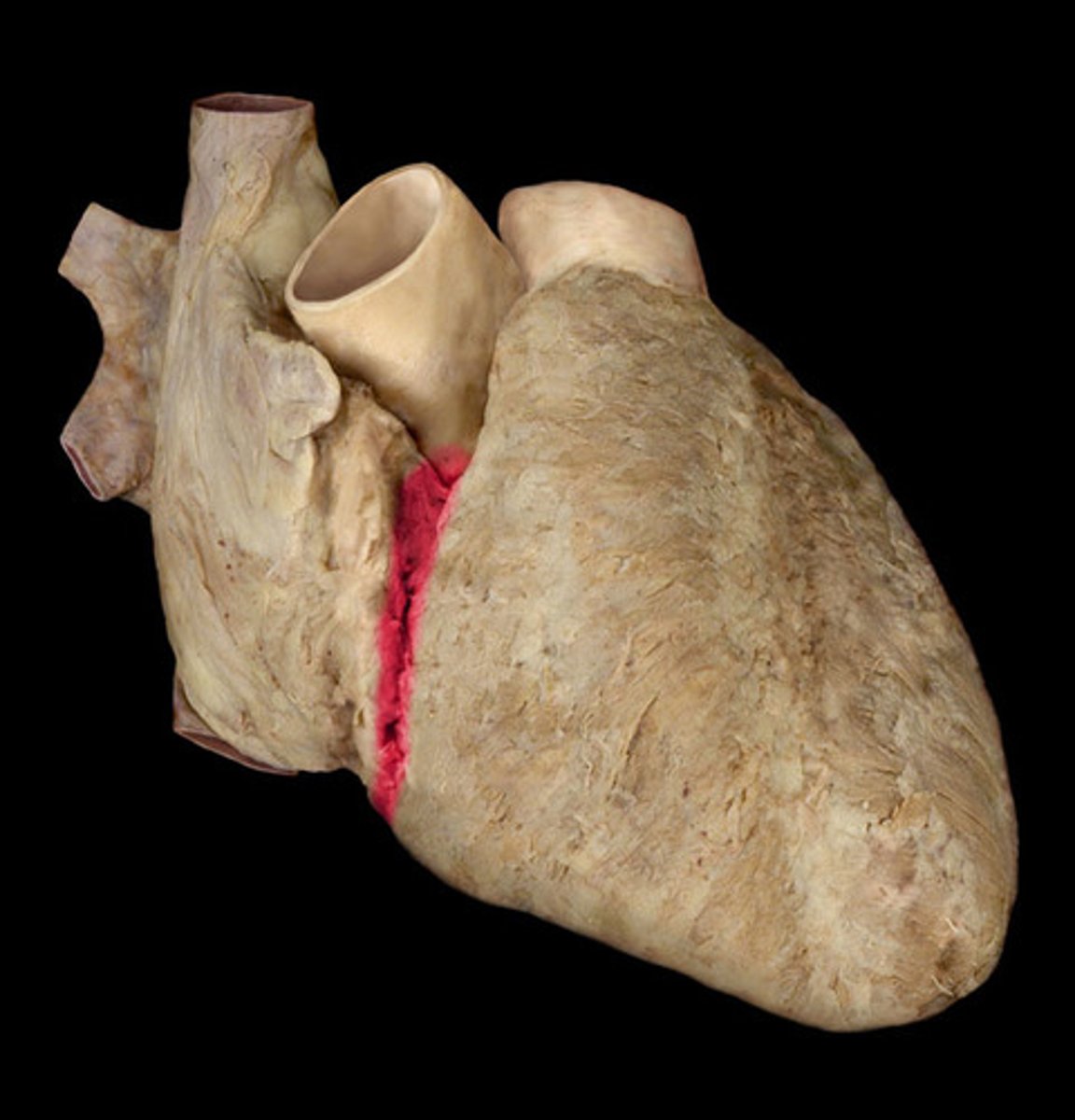
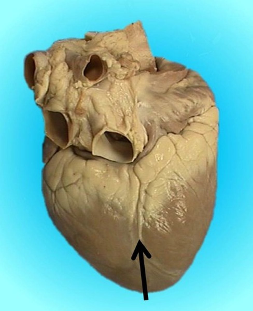
anterior interventricular sulcus
Depression; divot on right side, anterior, by right ventricle
H1
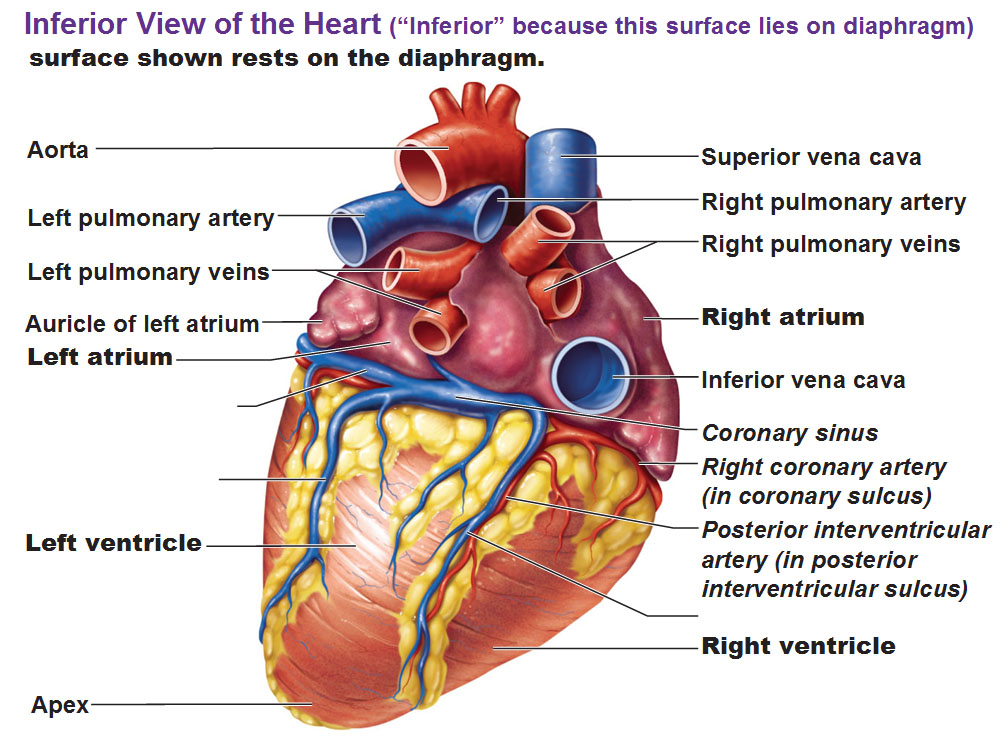
posterior interventricular sulcus
Depression; divot on right side, posterior, by right ventricle
H1
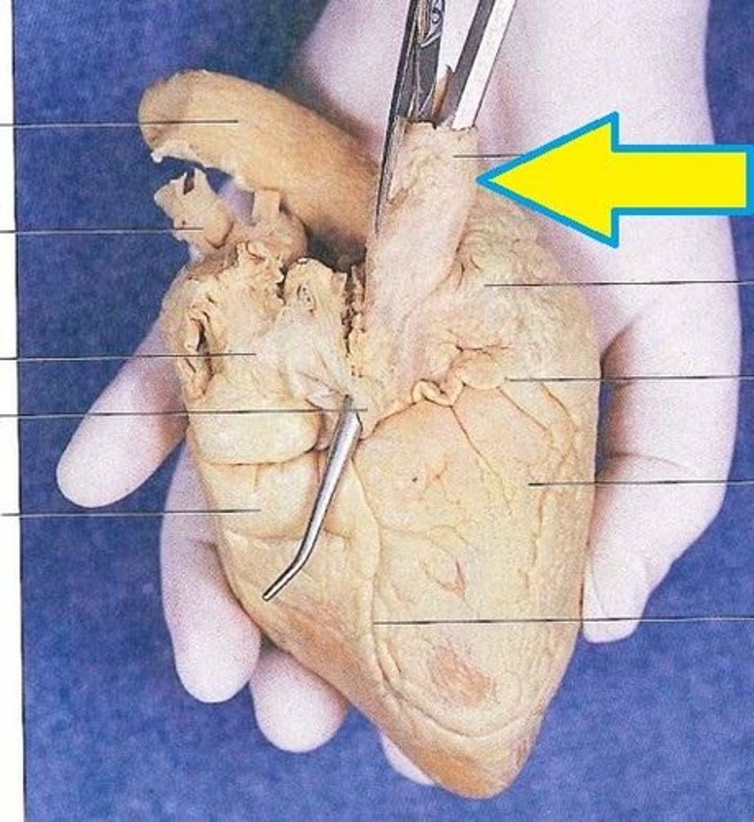
Aorta
Structure, big tube off heart with the “fingers”
light color
H1
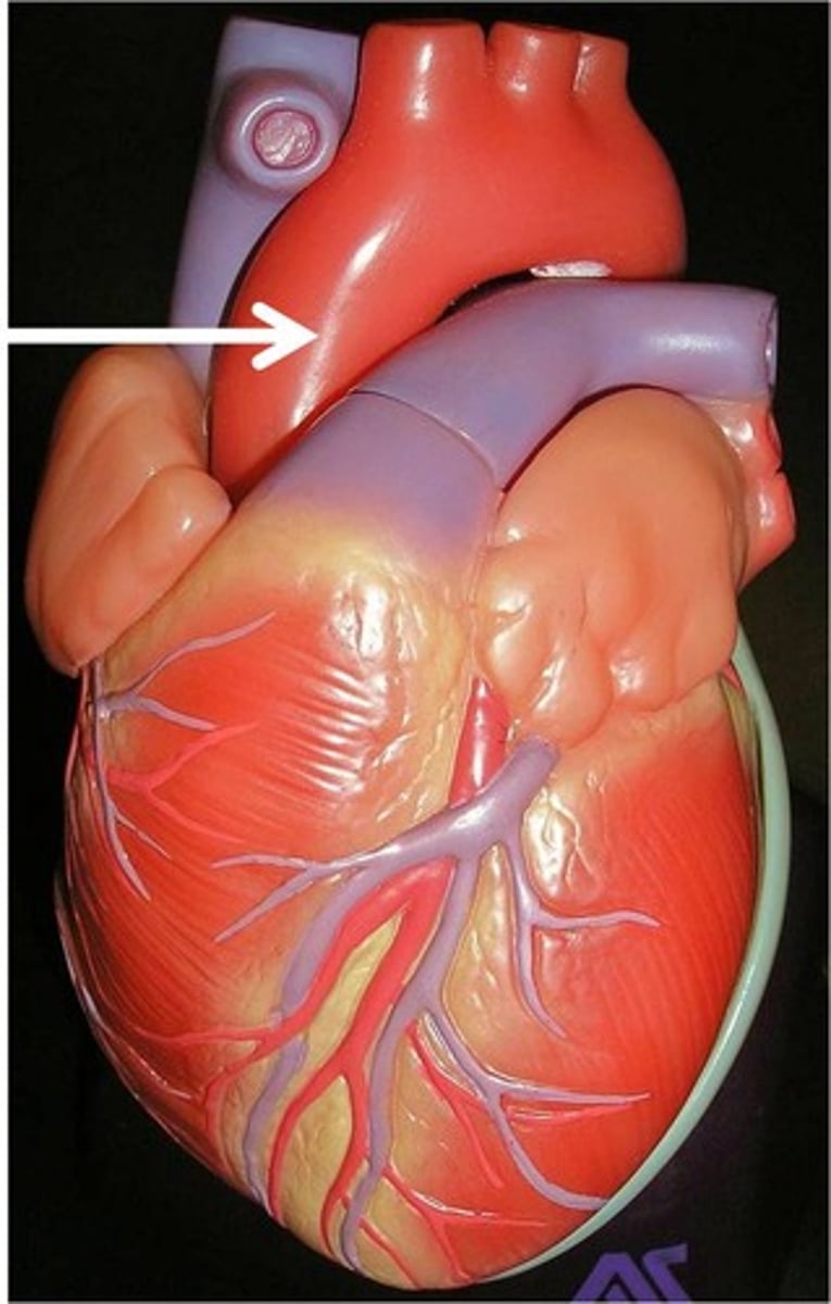
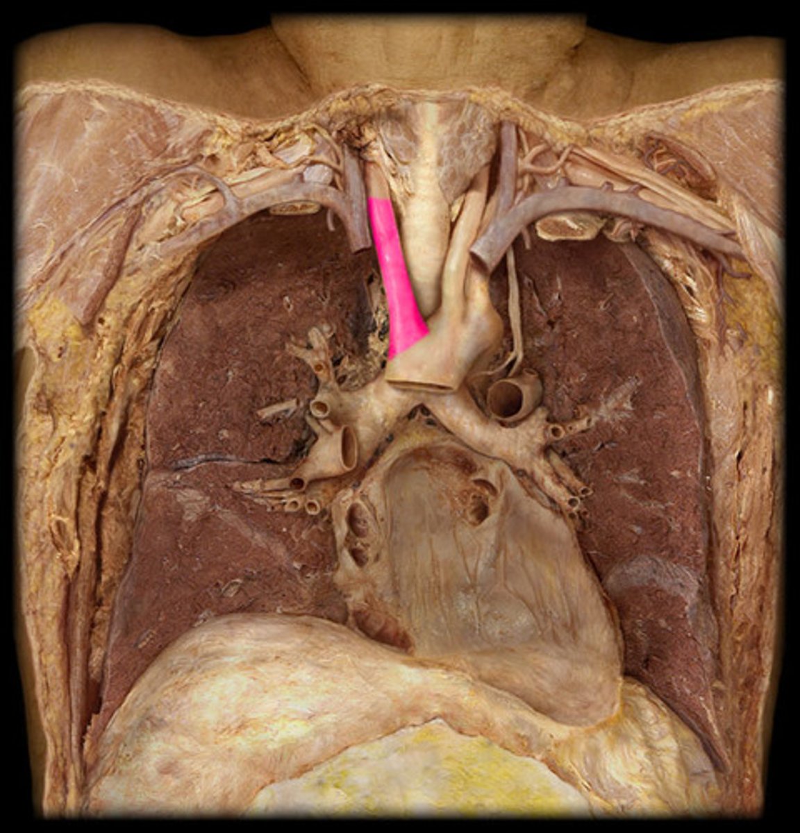
ascending aorta
Portion; start of aorta, before curve
H1
7
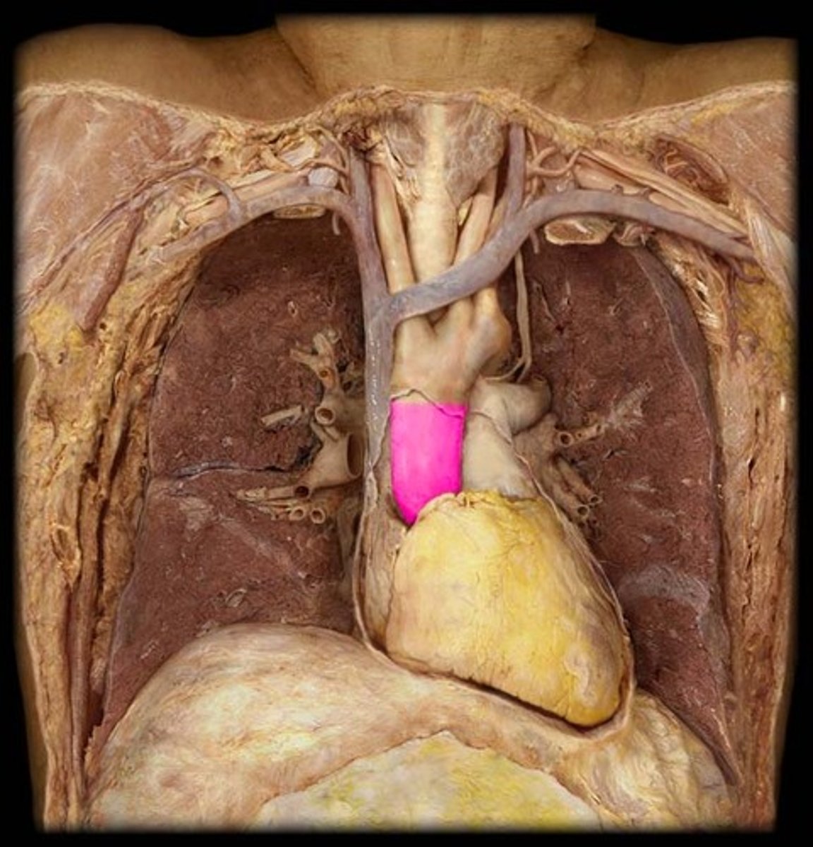
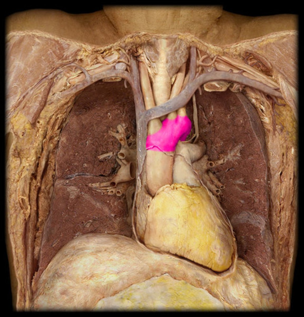
Arch of aorta
Portion; curve upper pt
“rainbow” part
H1
8
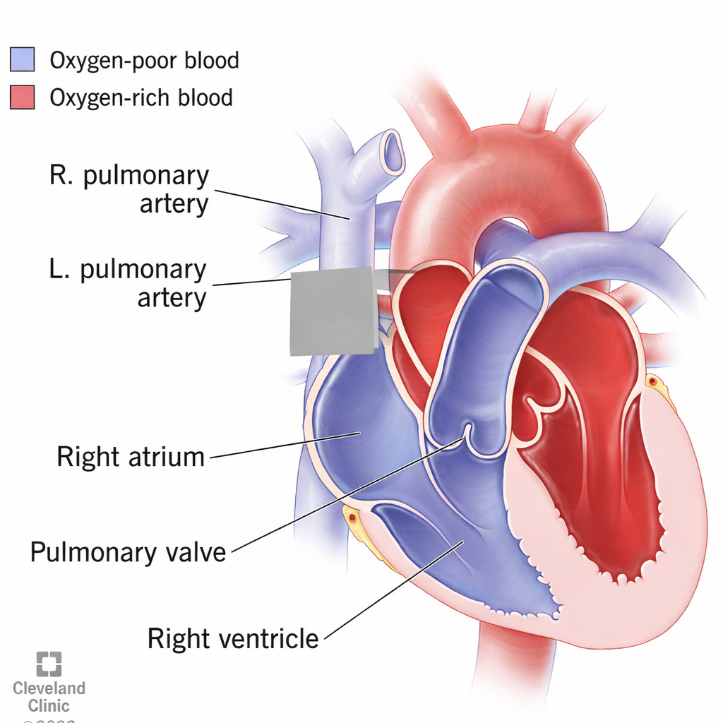
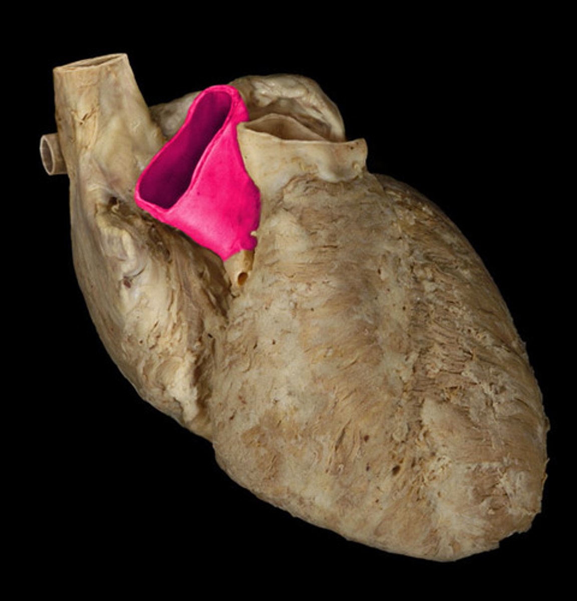
Pulmonary trunk
Collective structure; under aorta, splits into 2
6
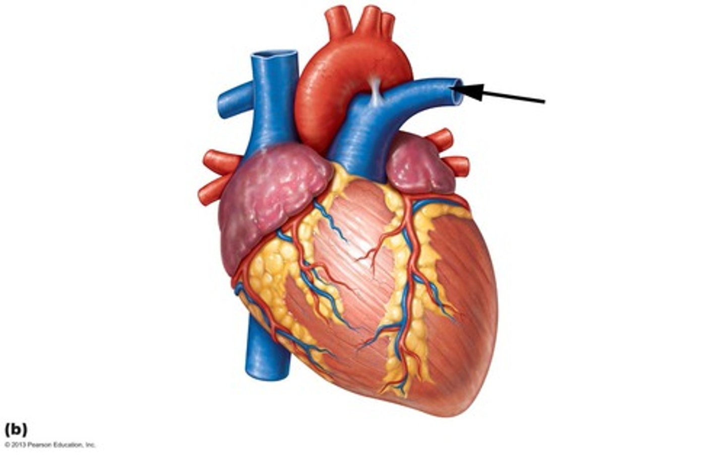
L pulmonary vein
Structure; lateral spit off trunk
H2
32
R pulmonary vein
Structure; medial split off trunk
34
ligamentum arteriosum
webbing connecting aorta and pulmonary trunk
12
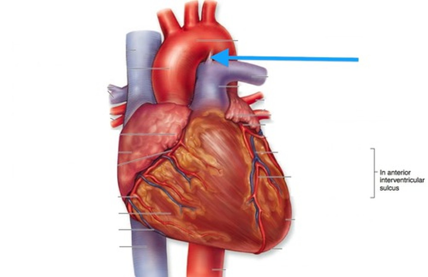
Ligamentum arteriosum
BQ: vestige of the fetal ductus arteriosus, which shunted blood in the pulmonary trunk away from the lungs in the fetus
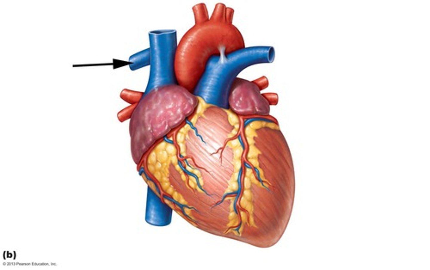
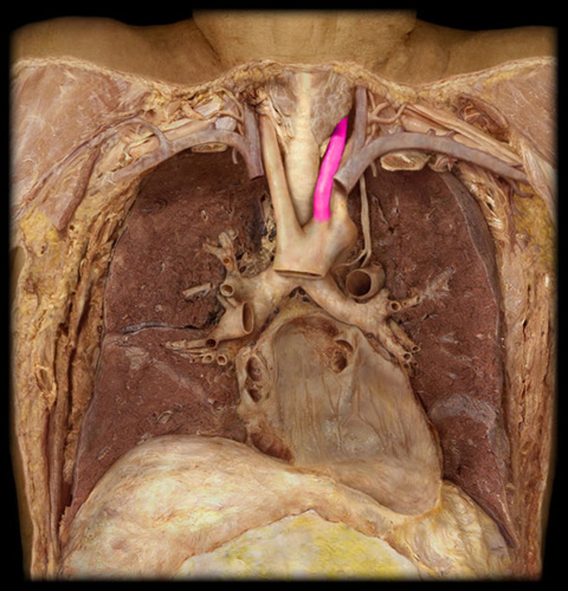
Superior vena cava
st
Posterior side, probe from above on right
H3
3
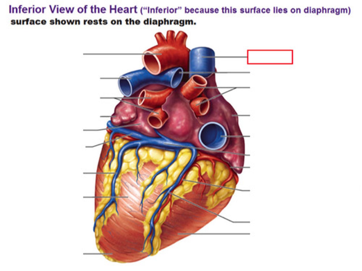
inferior vena cava
st
Posterior side, probe from below on right
H3
4
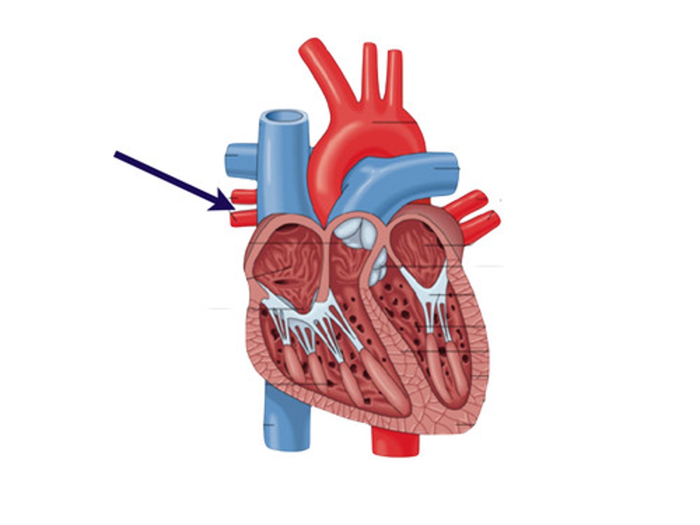
pulmonary veins
collective st
2 holes pipe cleaners go through
H3
L superior pulmonary vein
Posterior side, stick 2 probes through horizontally
red
H3
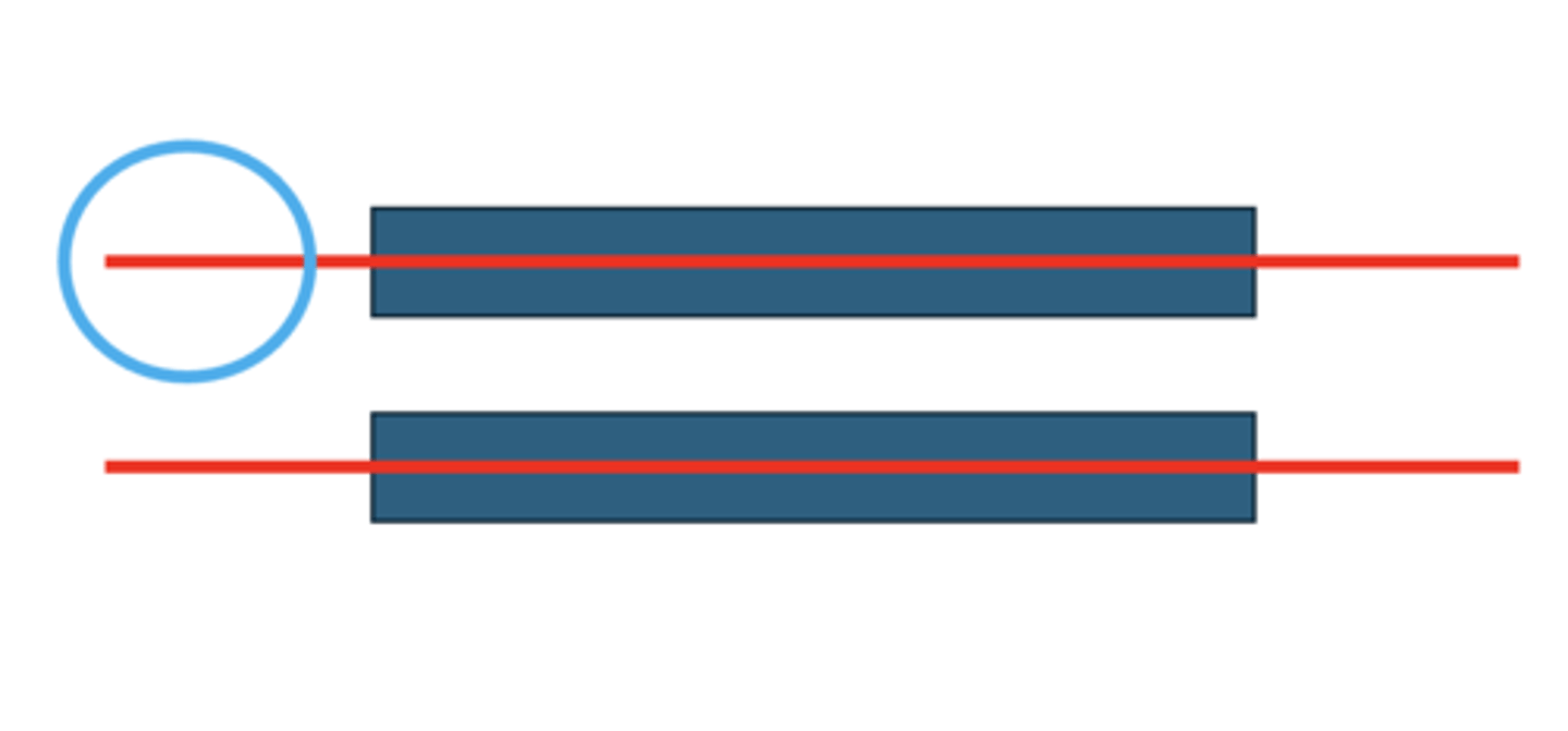
R superior pulmonary vein
Posterior side, stick 2 probes through horizontally
purple
H3
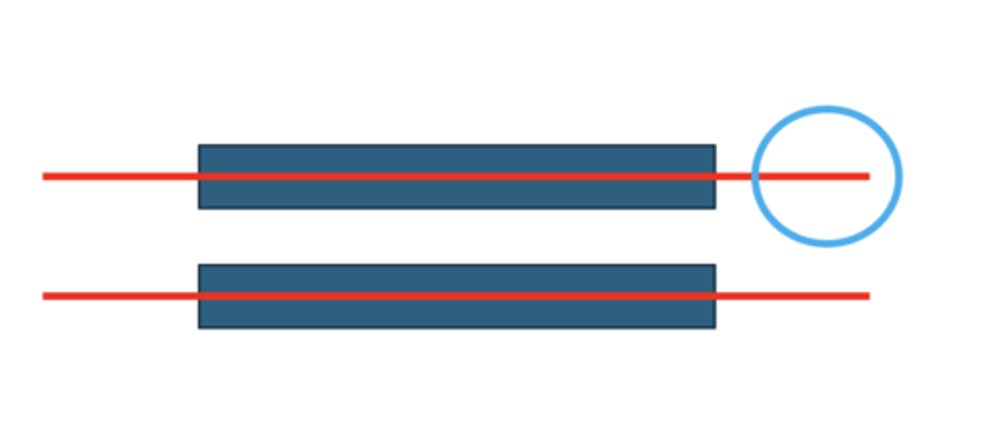
L inferior pulmonary vein
Posterior side, stick 2 probes through horizontally
brown
H3
R inferior pulmonary vein
Posterior side, stick 2 probes through horizontally
brown
H3
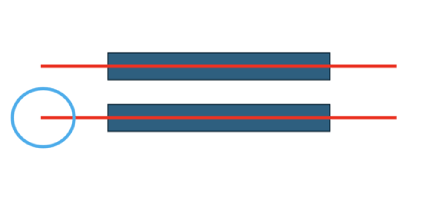
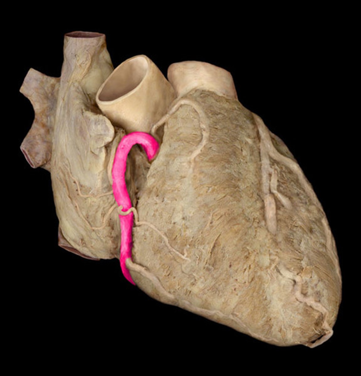
R coronary artery
st
pull right auricle back to show tube on anterior side
H1
16
R marginal artery
st
forms Y-shape off of coronary artery
1st branch off coronary artery
H1
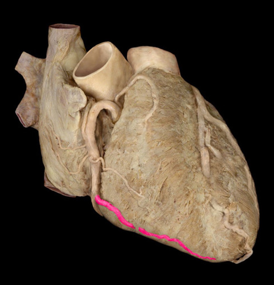
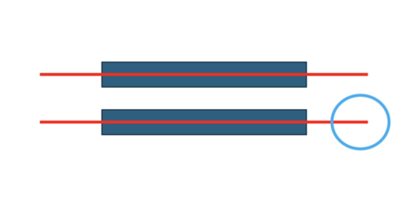
posterior interventricular artery
Structure; posterior side, crinkled ribbon in sulcus
H1
22
L coronary artery
st
pull vessels back to show tube
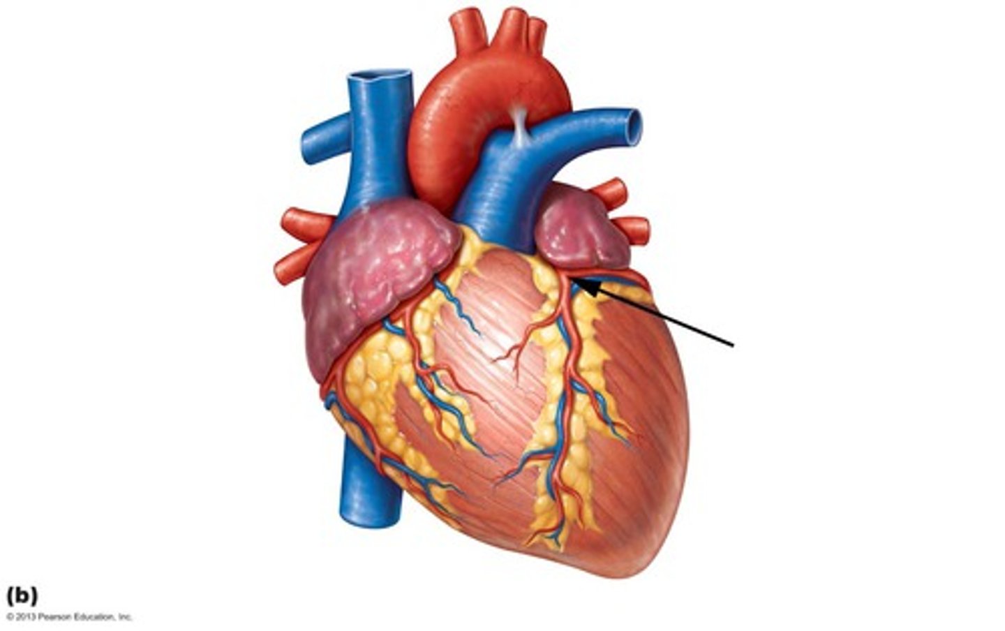
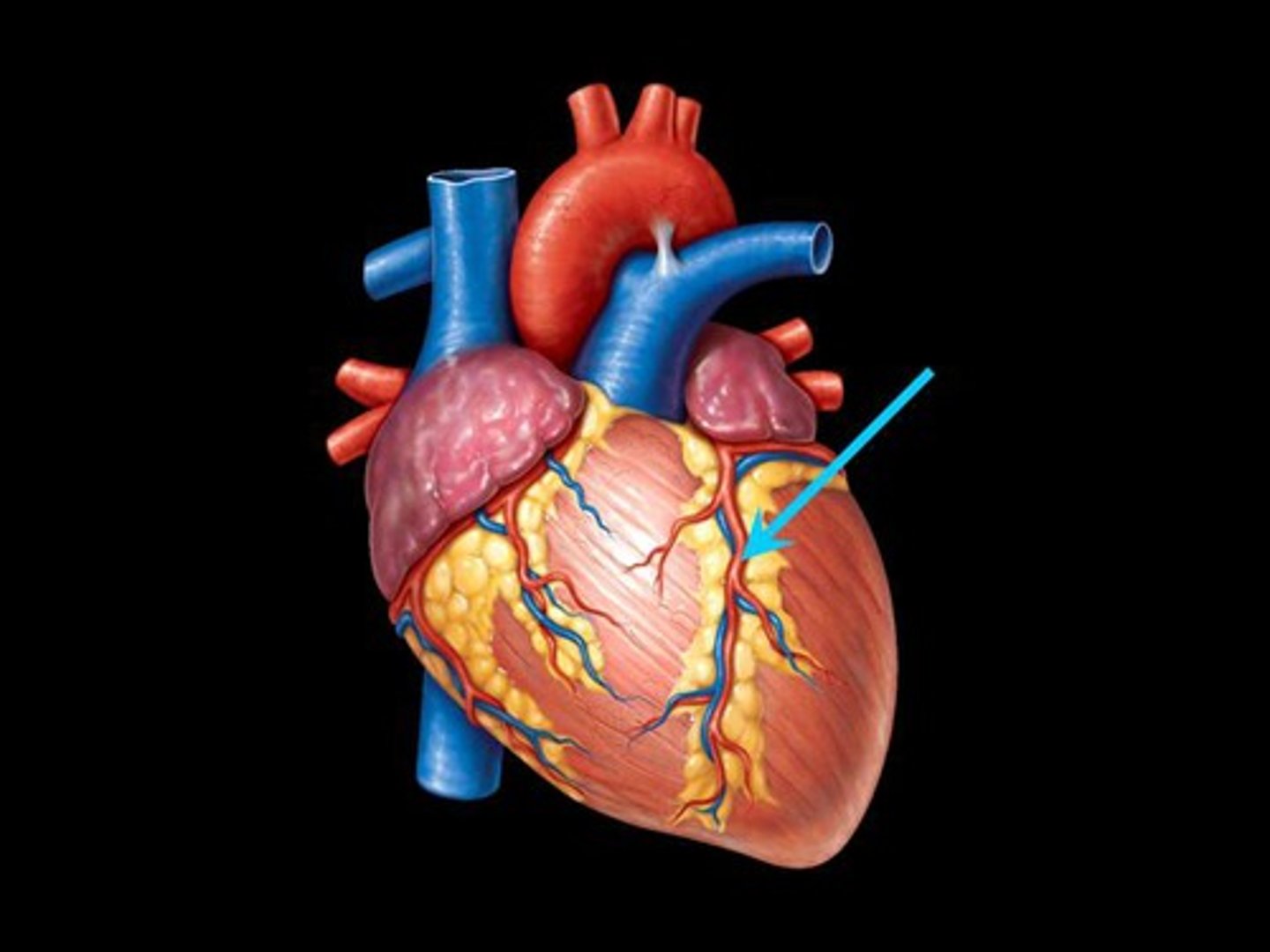
anterior interventricular artery
Structure; sits in anterior sulcus, anterior side, curly tube
H1
17
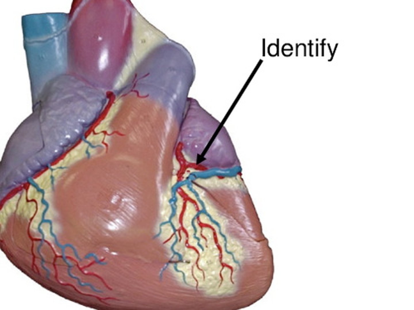
Circumflex artery
st
on left border of heart, curly
comes off L coronary arter
H1
18
right atrium
most of right ventricle
part of the left ventricle
the SA and AV nodes
BQ: know 2/4 ….the right coronary artery supplies
left atrium
most of left ventricle
part of the right ventricle
interventricular septum
BQ know 2/4 …the left coronary artery supplies
Coronary sinus
Space;
posterior central hole that probe goes into
H2
23
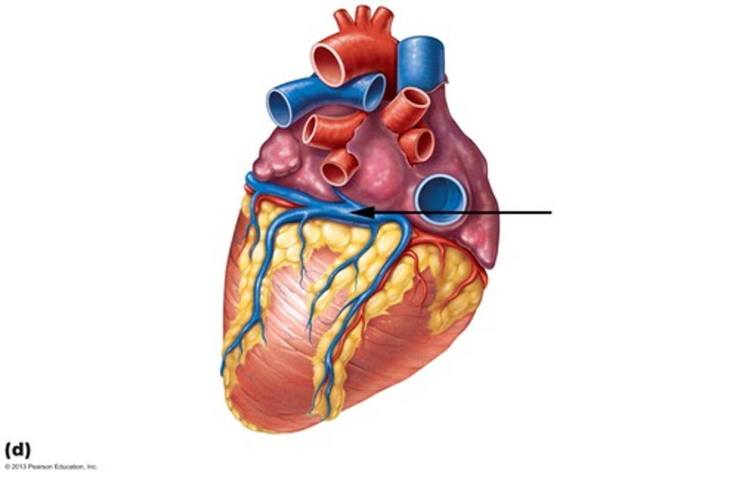
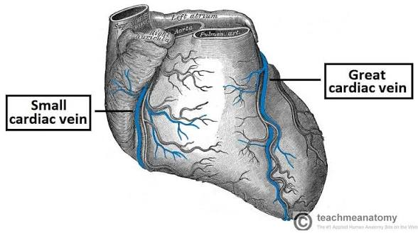
Great cardiac vein
st
left posterior flap
follow coronary sinus to opposite end
H2
19
middle cardiac vein
st
runs up and down posterior side
H2
20
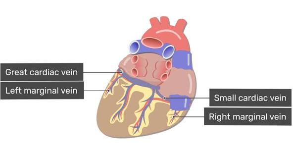
small cardiac vein
st
small posterior side, on my R
H2
21

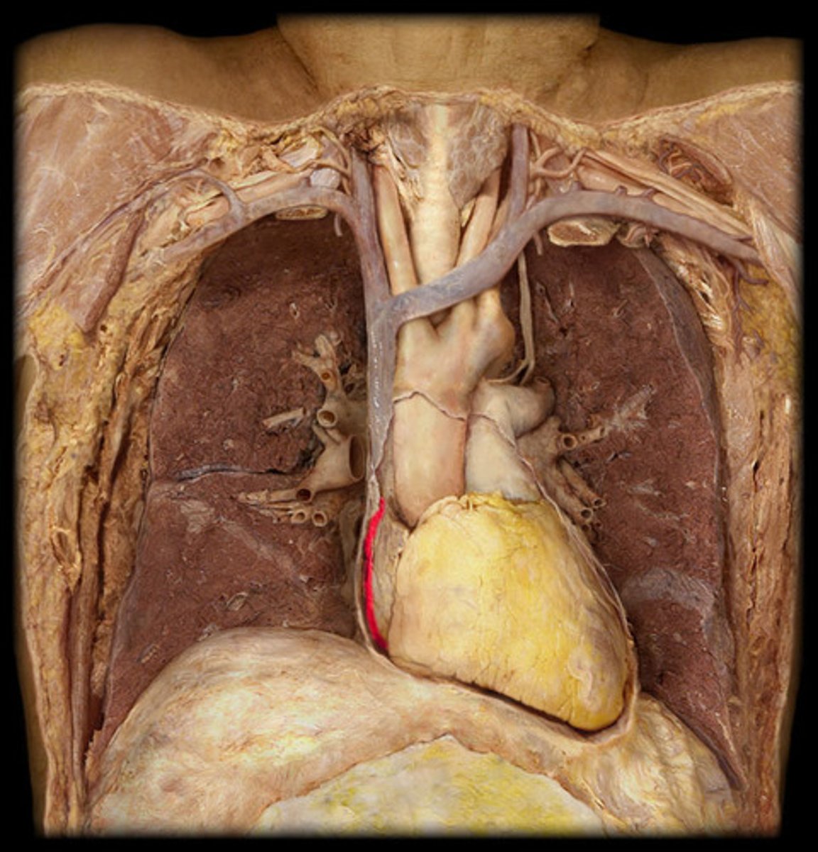
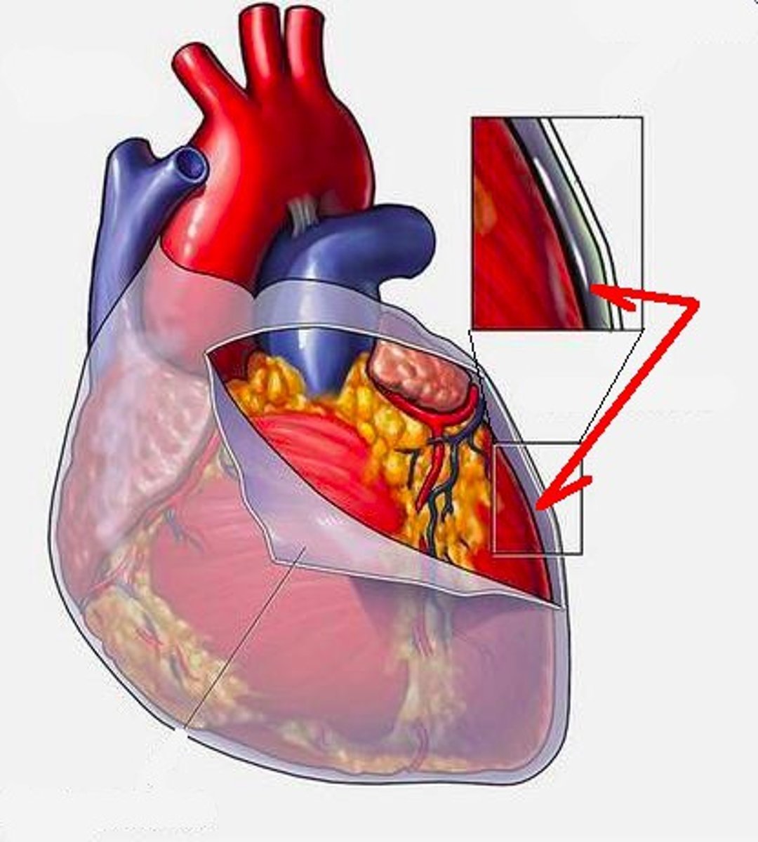
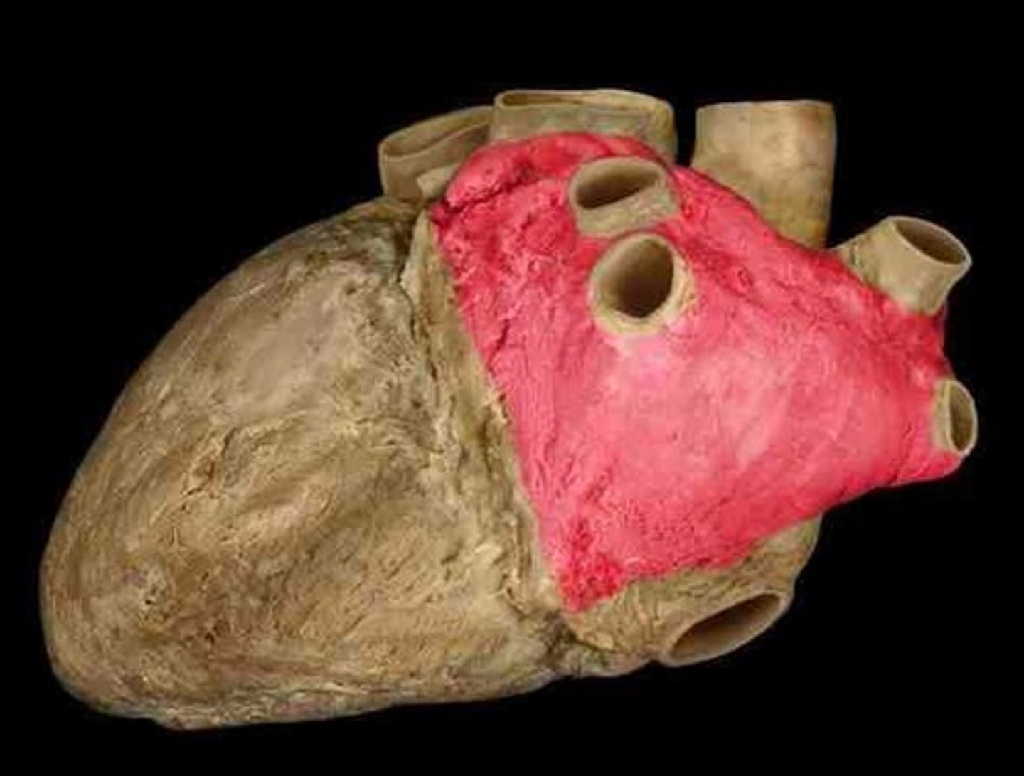
Fibrous pericardium
Covering; on outside of big heart A
H4
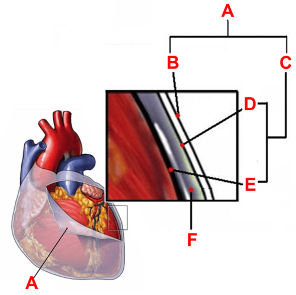
serous pericardium
Collective covering; flip fibrous pericardium back, both sides
C

parietal layer of serous pericardium
layer on the backside of the fibrous pericardium
D
H4
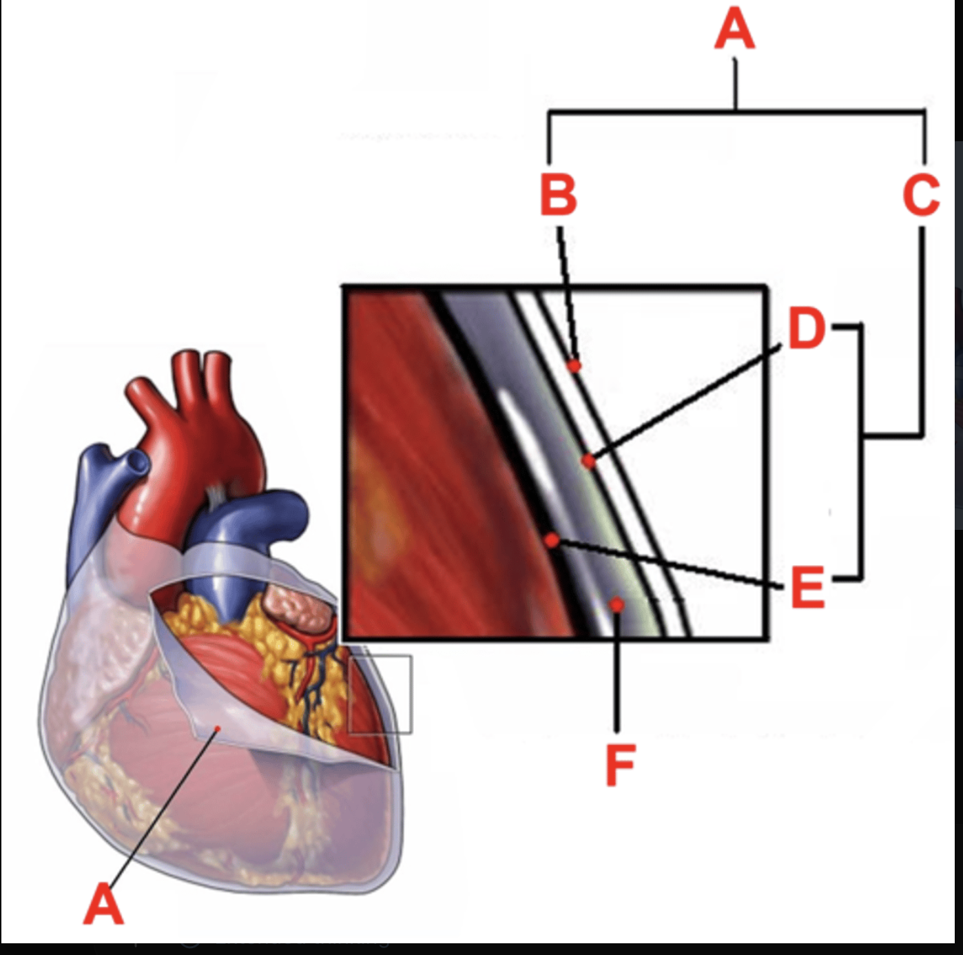
visceral layer of serous pericardium
layer directly on the heart
E
H4

Pericardial cavity
probe between the 2 layers
space
transverse pericardial sinus
BQ only: connects the left and right sides of the pericardial cavity
oblique pericardial sinus
BQ only: a blind "cul-de-sac" posterior to the heart - surrounded by the pulmonary veins
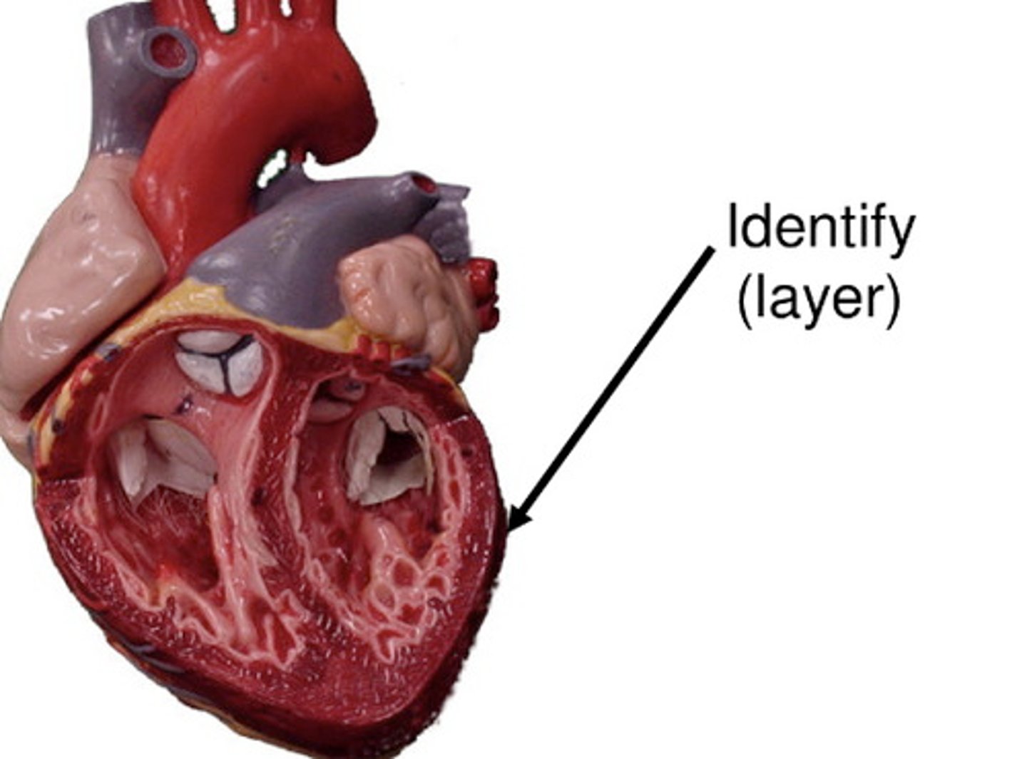
endocardium
Layer; layer inside the donut
cross section with circle
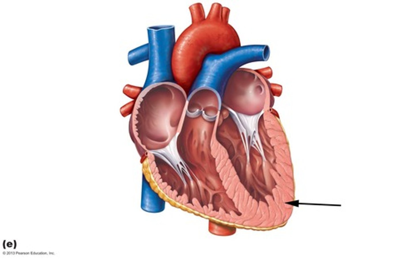
Myocardium
Layer; thick band, middle of donut
Epicardium
Layer; most superficial, cross section with circle
Interatrial septum
collective Structure; pinch between left and right atrium
H5
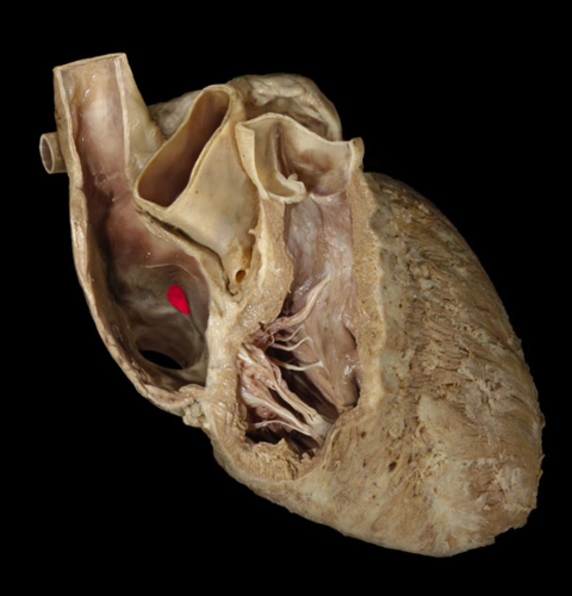
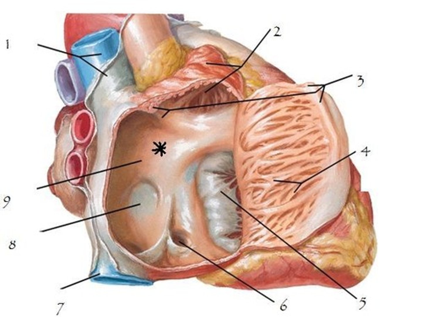
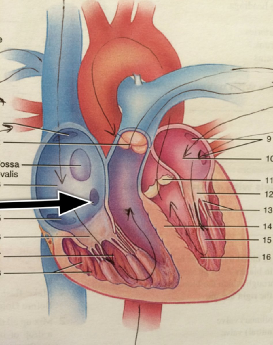
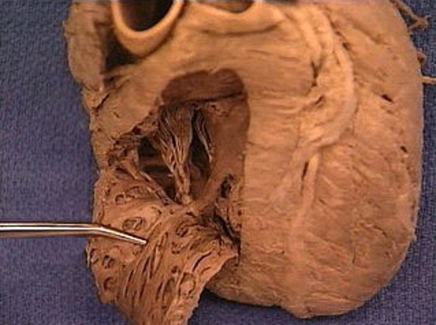
Fossa ovalis
depression
in R atrium, pinto bean buried in R atrium
H5
24
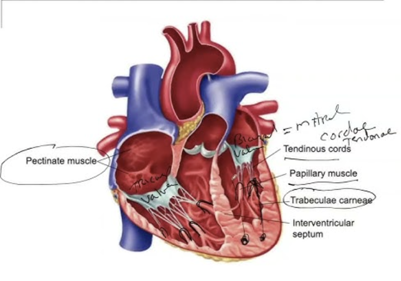
Pectinate muscle
st
stretch jeans on inside of right atrium
webbing on wall of R atrium
H5
Sinus venarum
surface; on inside of right atrium, smooth part, by fossa ovalis
H5
opening of the superior vena cava
opening; tip of probe, posterior side, smaller opening at top
H6
opening of the inferior vena cava
opening; posterior side, end of probe, bottom of shoe, smaller opening at back
H6
Opening of the coronary sinus
opening: Hole below fossa ovalis, just stick tip of probe in
H6
openings of the pulmonary veins
collective space; 2 probes in, parallel, across 2 loops, across left atrium
H6
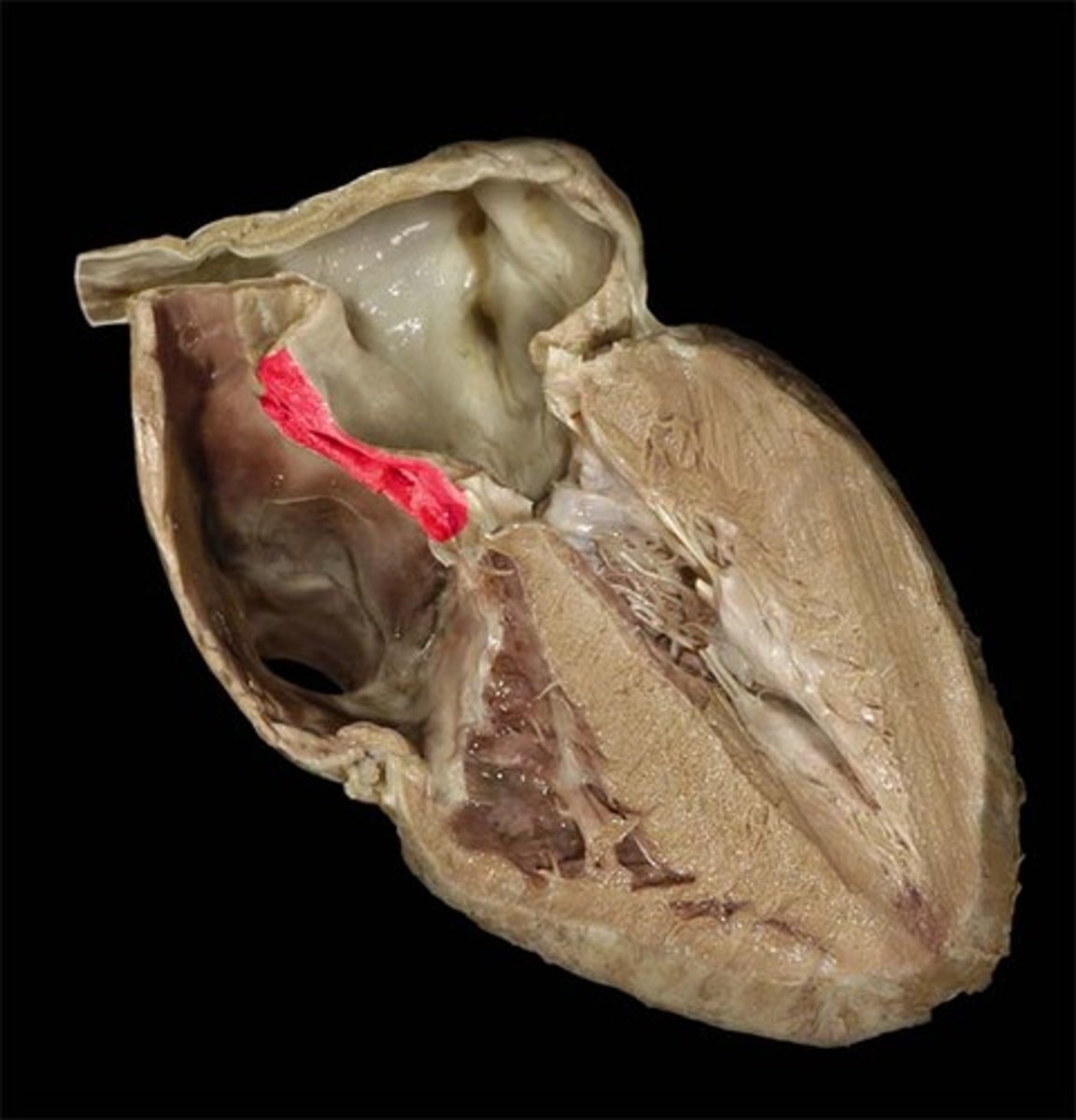
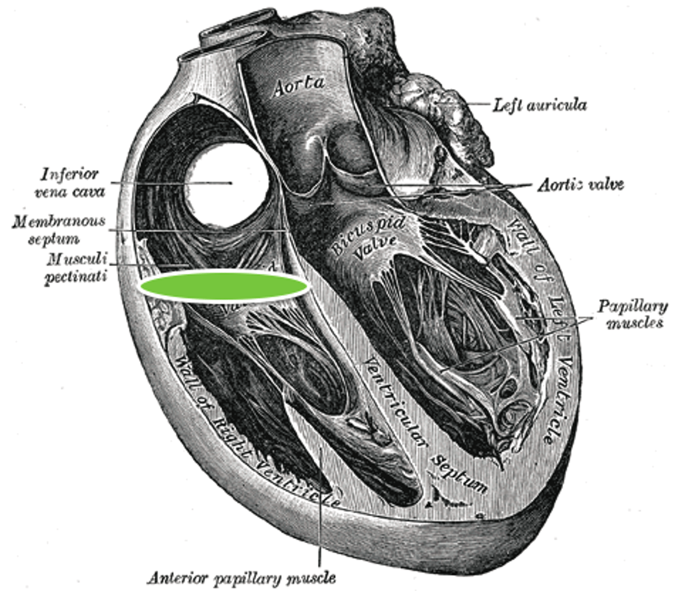
interventricular septum
Structure; separates the ventricles, pinch
H7
muscular part of the inter ventricular septum
THICK part of the septum
upper portion
H7
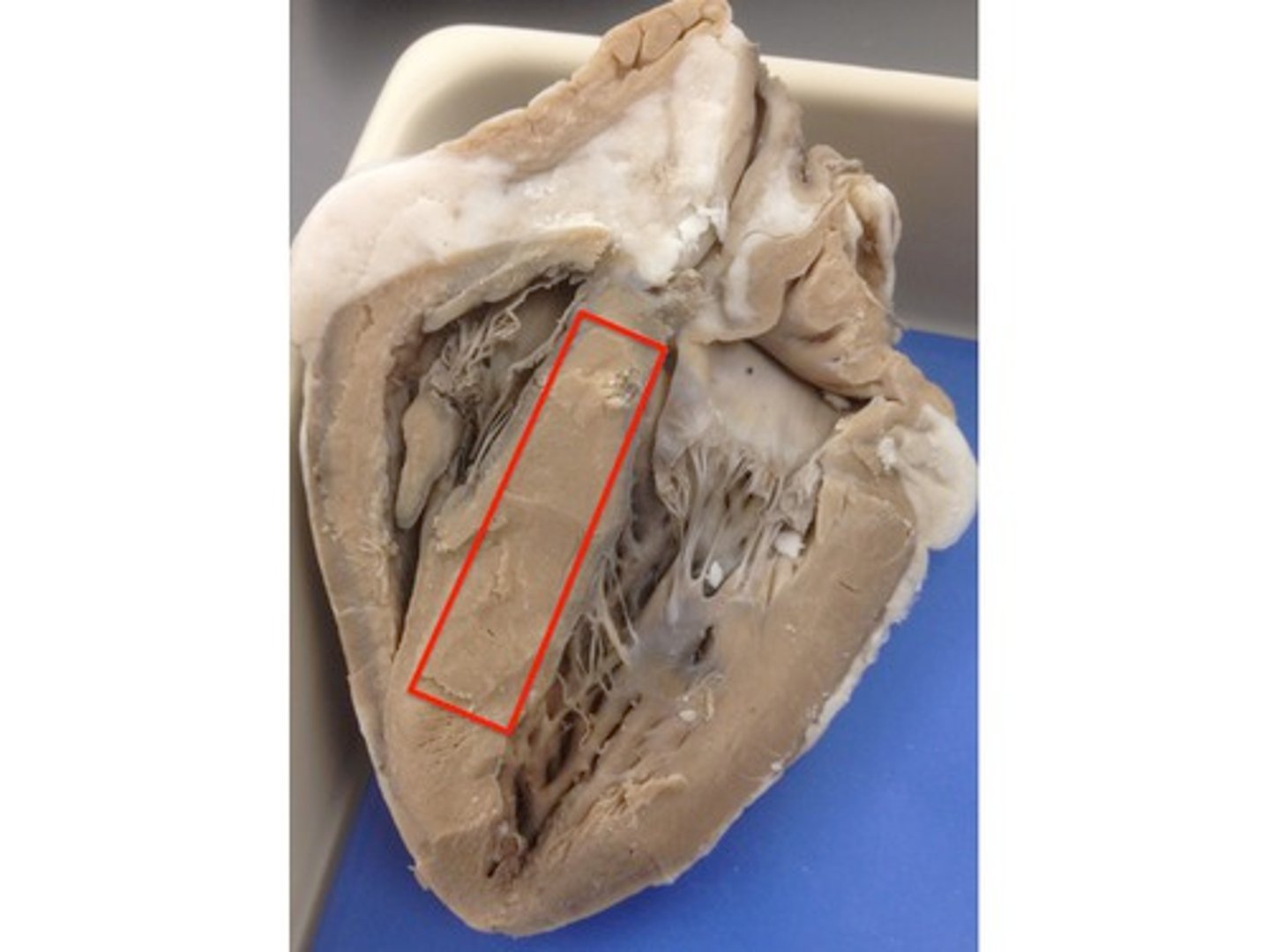
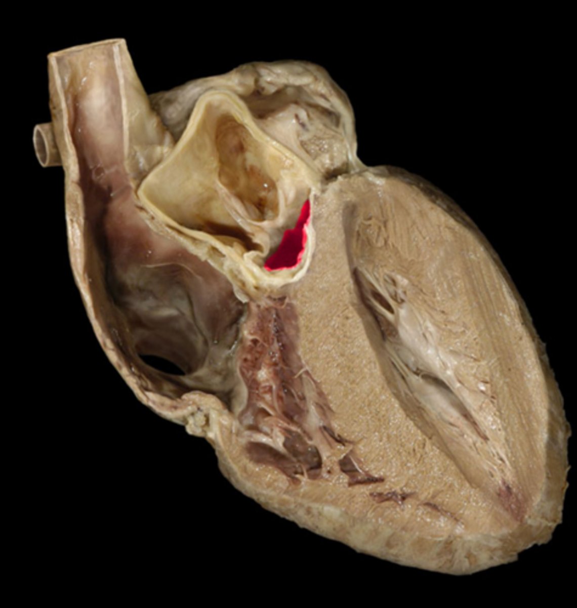
membranous part of the inter ventricular septum
THIN part of the septum
lower portion
H7
membranous part of the inter ventricular septum
BQ: thin-uppermost part the site of ventricular septal defects (common birth defects)
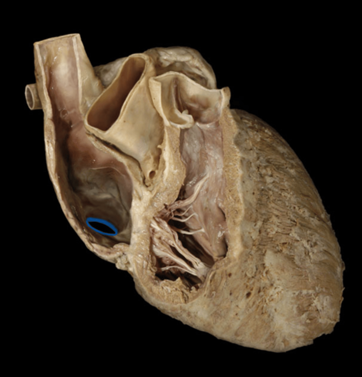
Right atrioventricular orifice
Space, stick probe from atrium to ventricle to open doorway
H5
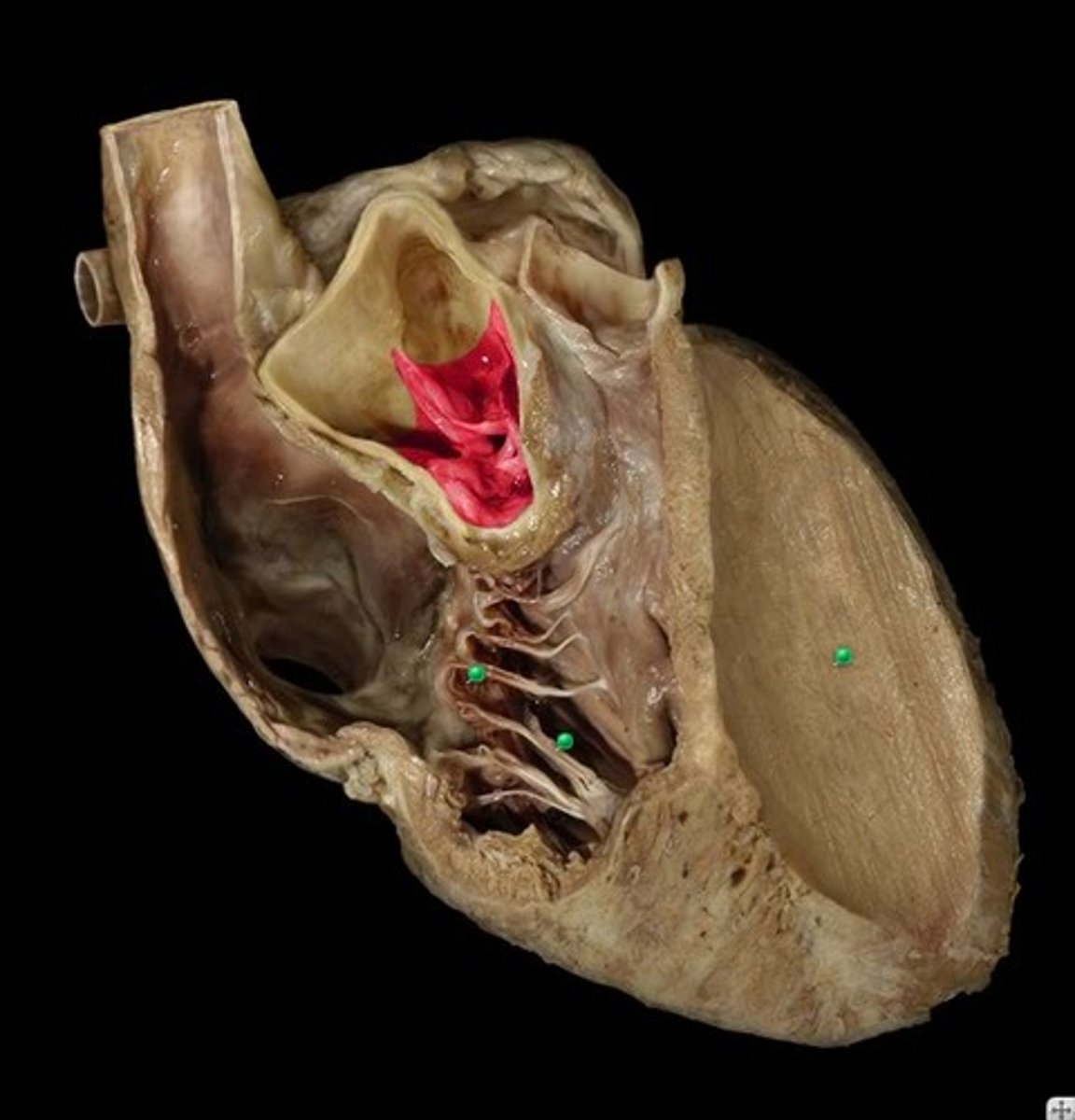
tricuspid valve
Structure; closed door, three flaps
H5
26
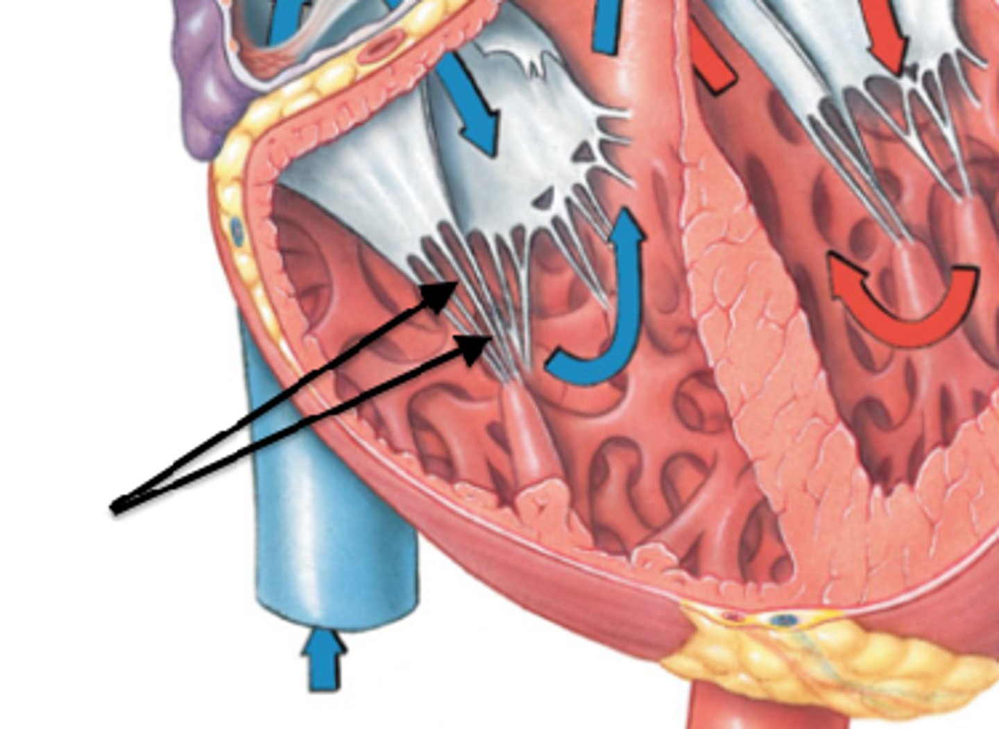
R papillary muscles
Structure; right ventricle,
orange part of the carrot
H5
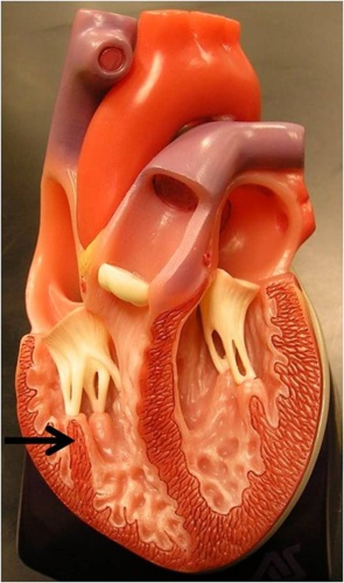
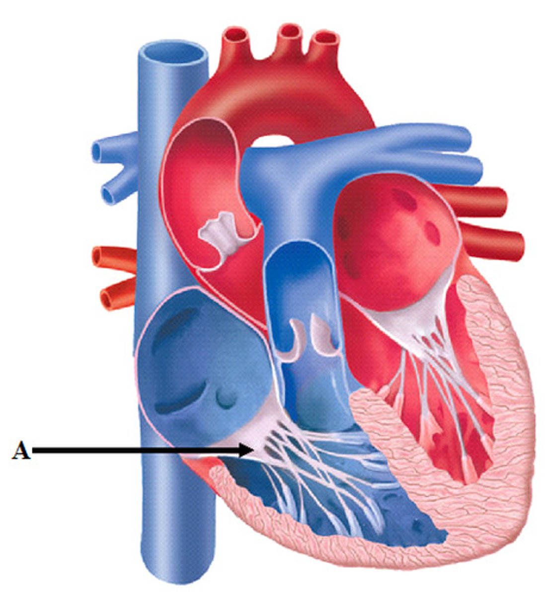
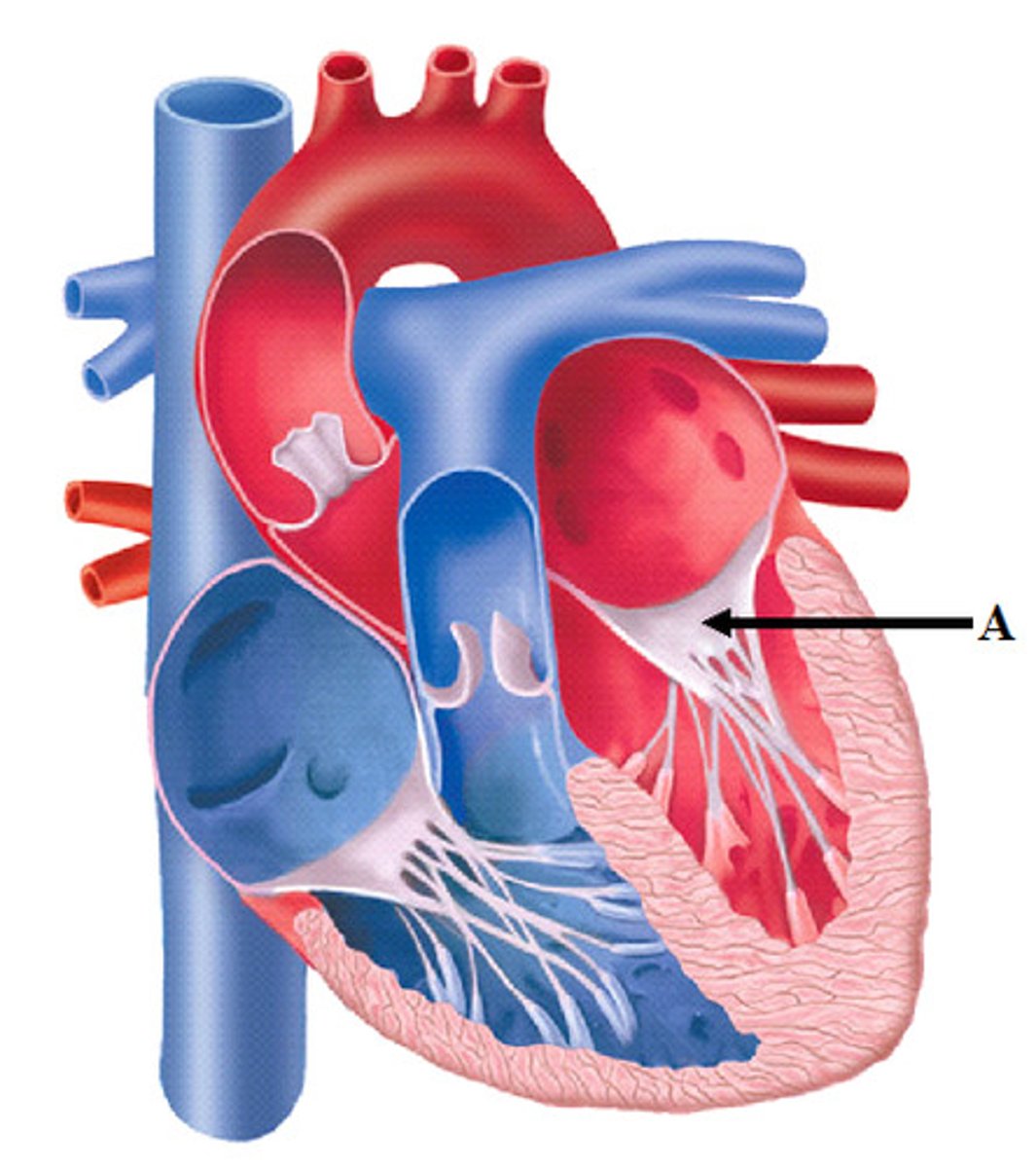
tendinous cords
Feature; right ventricle
leaves of carrot
trabeculae carneae
Structure; in right ventricle
dirt of carrot
R/L
H8
conus arteriosus
Narrowing; v-shape before tube
H8
opening of the pulmonary trunk
Space; probe into big tube that goes into right ventricle
H8
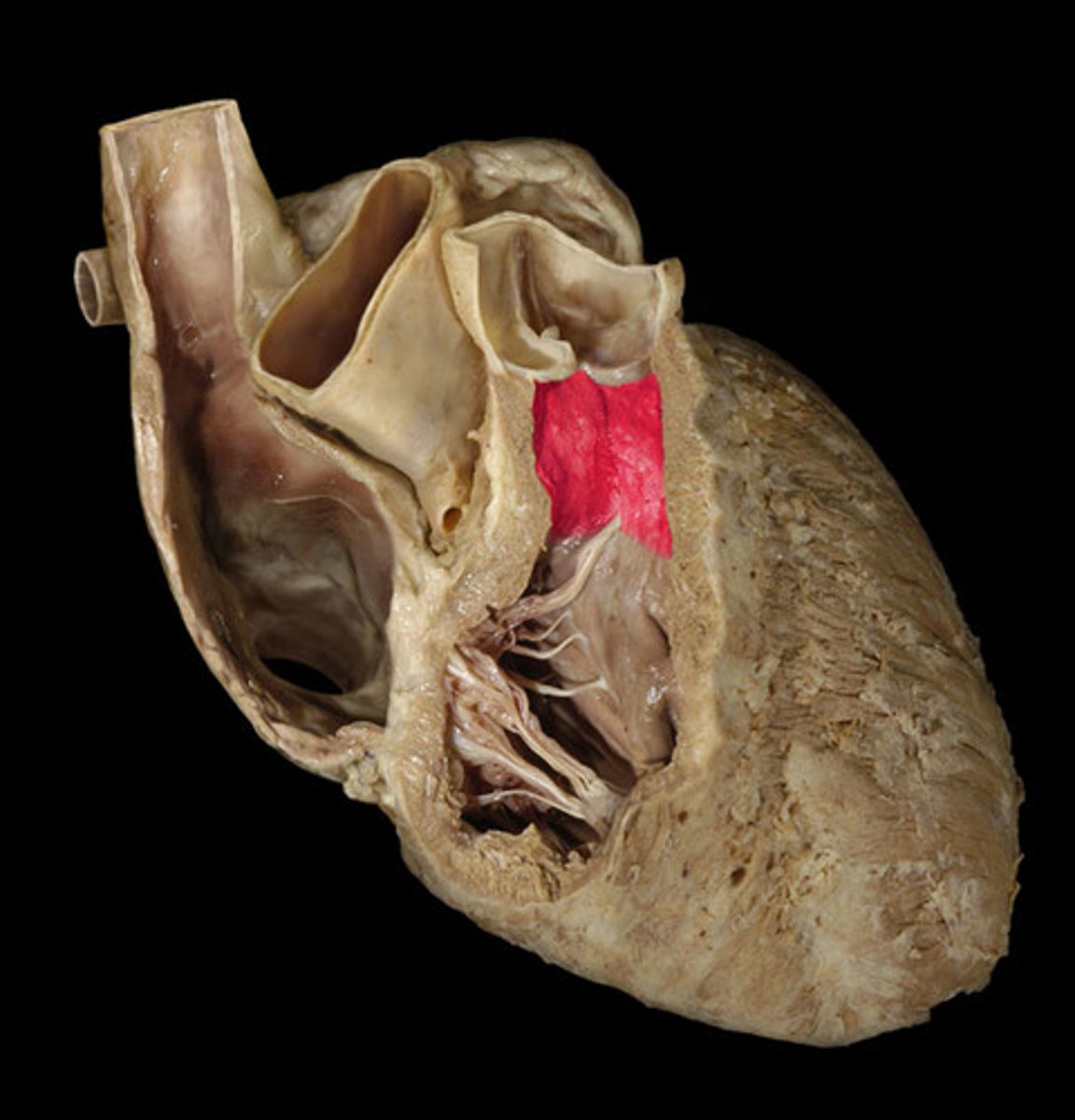
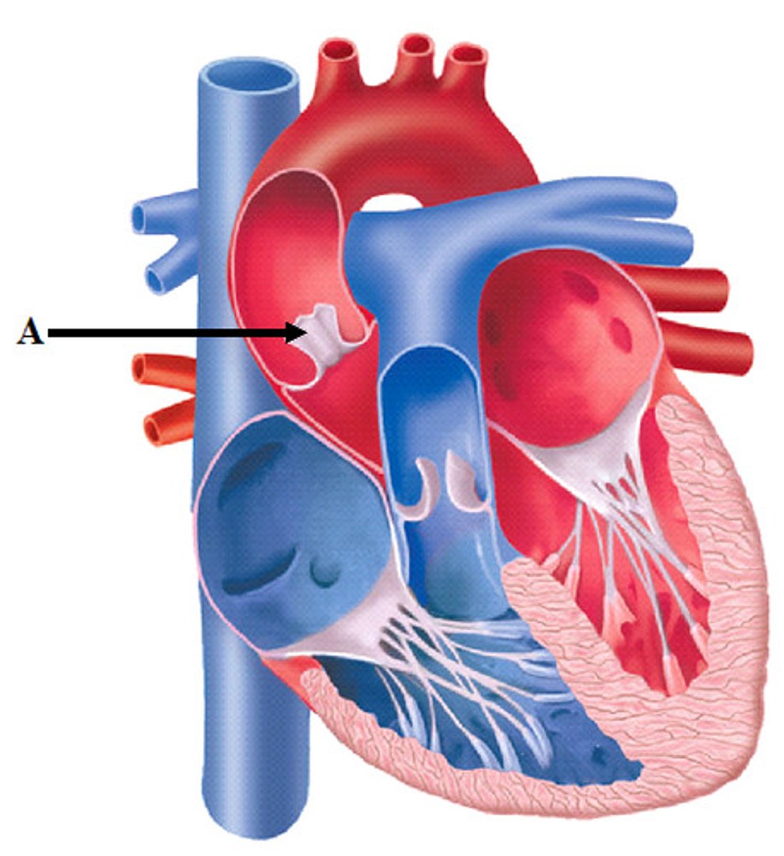
pulmonary valve
Entire structure;
all 3 drapes inside valve, right ventricle
H8
27
semilunar cusps of the pulmonary valve
Components; only 1 of the 3 drapes of the pulmonary valve
H8

left atrioventricular orifice
Space; probe in atrium to ventricle, open doorway
the hotdog bun
ta will shove probe in space
H8

mitral valve (bicuspid valve)
Structure; door in between LA and LV
2 flapped valve
H8
29
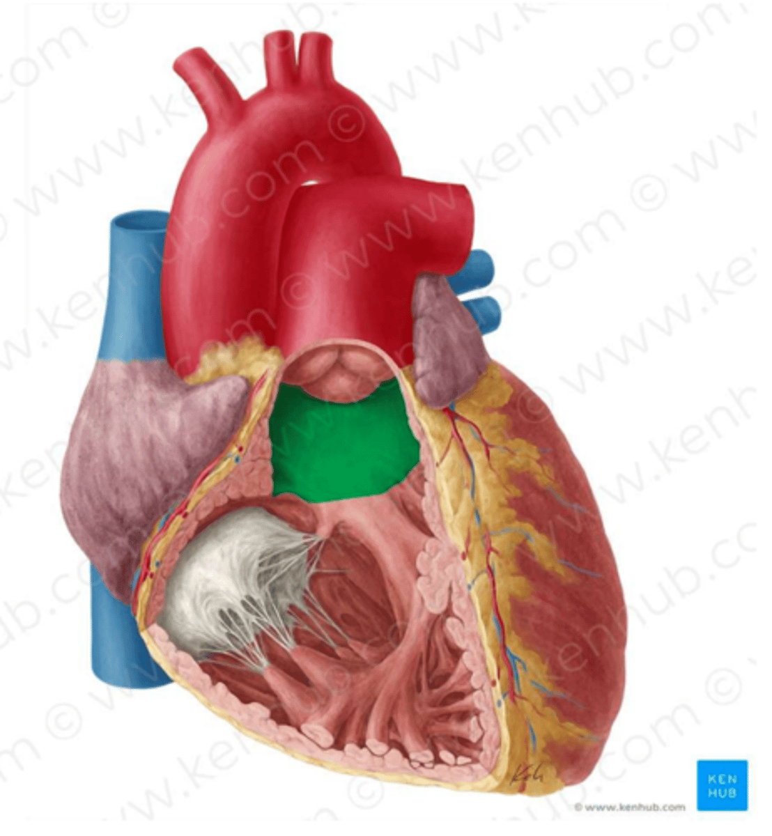
aortic vestibule
Narrowing; open heart in half, narrowing below the flaps
H8
opening of the ascending aorta
giant space at top of aorta
H8
space
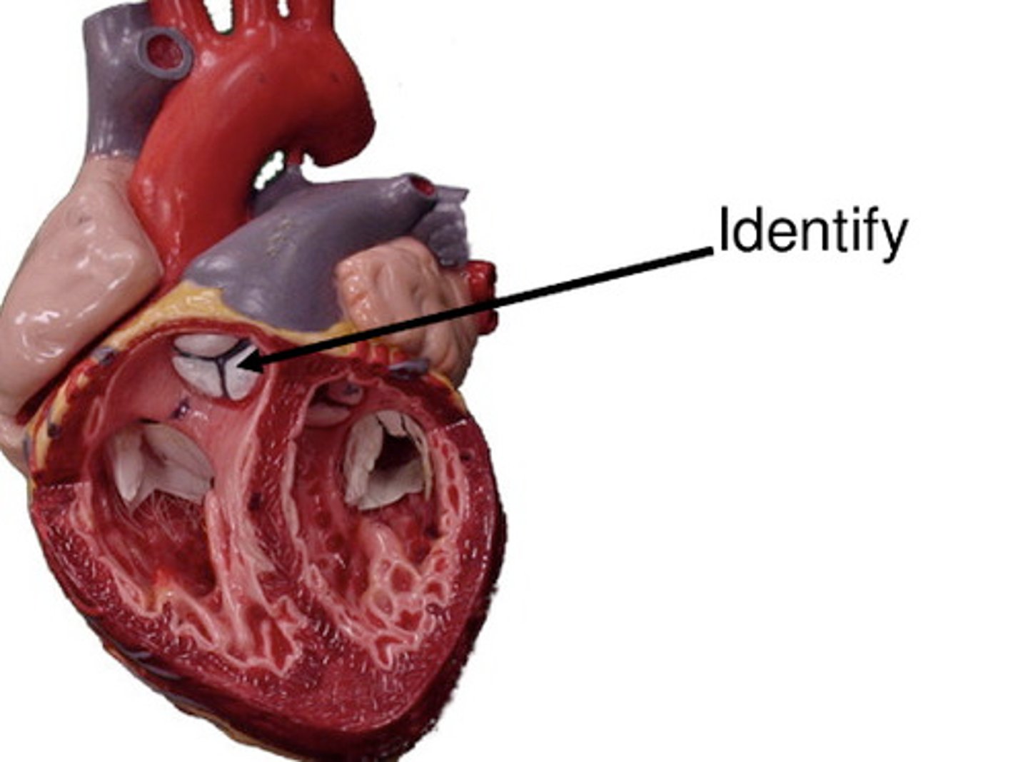
Aortic valve
Entire structure; all 3 of the drapes above the narrowing in LV
H8
30
semilunar cusps of the aortic valve
Component; only 1 of the 3 drapes
Sino-atrial node
pacemaker of the heart
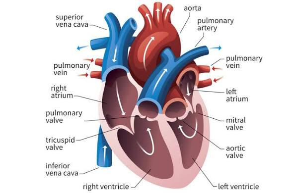
Superior and inferior vena cava -> right atrium -> tricuspid valve -> right ventricle -> pulmonary valve -> pulmonary artery -> lungs -> left atrium -> mitral valve -> left ventricle -> aortic valve -> aorta -> rest of body
the circulation of blood through the heart
Deoxygenated until lungs
Oxygenated after lungs
oxy/deoxy status of blood as it circulates through the heart
ascending aorta
portion
attached portion of aorta
7
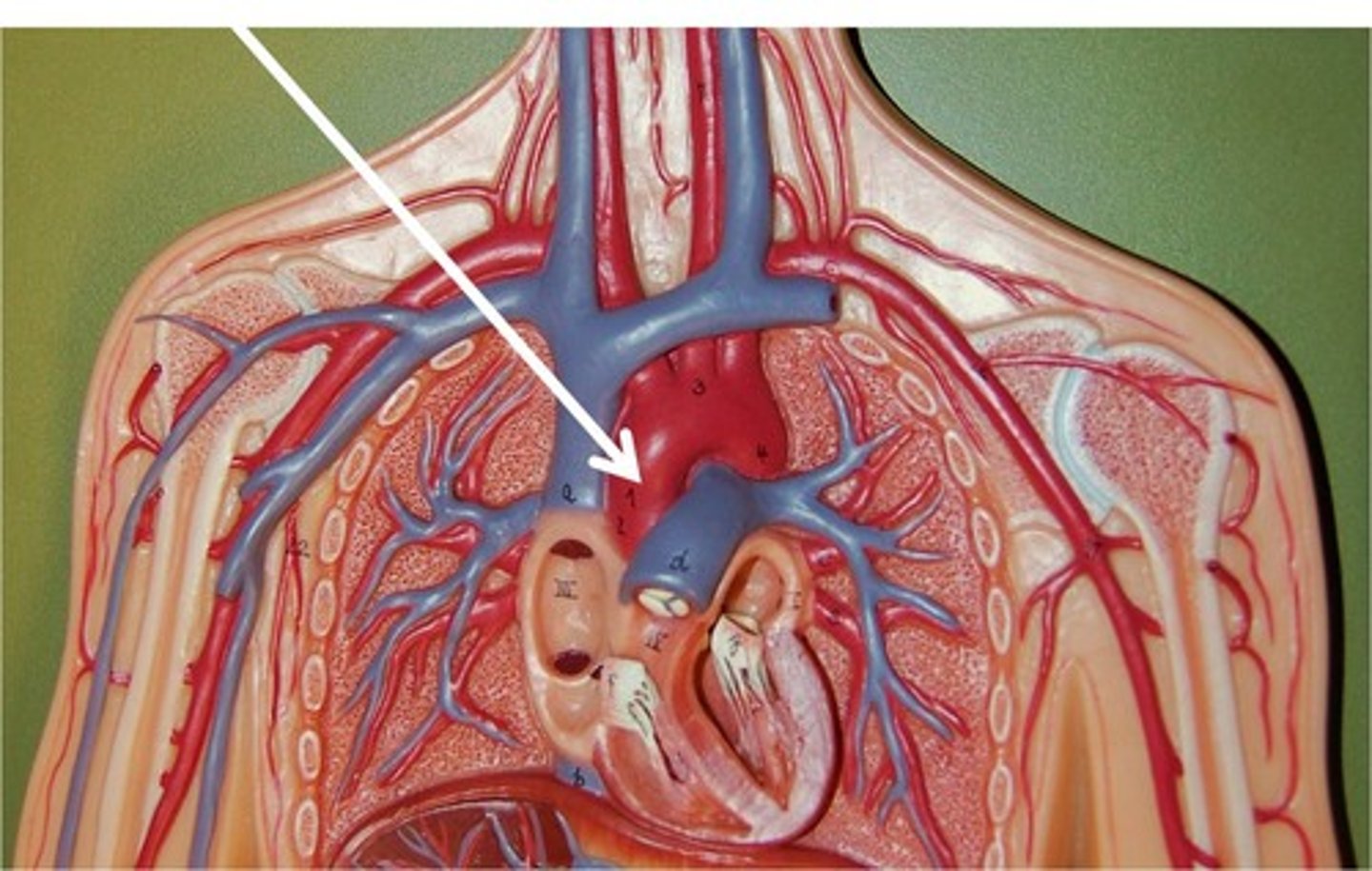
arch of aorta
curvature
into side (body of candycane)

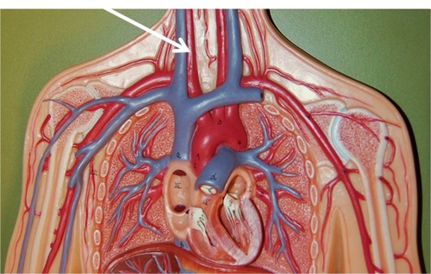
brachiocephalic artery
st
flat tube before curve on top of heart
9
R common carotid artery
st
medial split off brachiocephalic artery
10
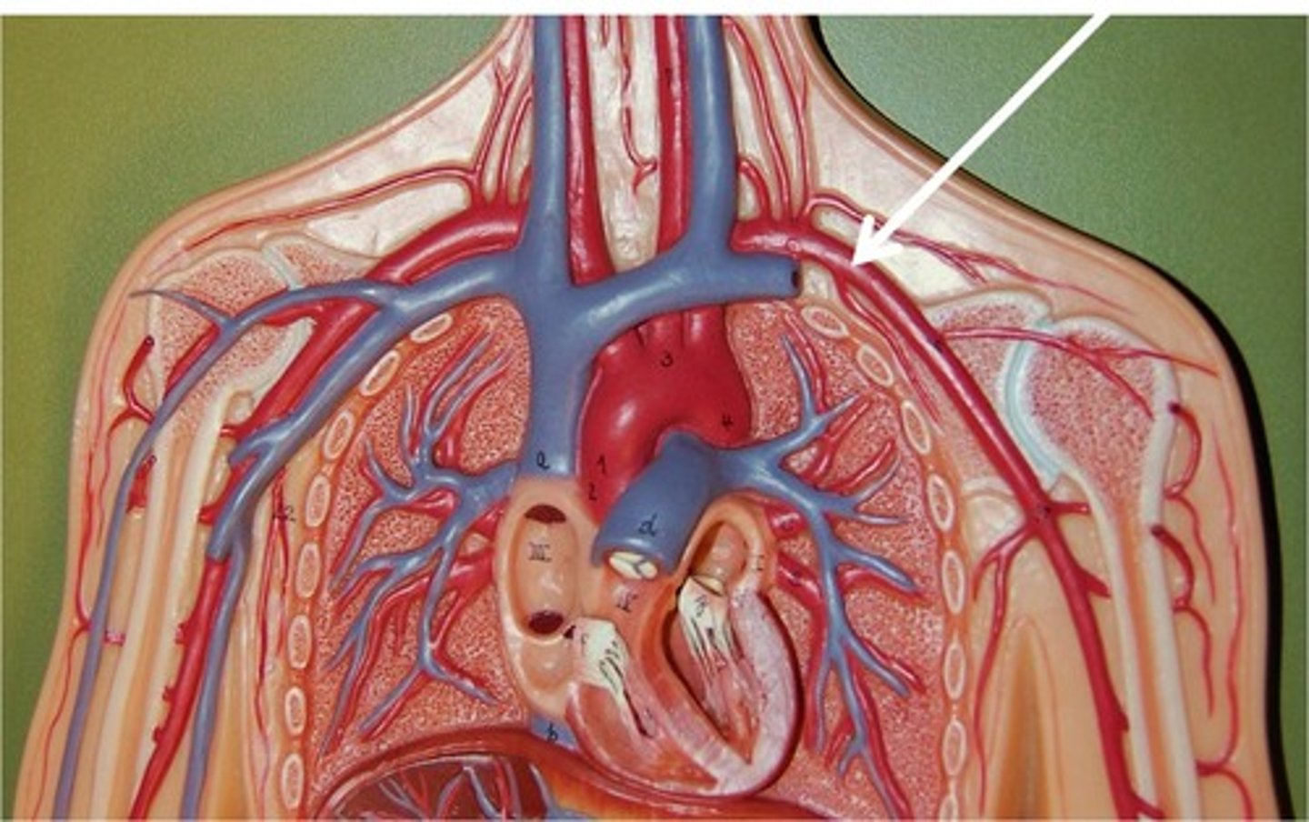
R subclavian artery
st
lateral split off brachiocephalic artery, goes under clavicle
11
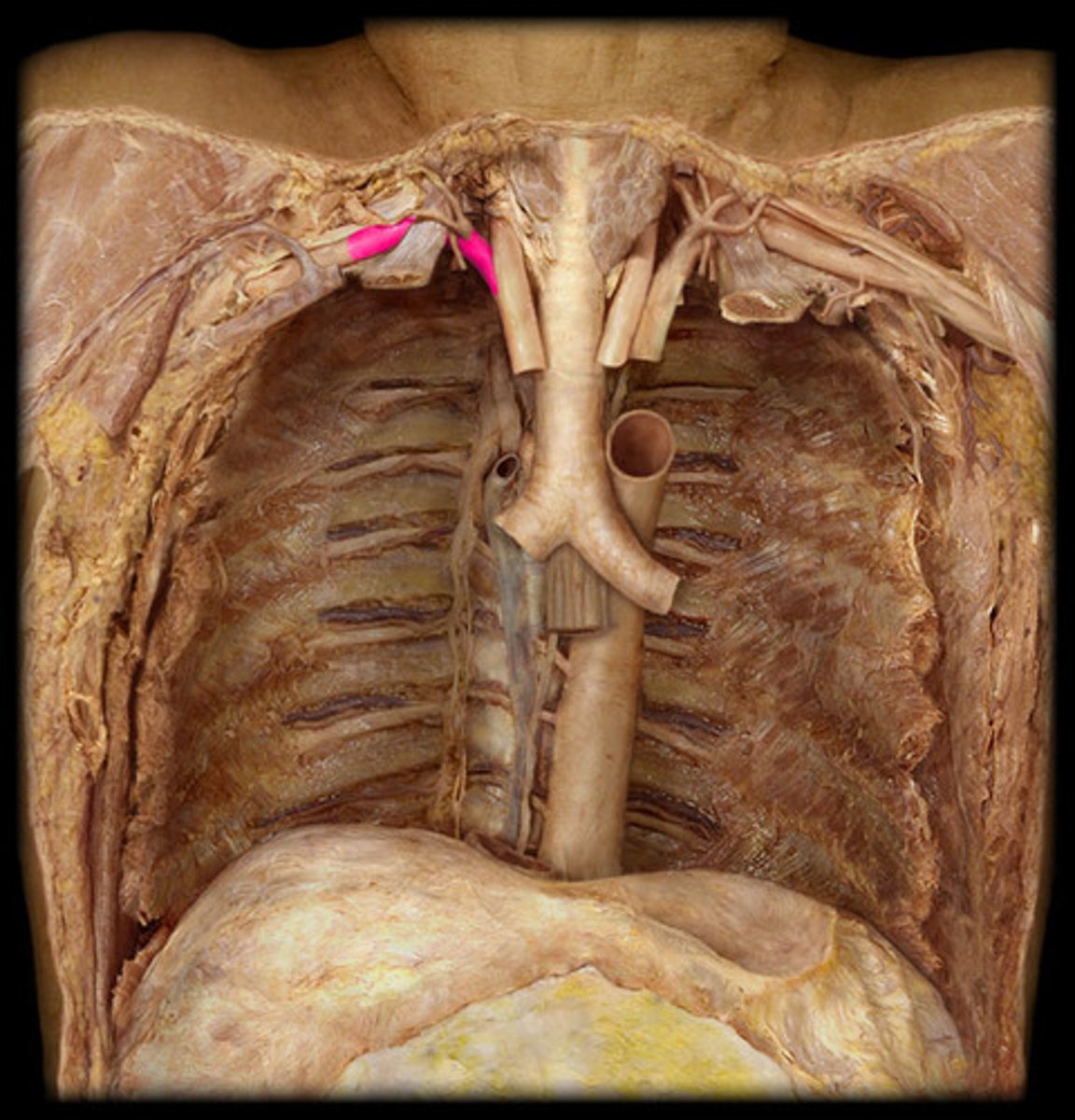
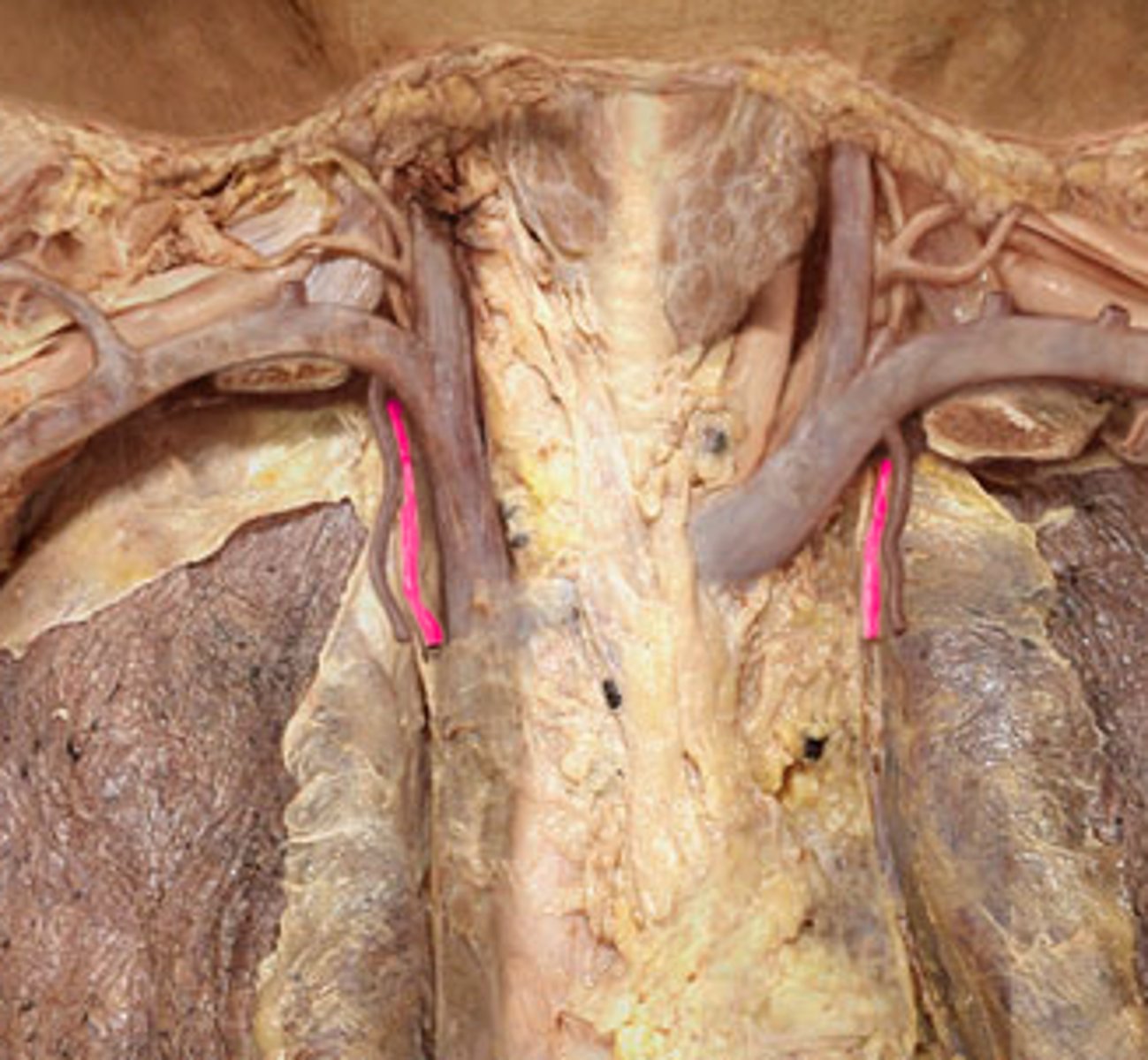
R internal thoracic artery
st
little cut one by phrenic nerve Right
L common carotid artery
st
2nd split off arch
10
left subclavian artery
st
3rd split off arch, goes directly left
leftmost tube of aorta
11
L internal thoracic artery
cut one hanging down by phrenic nerve Left

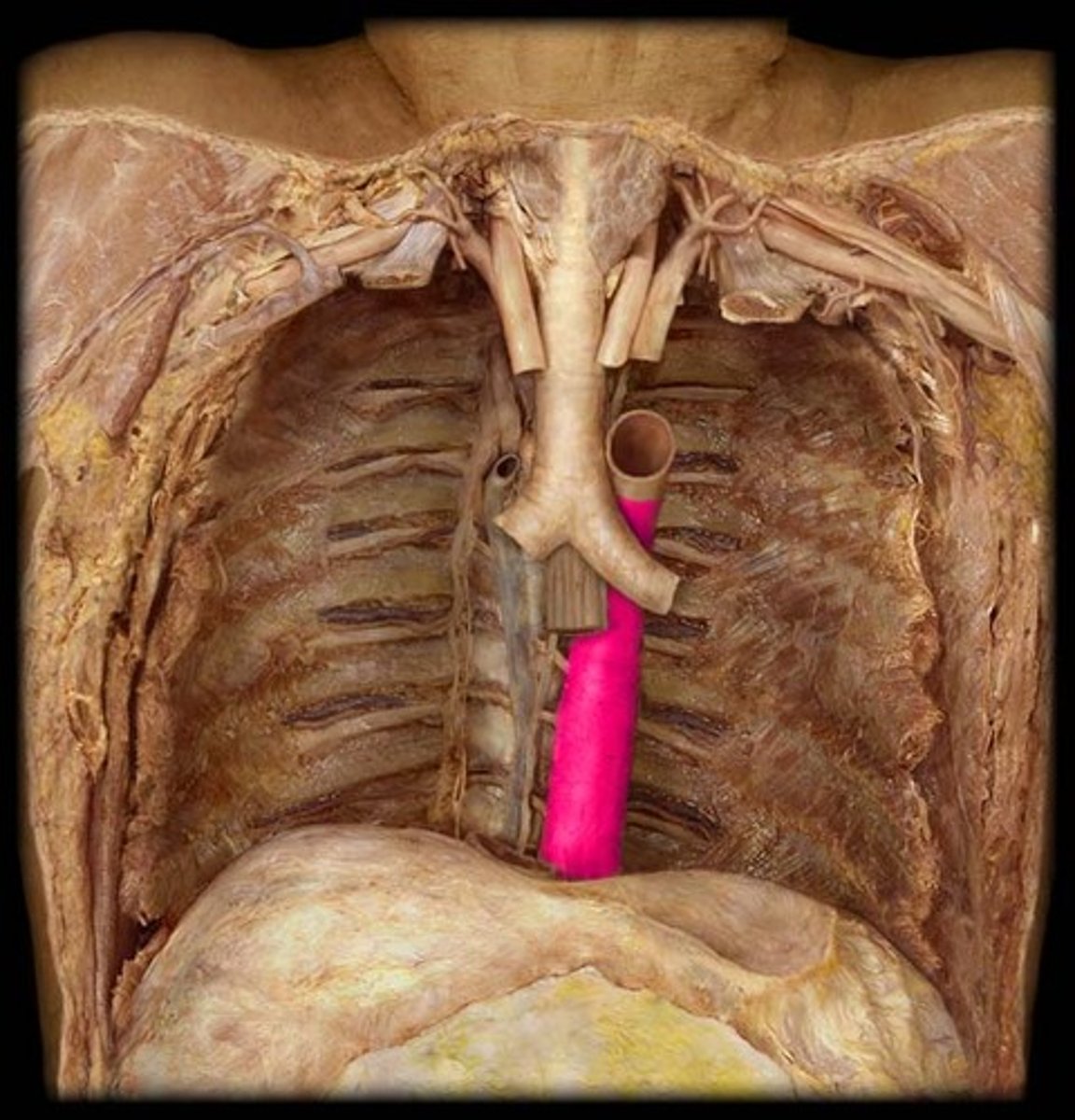
descending aorta
thoracic aorta and abdominal aorta
13
thoracic aorta
Portion, straight part on descending aorta, further down
*ta will run probe down pt. L side of heart
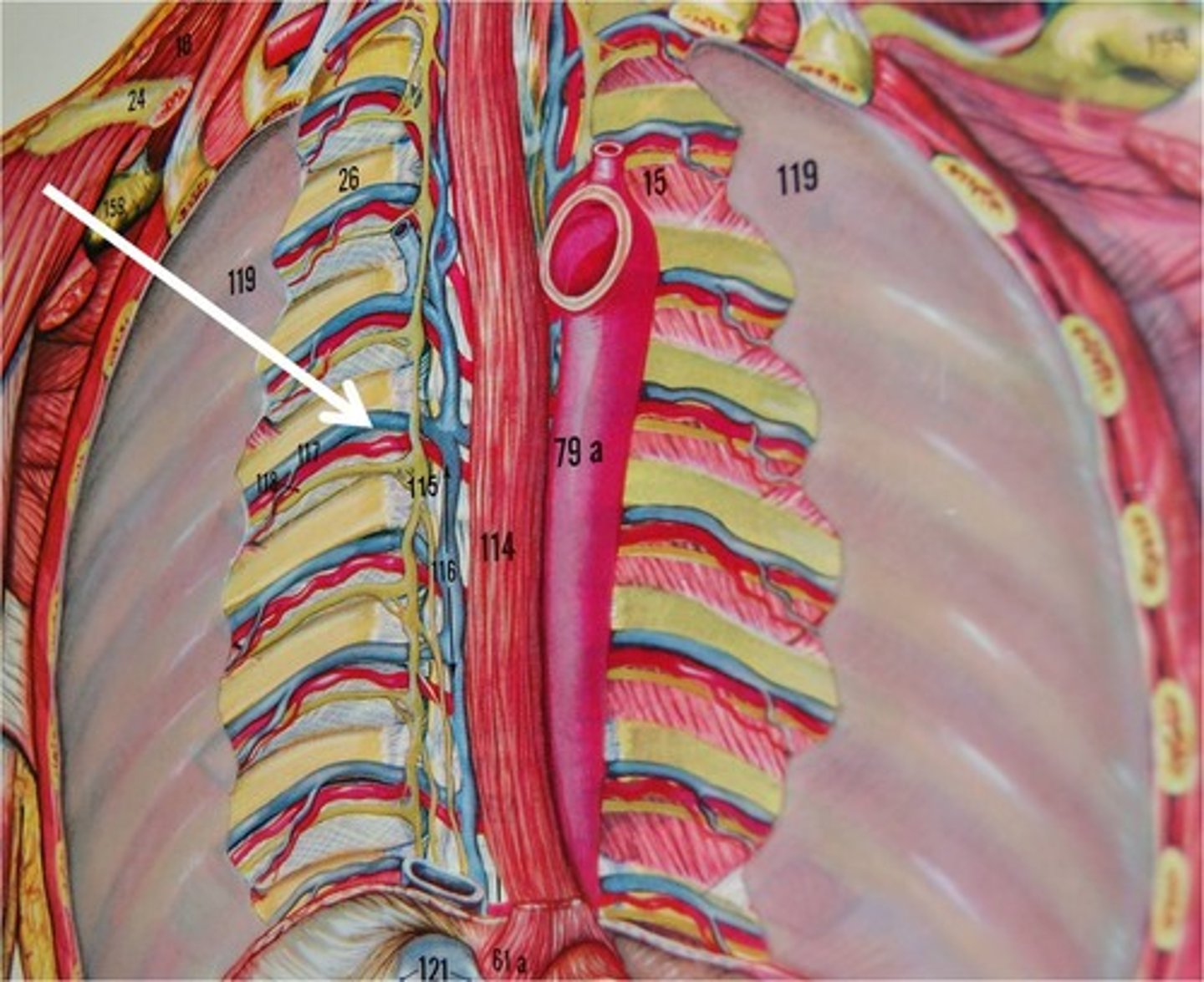
R. posterior intercostal artery
st
R ONLY
lower of the two white pins in ab aorta area
abdominal aorta
Portion; part of descending aorta, below the diaphragm
body: 2352
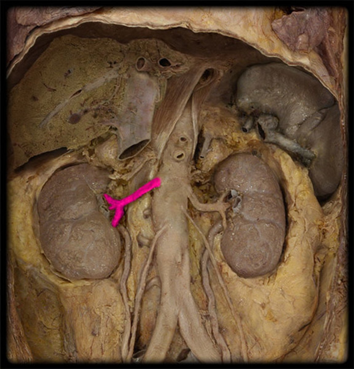
L renal artery
st
LEFT
only thing TA will lift kidney for