Looks like no one added any tags here yet for you.
reversible injury examples
swell, fatty change
Necrosis process
swell → membrane fall apart → enzymes leak → self digest
Necrosis characteristics
increased eosinophilia
Nuclear change in necrosis
pyknosis (shrink), karyorrhexis (fragment), karyolysis (loss)
types of necrosis
coagulative, liquefactive, gangrenous, fibrinoid, fat, caseous
coagulative necrosis characteristics
architecture preserved but no nucleus because structural protein denatured, preventing proteolysis of dead cells
coagulative necrosis cause
loss of blood supply
infarcts
necrosis caused by ischemia
fat necrosis cause
pancreatic lipase destroys fat
fat necrosis examples in disease
acute pancreatitis
fat necrosis characteristics
undefined border between fat
liquefactive necrosis cause
ischemia → coagulative necrosis → infections → inflammation → enzyme digest tissue (liquefaction) → pus
liquefactive necrosis characteristic
hemorrhage
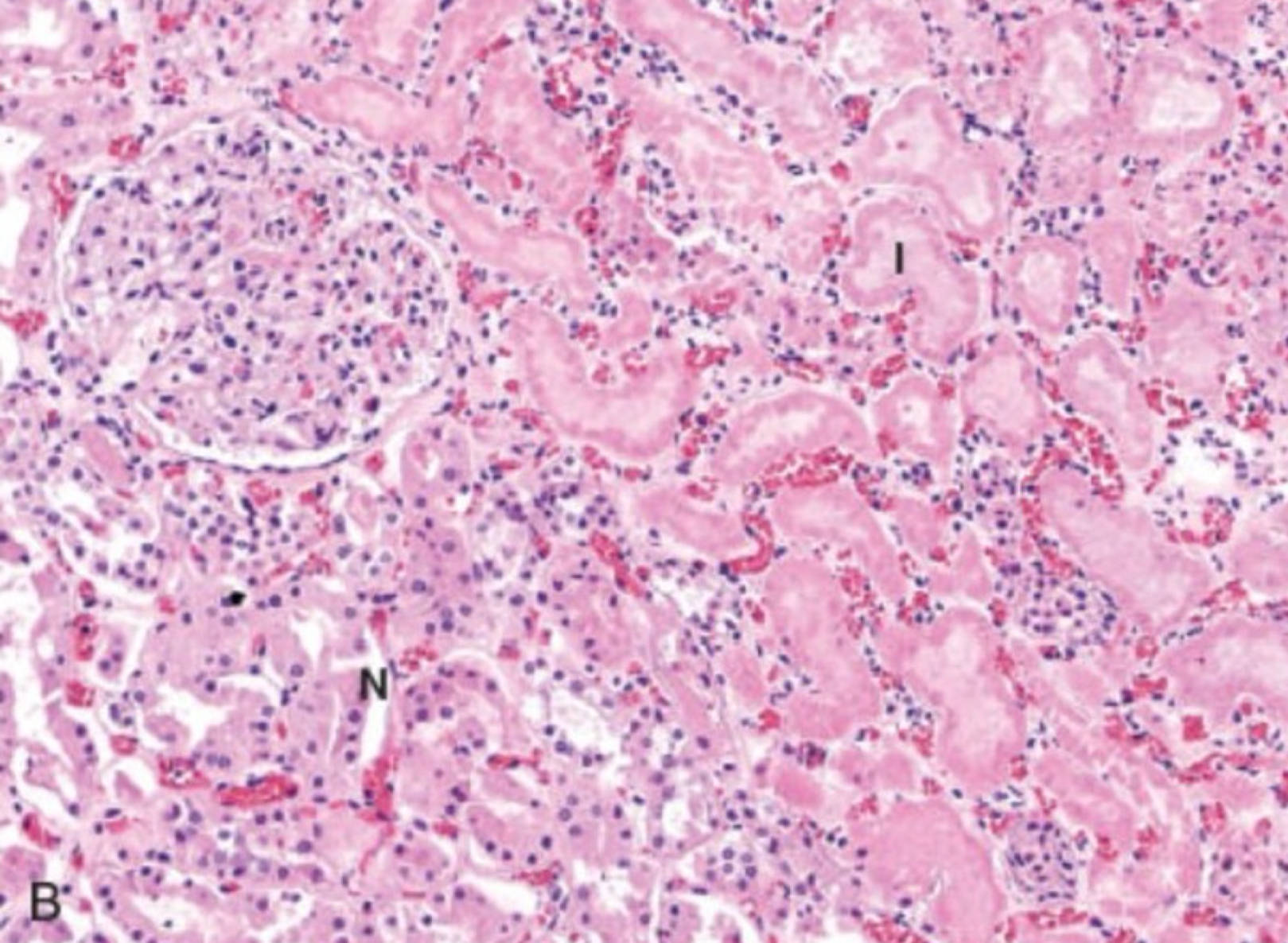
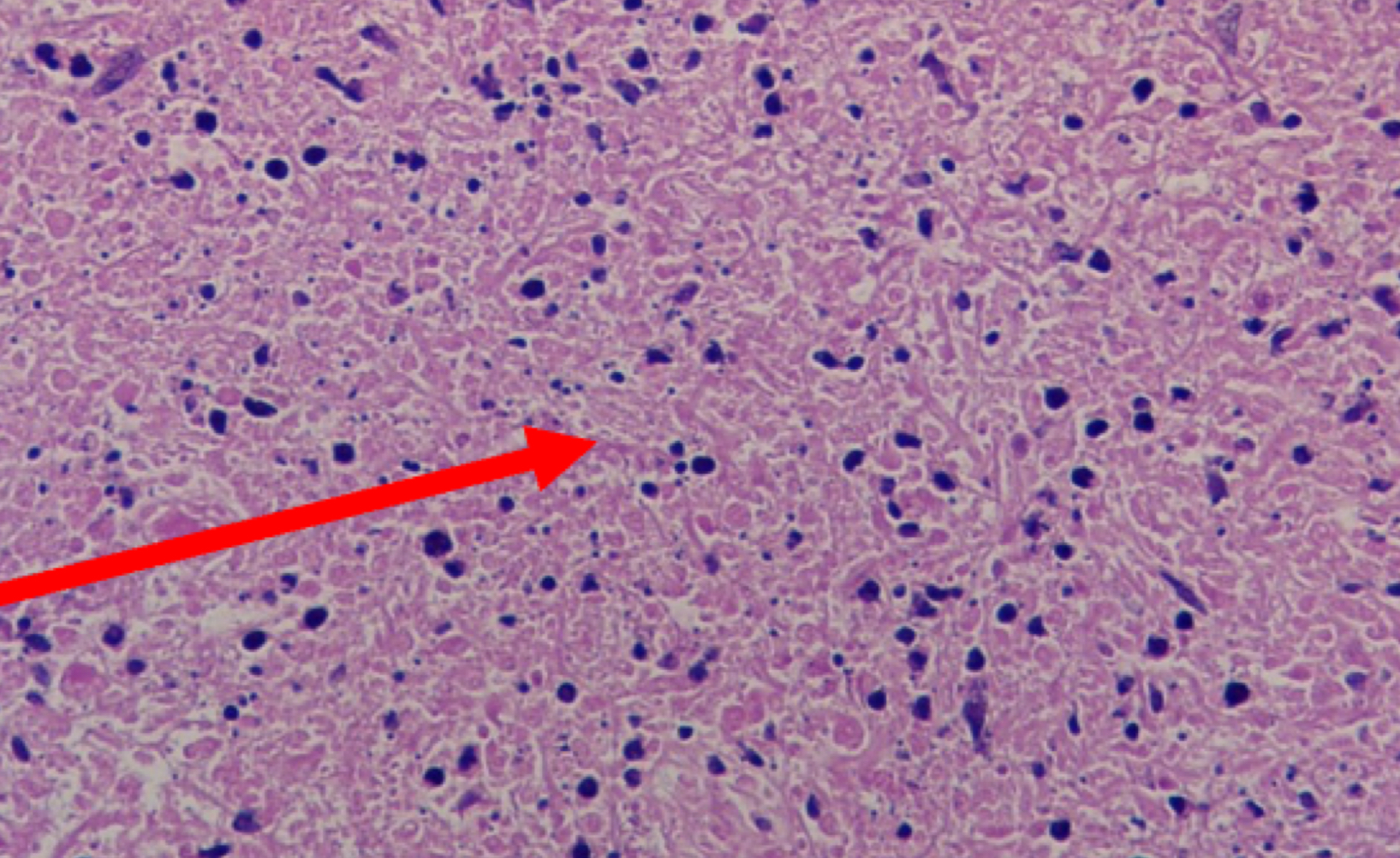
what type of necrosis
coagulative necrosis
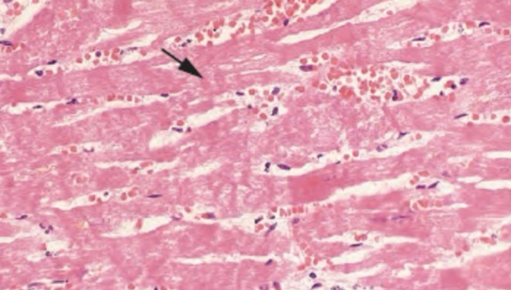
what type of necrosis
coagulative necrosis
abscess
pus
what type of necrosis
liquefactive necrosis
what type of necrosis
liquefactive necrosis
gangrenous necrosis
coagulative on limb
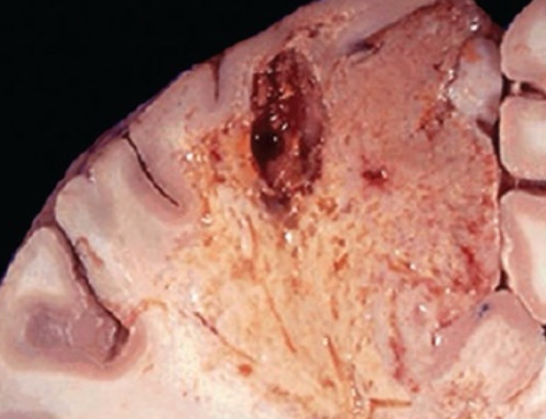
caseous necrosis disease
tuberculosis
caseous necrosis characteristic
lysed cells, cheese-like
what type of necrosis
caseous necrosis
what type of necrosis
caseous necrosis
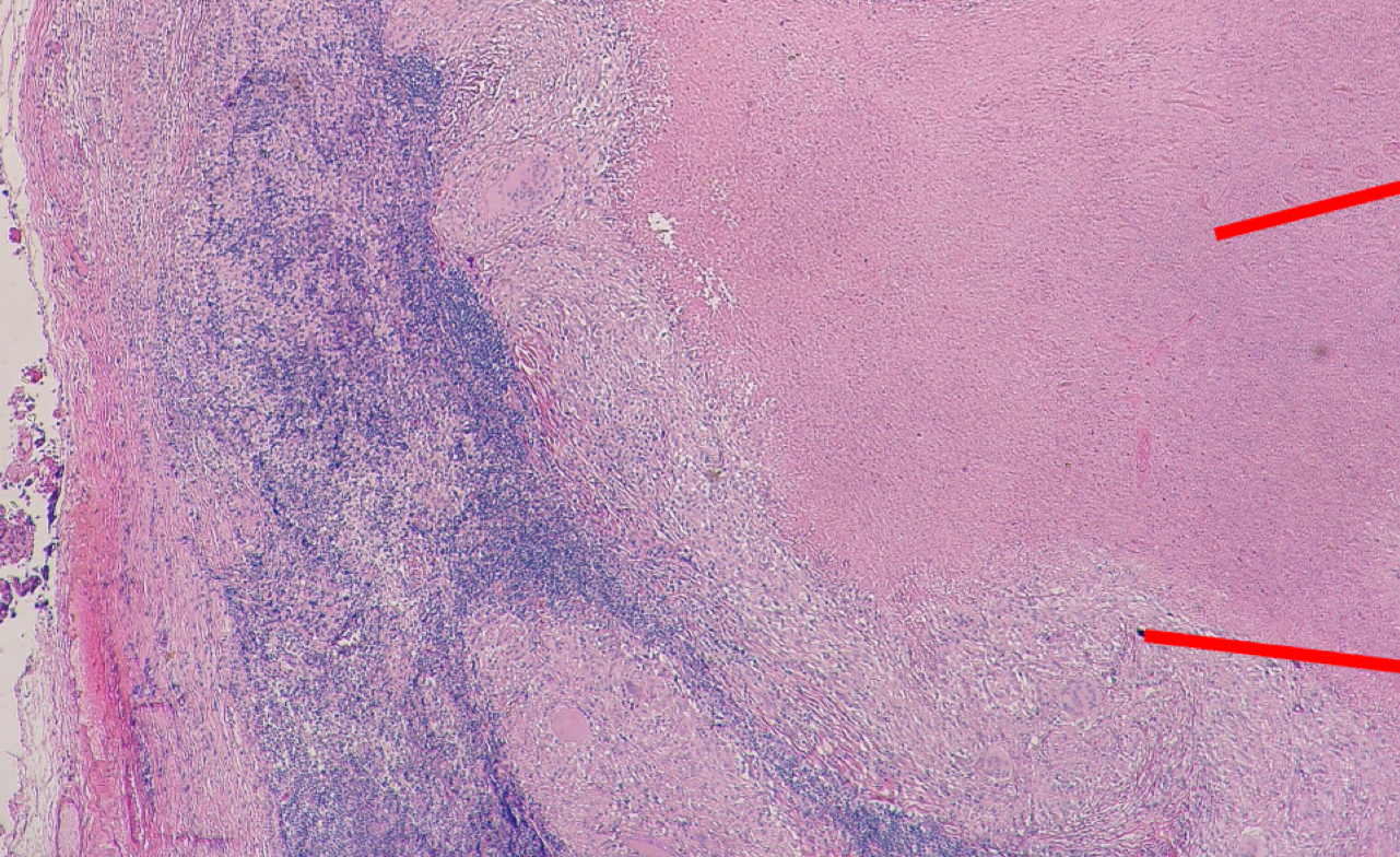
granulomatous inflammation characteristics
caseous necrosis, epithelioid, giant cell
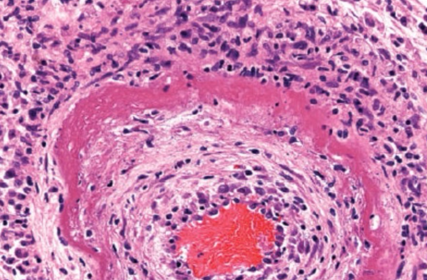
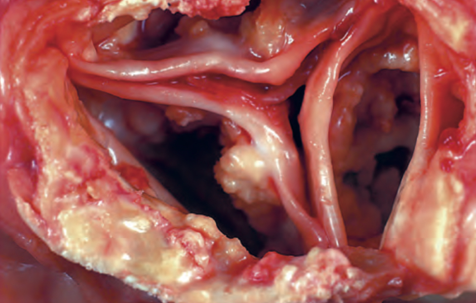
fibrinoid necrosis cause
antigen and antibody deposit in blood vessel wall
conditions linked to fibrinoid necrosis
vasculitis, hypertension
fibrinoid necrosis characteristic
blood vessel not empty inside
what type of necrosis
fibrinoid necrosis
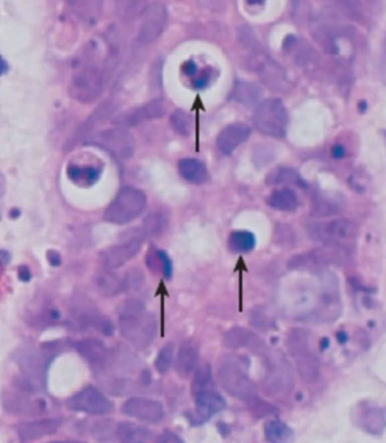
apoptosis process
cells activate enzyme → shrink → digested
what is shown in the picture
apoptosis
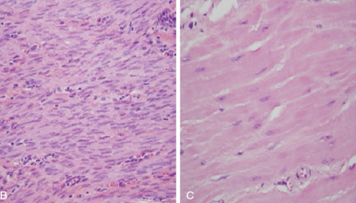
hypertrophy
increased cell and organ size
hypertrophy cause
increased workload induced by GFs
hypertrophy happens in
cells that can’t divide (heart, uterus, brain, muscle)
what type of cell adaptation is depicted
hypertrophy
hyperplasia
increased cell number
hyperplasia happens in
cells that can divide
physiologic change
due to hormone
pathologic change
due to abnormalities
atrophy
decreased cell or organ size
atrophy cause
decreased workload or nutrient
what cell adaptation is depicted
atrophy
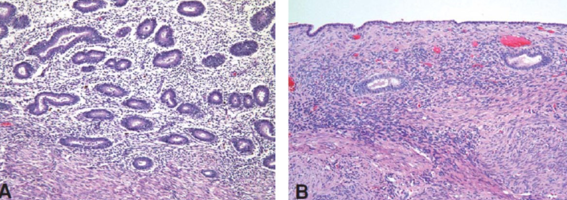
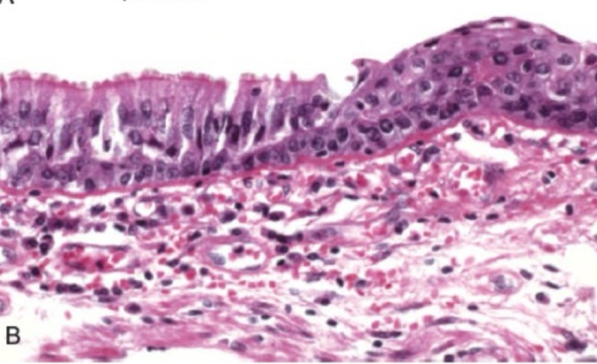
metaplasia
change in cell type
metaplasia cause
irritation induced by altered differentiation
metaplasia can become
reduced function, malignant
metaplasia examples
squamous metaplasia in smoker, barrett esophagus
what type of cell adaptation is depicted
metaplasia
types of pathogenic calcification
dystrophic, metastatic
dystrophic
normal calcium level, but deposit in injured/dead
metastatic
hypercalcemia in normal tissues
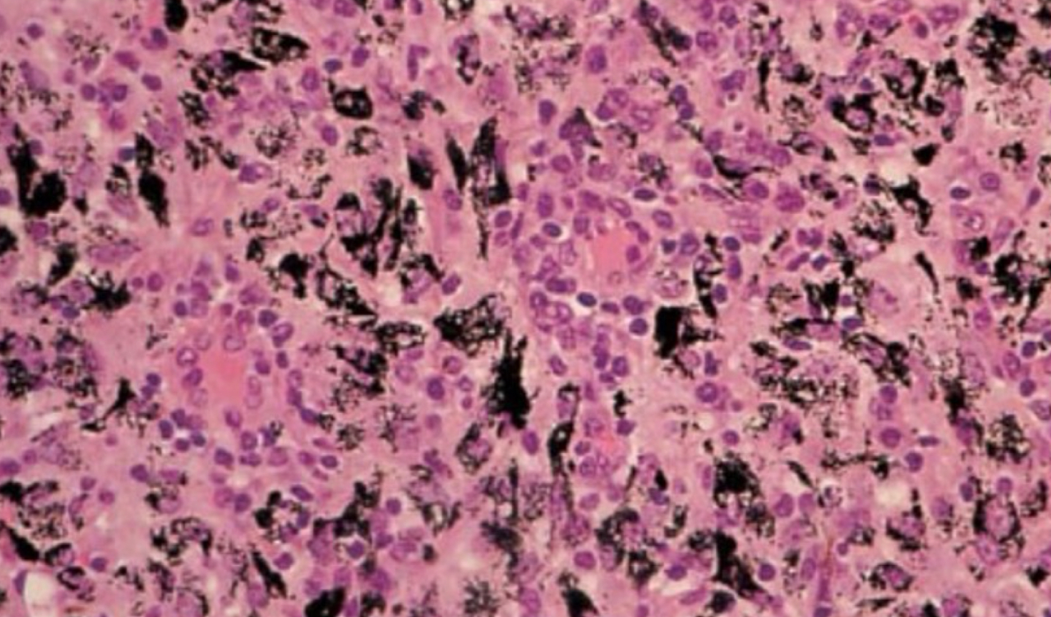
exogenous pigment examples
carbon, pollutant
exogenous pigment disease
antracosis
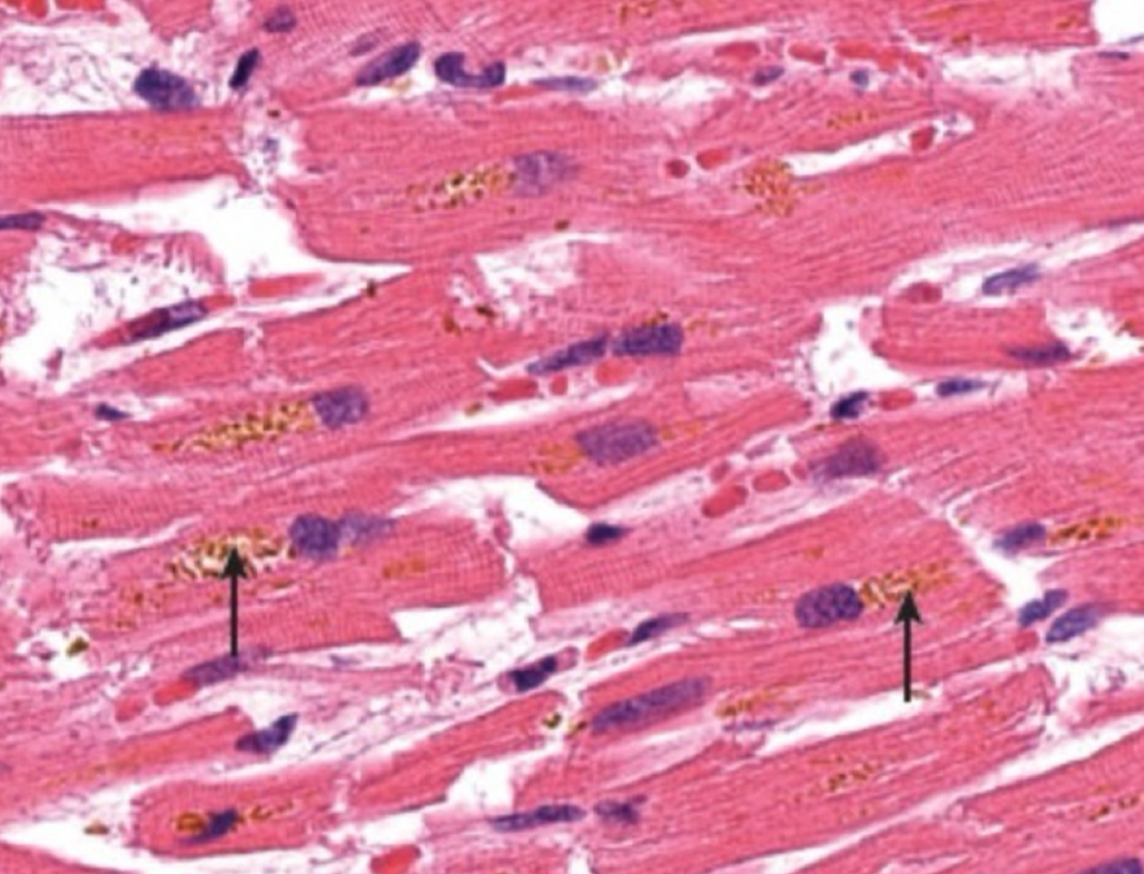
endogenous pigment example
lipofuscin, melanin, hemosiderin
lipofuscin cause
aging or atrophy
hemosiderin cause
excess iron
what type of pigment is depicted
exogenous (carbon)
what type of pigment is depicted
lipofuscin
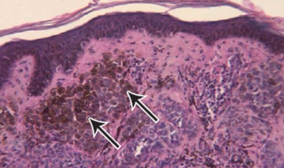
what type of pigment is depicted
melanin
what type of pigment is depicted
hemosiderin
what condition is depicted
calcification