Neuroscience anatomy - The meninges and the cerebrospinal fluid
1/12
There's no tags or description
Looks like no tags are added yet.
Name | Mastery | Learn | Test | Matching | Spaced | Call with Kai |
|---|
No analytics yet
Send a link to your students to track their progress
13 Terms
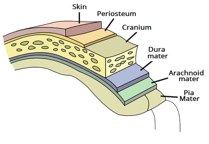
What is the meninges? What are its layers called? What are the functions of it?
3 protective membrane layers that envelop the brain and spinal cord
Dura mater (outermost)
Thick, fibrous layer
Adhering to skull/vertebrae
Anchors CNS
Contains venous sinuses for blood/CSF drainage
Arachnoid mater (middle)
Thin, avascular layer
Trabecular bridging to pia
Forms subarachnoid space filled with CSF for shock absorption/nutrient delivery
Pia mater (innermost)
Thin layer
Hugging CNS surface
Nourishes tissue
Produces some CSF
Supports blood vessels
Main functions → Protect against trauma, stabilize CNS, and facilitate circulation
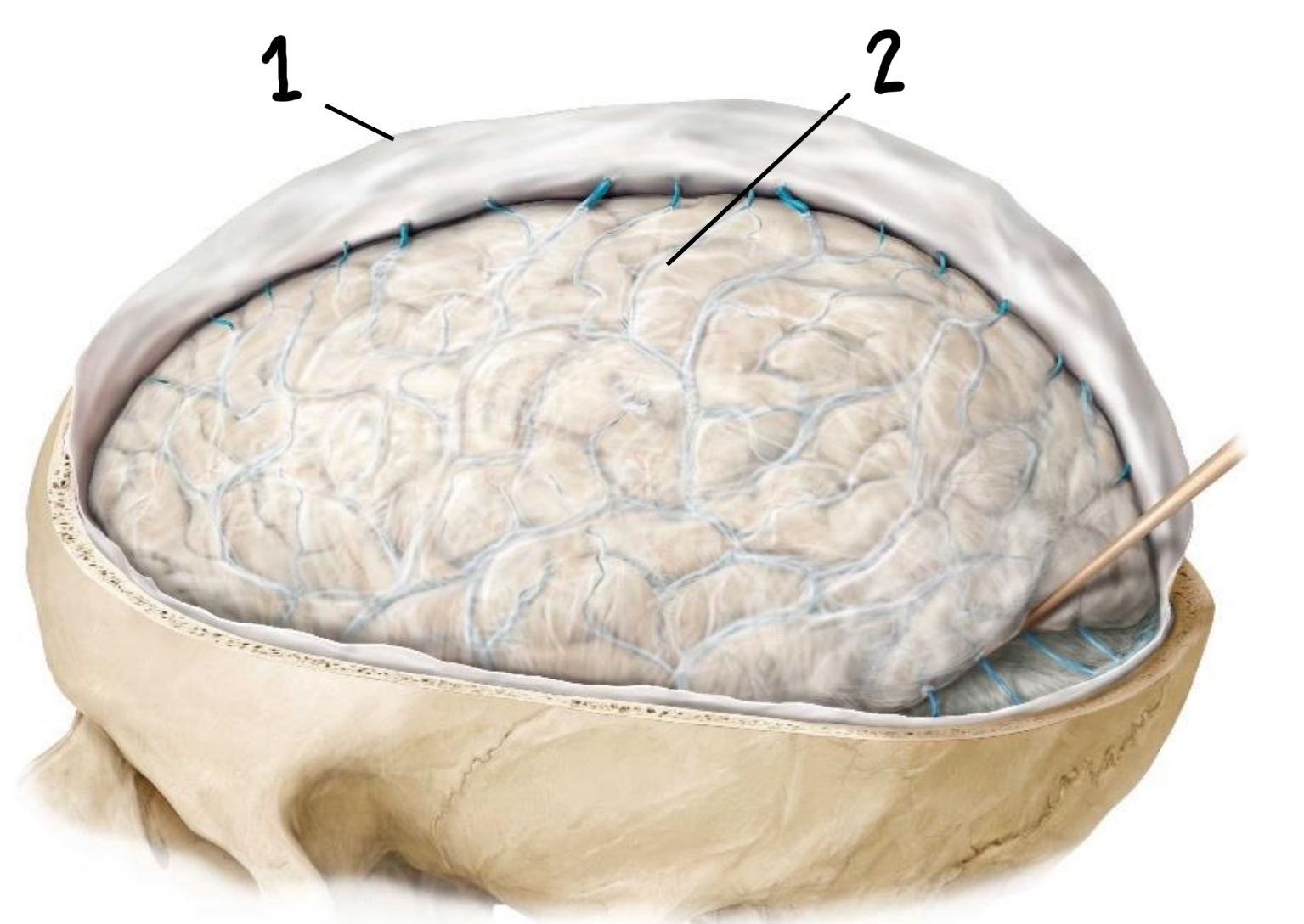
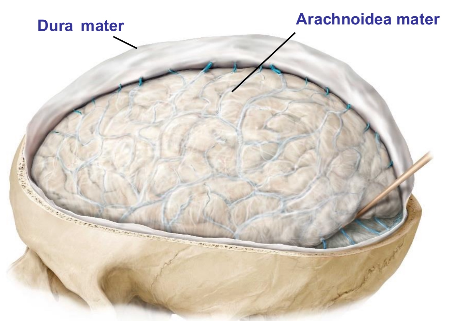
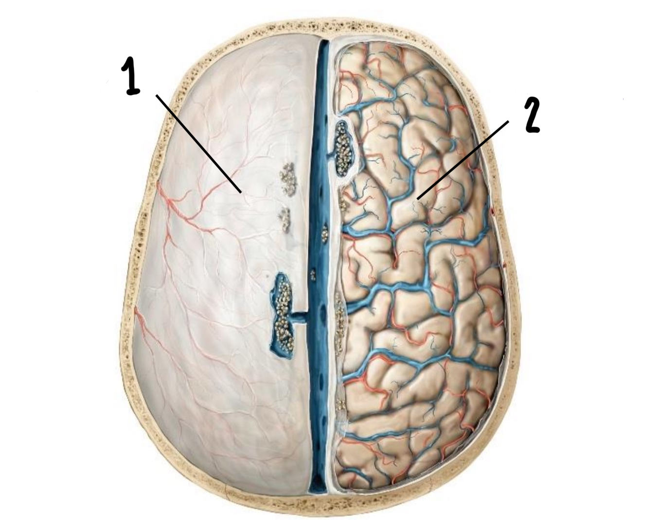
Which are these 2 layers of the meninges?
Dura mater
Arachnoidea mater
Which are these 2 layers of the meninges (outermost and innermost)?
Dura mater
Pia mater
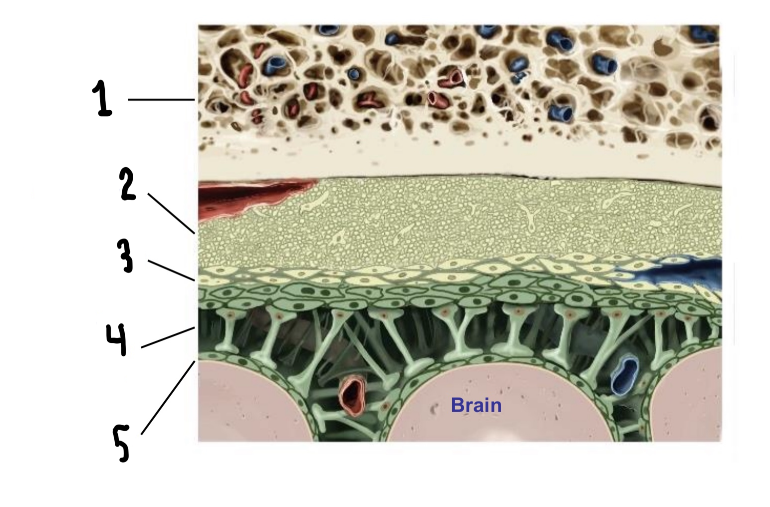
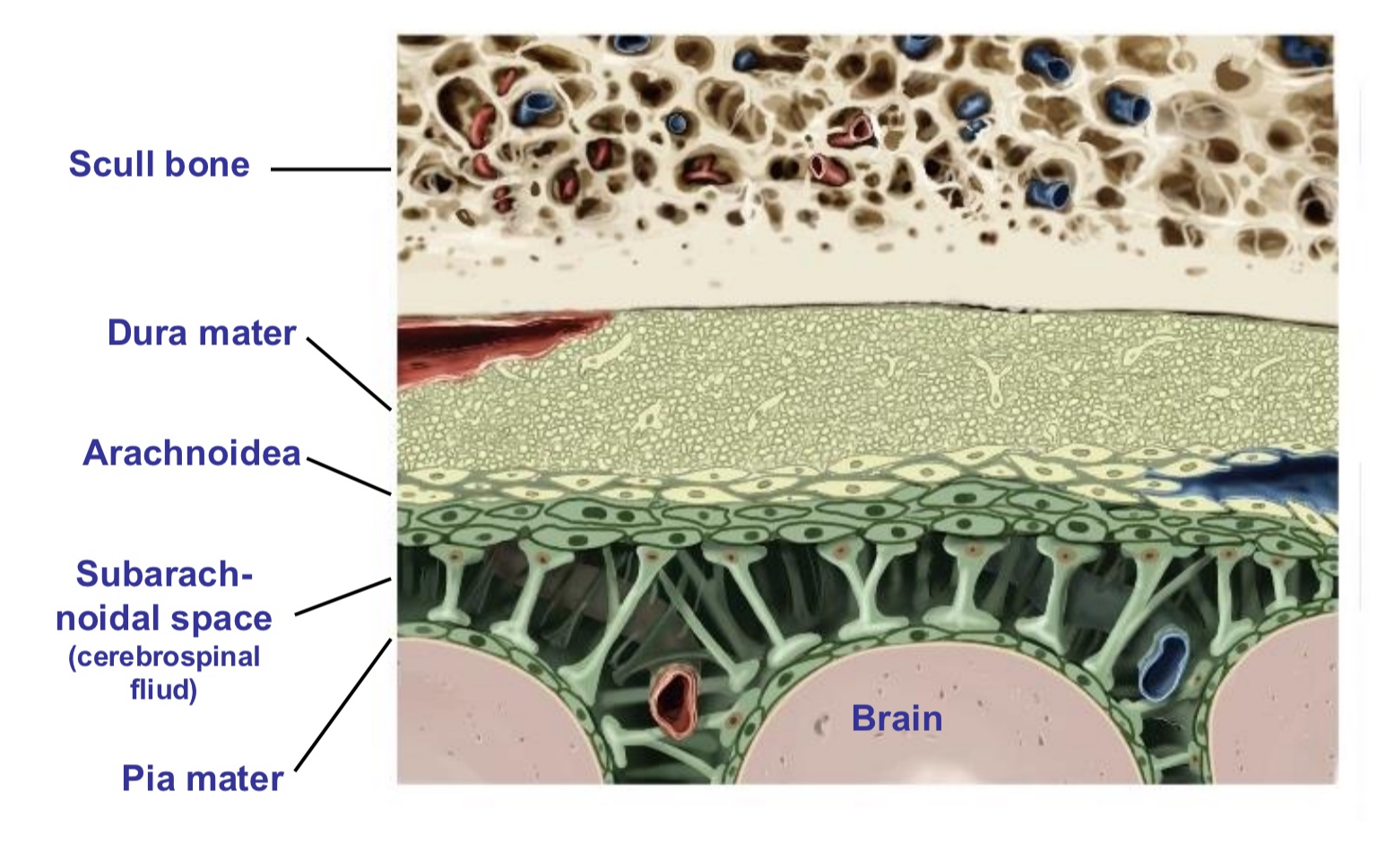
Which are these layers of the meninges?
Scull bone
Dura mater
Arachnoidea
Subarachnoidal space
Below arachnoidea
Filled with cerebrospinal fluid
Pia mater
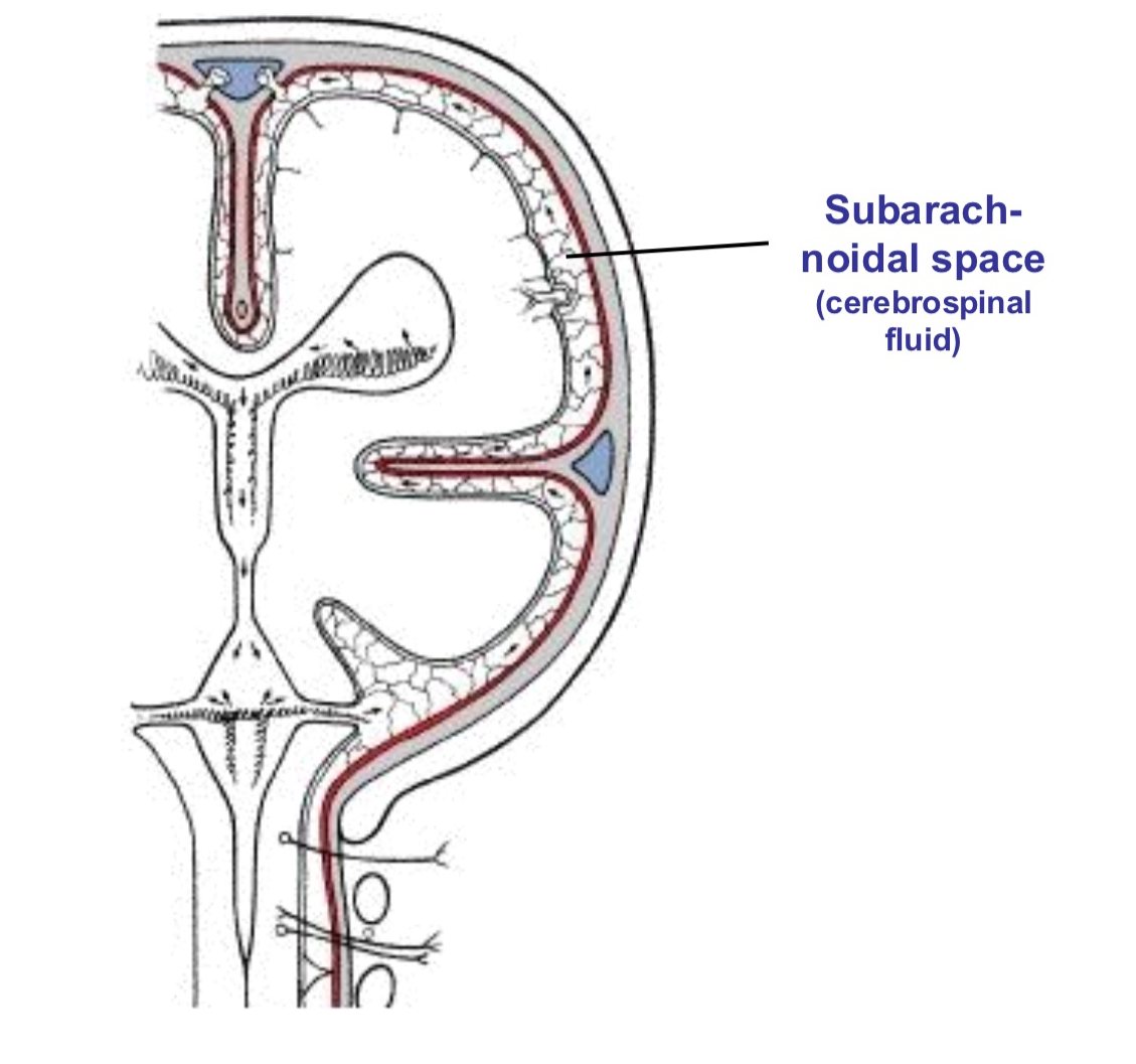
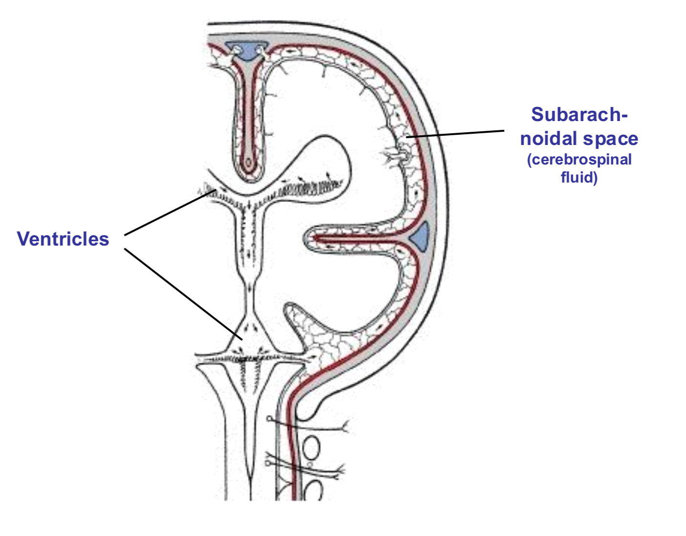
What compartment in the meninges is this? What are its functions?
The subarachnoid space
The fluid-filled compartment between the arachnoid mater and Pia mater
Surrounds the brain and spinal cord
Contains cerebrospinal fluid (CSF)
Functions:
Provides mechanical shock absorption
Maintains intracranial pressure
Circulates CSF
Supports neuromuscular structures
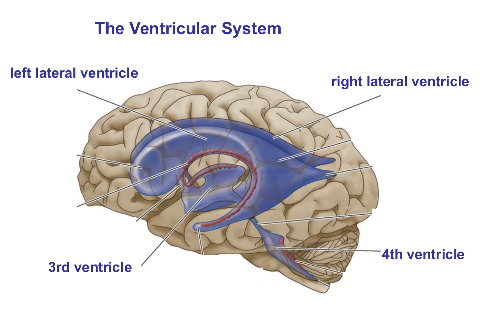
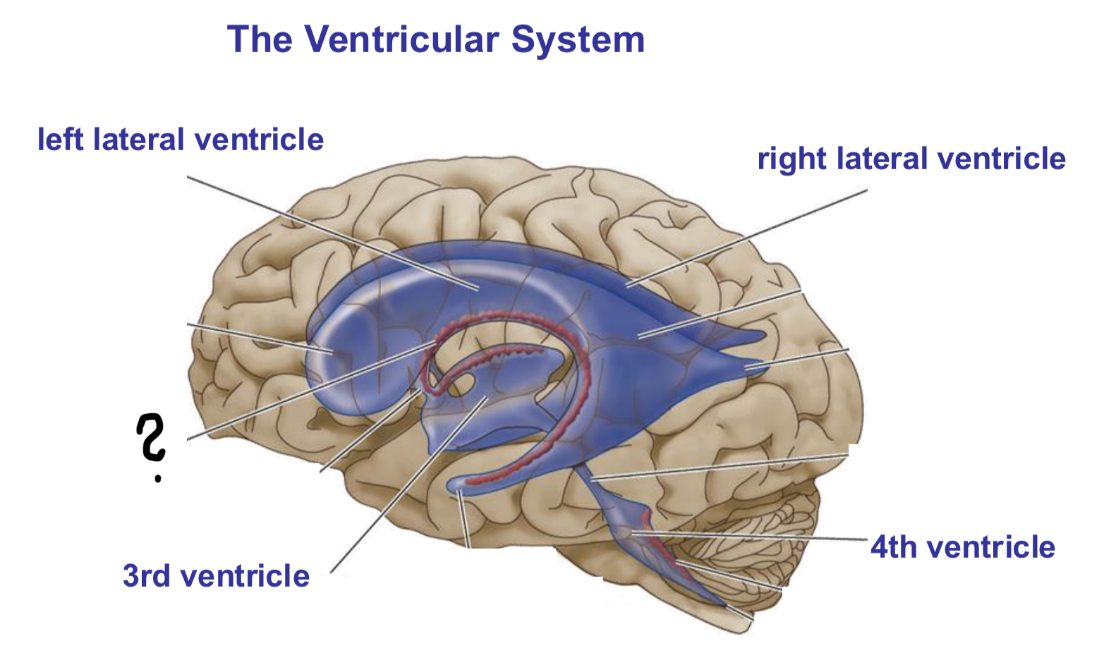
What is the ventricular system?
A network of 4 interconnected, CSF-filled cavities within the brain that produce, circulate, and house cerebrospinal fluid (CSF)
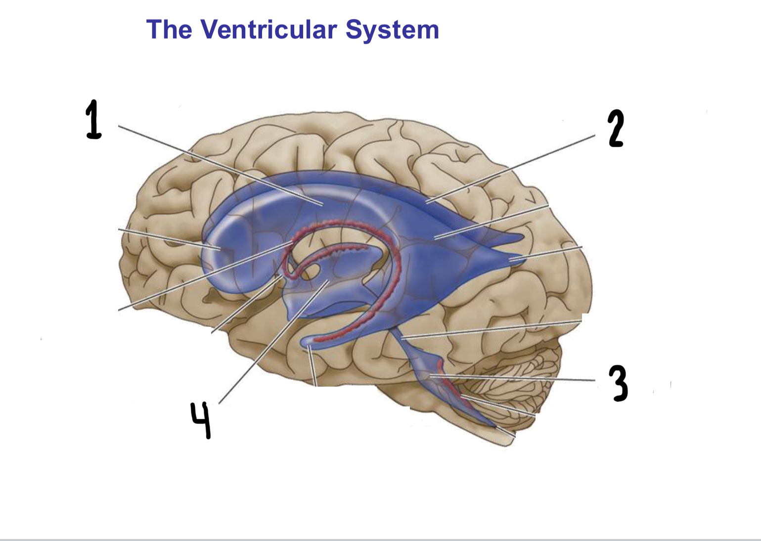
What are the 4 ventricles of the ventricular system called?
Left lateral ventricle (in left hemisphere)
Right lateral ventricle (in right hemisphere)
3rd ventricle
4th ventricle
What is this structure called? What is its main function?
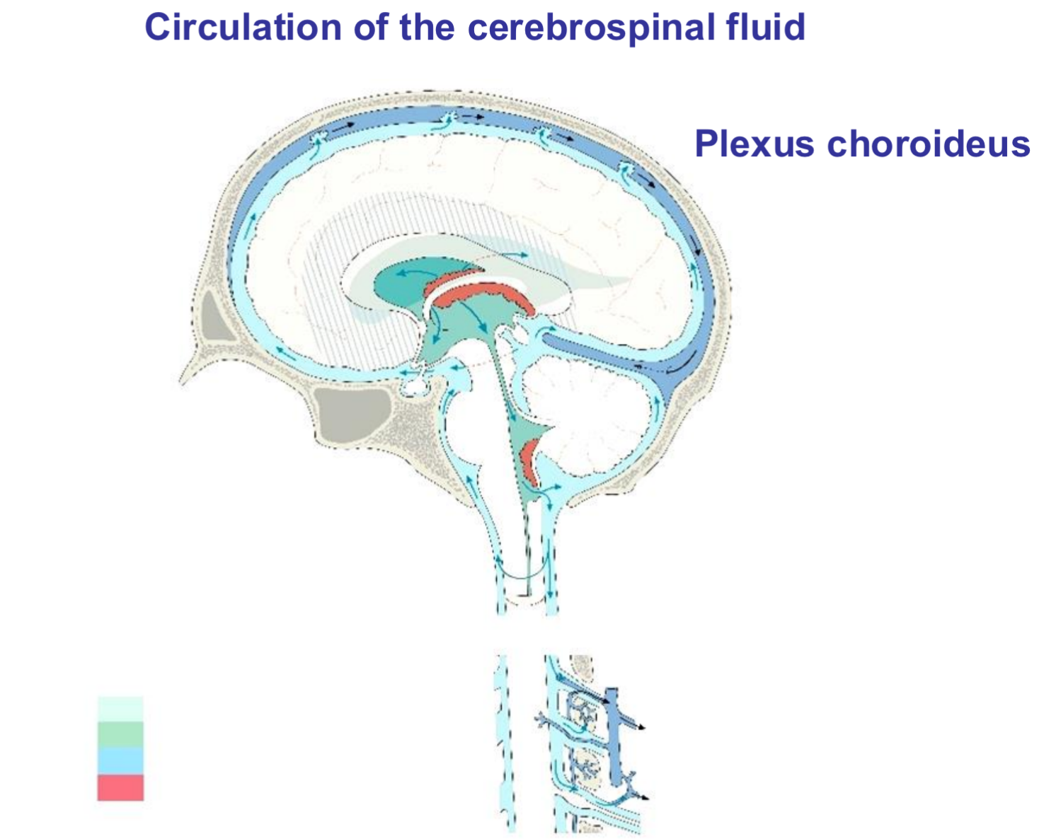
Choroid plexus
A highly vascularised network of capillaries and specialized ependymal cells located within the brain’s ventricles
Responsible for producing ~70-80% of CSF

Which ventricles are these (horizontal section)?
Lateral ventricles
How does the CSF circulate?
CSF circulates through the brain's ventricular system and subarachnoid space in a unidirectional flow driven by choroid plexus production (~500 mL/day), arterial pulsations, respiration, and posture
Circulation Pathway
Produced in lateral ventricles by choroid plexus
Flows from lateral ventricles to 3rd ventricle
Flows from 3rd ventricle to 4th ventricle
Exits 4th ventricle into cisterna magna and subarachnoid space around brain/spinal cord
Circulates multidirectionally over brain convexities, basal cisterns, spinal subarachnoid space
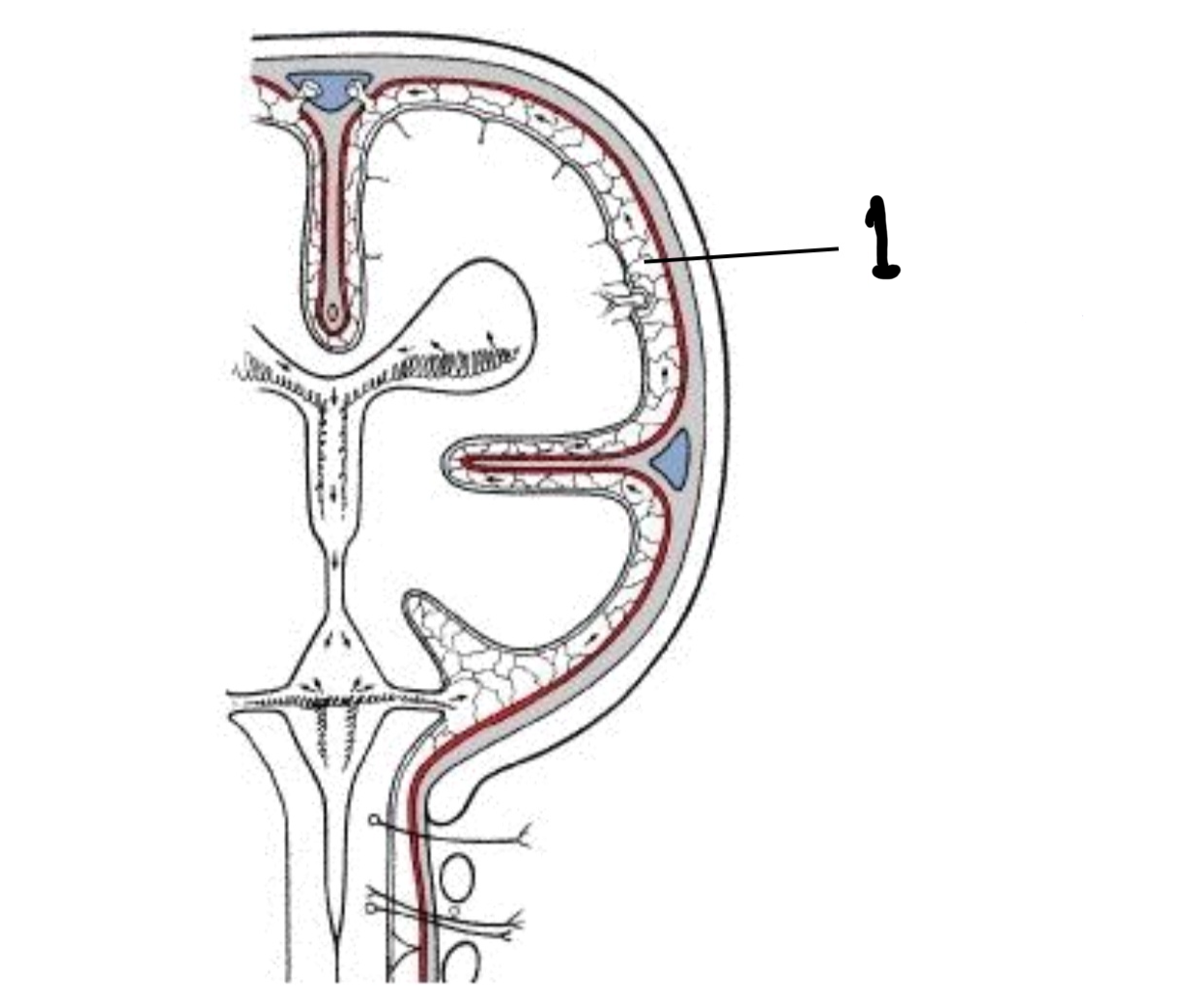
How is the CSF reabsorbed into the blood?
Reabsorbed into venous blood via extensions (villi) of the arachnoidea, which extends into large veins
Pressure gradient favors one-way flow
![<ul><li><p class="my-2 [&+p]:mt-4 [&_strong:has(+br)]:inline-block [&_strong:has(+br)]:pb-2">Reabsorbed into venous blood via extensions (villi) of the arachnoidea, which extends into large veins </p></li><li><p class="my-2 [&+p]:mt-4 [&_strong:has(+br)]:inline-block [&_strong:has(+br)]:pb-2">Pressure gradient favors one-way flow </p></li></ul><p></p>](https://knowt-user-attachments.s3.amazonaws.com/eb7e35ad-3a57-4990-8227-bccdc7c77aec.png)
What are the main functions of the CSF?
Mechanical protection
Acts as a shock absorber, protecting the brain and spinal cord against blows or sudden movements
Buoyancy
By suspending the brain in fluid, CSF effectively reduces the brain’s weight from about 1.5 kg to a few tens of grams → Preventing it from compressing its own blood vessels and tissue
Homeostasis, nutrion, and waste removal
Maintain a stable chemical environment (pH, electrolytes) necessary for proper neuronal function and regulation of blood flow and hormone signaling
Contributes to nutrient delivery and waste clearance, carrying metabolic by-broducts away from brain tissue and into venous or lymphatic circulation
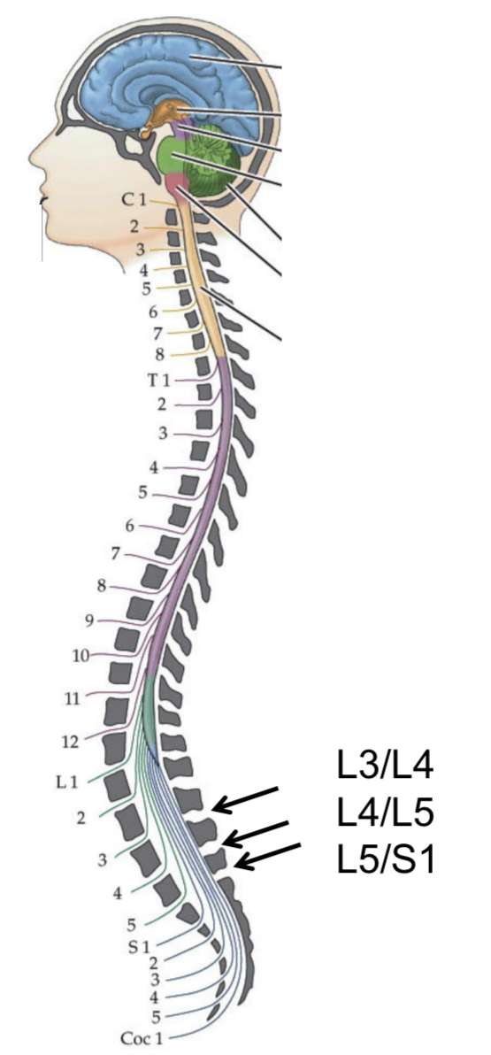
How are diagnostic CSF samples taken? What can they detect?
Collected via lumbar puncture (spinal tap)
A needle is inserted between the 3rd, 4th, or 5th lumbar vertebrae (below the spinal cord’s end) into the subarachnoid space to withdraw CSF for analysis of cells, proteins, glucose, infections, or biomarkers
Analysis detects meningitis, haemorrhage, MS, tumours