Muscles and Tendons of the Distal Limb (Week 2, Mod 7)

0.0(0)
studied byStudied by 0 people
0.0(0)
full-widthCall with Kai
GameKnowt Play
New
learnLearn
examPractice Test
spaced repetitionSpaced Repetition
heart puzzleMatch
flashcardsFlashcards
Card Sorting

1/17

encourage image

There's no tags or description

Looks like no tags are added yet.

Study Analytics
Name
Mastery
Learn
Test
Matching
Spaced

No study sessions yet.

18 Terms

1
New cards

What are some general features of the muscles of the antebrachium? Keep these in mind as you go along, as these will remain constant (there are 5)

1) Will affect the joints of the carpus and digits

2) Origin - epidcondyles of the humerus

3) Muscle belly located in the antebrachium ; extend as tendons to the carpus and digits

4) Tendon of insertion → distal to carpus

5) EXTENSOR = DORSAL ASPECT OF CARPUS 

FLEXOR = PALMAR ASPECT OF CARPUS 

2
New cards

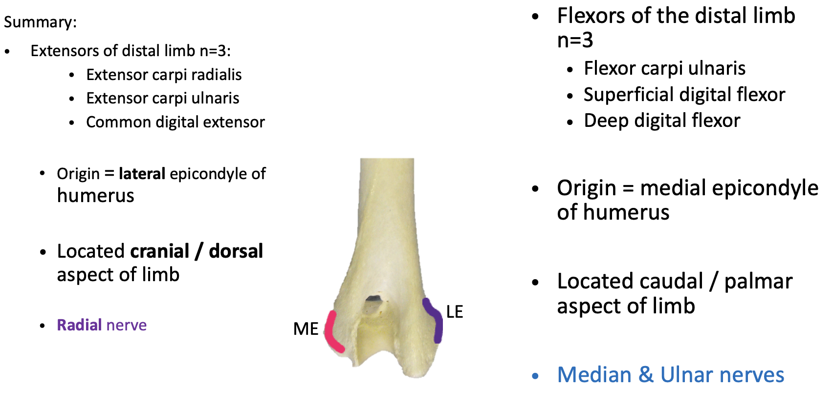
Describe the extensor carpi radialis muscle… origin, insertion, location, function, nerve supply, and any unique characteristics for identification

Origin - Lateral epicondyle of humerus

Insertion - the metacarpal bones

Location - CRANIAL aspect of antebrachium, becomes a tendon once reaching the carpus

Function - in the name… extensor carpi = CARPAL EXTENSOR 

Nerve supply - also in the name; Radial nerve 

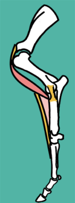
Unique feature - has a small, oblique muscle that runs over the tendon portion of the carpi radialis (see image)

<p><strong>Origin - </strong>Lateral epicondyle of humerus </p><p></p><p><strong>Insertion - </strong>the metacarpal bones</p><p></p><p><strong>Location - </strong>CRANIAL aspect of antebrachium, becomes a tendon once reaching the carpus </p><p></p><p><strong>Function -</strong> in the name… extensor carpi = CARPAL<strong> EXTENSOR&nbsp;</strong></p><p></p><p><strong>Nerve supply -</strong> also in the name; Radial nerve&nbsp;</p><p></p><p><strong>Unique feature -</strong> has a small,<u> oblique</u>&nbsp;muscle that runs over the tendon portion of the carpi radialis&nbsp;(see image)</p>
3
New cards

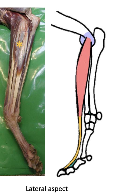
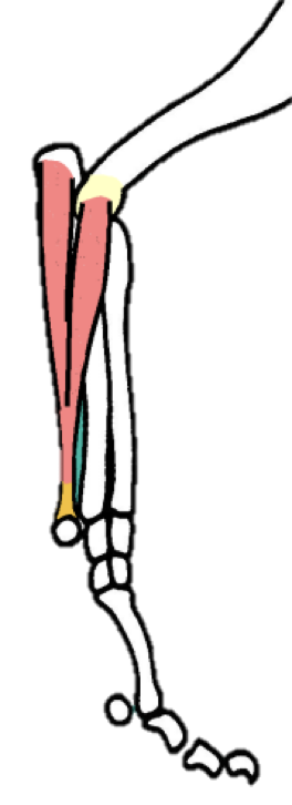
Describe the common digital extensor muscle… origin, insertion, location, function, nerve supply, and any unique characteristics for identification

IS THE MOST IMPORTANT EXTENSOR MUSCLE

Origin - lat epicondyle of humerus

Insertion - ALL DIGITS (at the distal phalanx, splits and inserts at each digit)

  • In the horse, is one single branch down to the distal phalanx

    • Protected by dorsal bursas from the associated joint capsules

    • RECEIVES suspensory ligament branches (see previous notecard set)

Location - Cranio - lateral aspect of antebrachium (see images); crosses over DORSAL carpus and DORSALLY over metacarpo-phalangeal and interphalangeal joints

Function - Carpal AND digital EXTENSOR

Nerve supply - Radial nerve

<p>IS THE MOST IMPORTANT EXTENSOR MUSCLE </p><p><strong>Origin - </strong>lat epicondyle of humerus </p><p></p><p><strong>Insertion - </strong>ALL DIGITS (at the distal phalanx, splits and inserts at each digit)</p><ul><li><p>In the horse, is one single branch down to the distal phalanx </p><ul><li><p>Protected by dorsal bursas from the associated joint capsules</p></li><li><p>RECEIVES <strong>suspensory ligament branches</strong> (see previous notecard set)</p></li></ul></li></ul><p></p><p><strong>Location - </strong>Cranio - lateral aspect of antebrachium (see images); crosses over DORSAL carpus and DORSALLY over metacarpo-phalangeal and interphalangeal joints </p><p></p><p><strong>Function - </strong>Carpal AND digital <strong>EXTENSOR </strong></p><p></p><p><strong>Nerve supply - </strong>Radial nerve </p>
4
New cards

Describe the extensor carpi ulnaris / ulnaris lateralis muscle… origin, insertion, location, function, nerve supply, and any unique characteristics for identification

Origin - Lat epicondyle 

Insertion - 5th Metacarpal bone + accessory carpal bone 

Location - LATERAL aspect of antebrachium (say ulnaris lateralis… gives location better);

crosses over the LATERAL aspect of the CARPUS 

Function - DEPENDS ON LIMB POSITION 

  • Supports both extension AND flexion, depending on limb positioning 

    • When limb is already EXTENDED, muscle SUPPORTS that extension

    • When limb is FLEXED, helps produce + maintain flexion via attachment to ACB (the “lever” of the carpal bones")

Nerve supply - Radial nerve

<p><strong>Origin -</strong> Lat epicondyle&nbsp;</p><p></p><p><strong>Insertion -</strong> 5th Metacarpal bone + accessory carpal bone&nbsp;</p><p></p><p><strong>Location -</strong> LATERAL aspect of antebrachium (say ulnaris lateralis… gives location better);</p><p>crosses over the LATERAL aspect of the CARPUS&nbsp;</p><p></p><p><strong>Function - DEPENDS ON LIMB POSITION&nbsp;</strong></p><ul><li><p>Supports both extension AND flexion, depending on limb positioning&nbsp;</p><ul><li><p>When limb is already EXTENDED, muscle SUPPORTS that extension </p></li><li><p>When limb is FLEXED, helps produce + maintain flexion via attachment to ACB (the “lever” of the carpal bones")</p></li></ul></li></ul><p></p><p><strong>Nerve supply - </strong>Radial nerve </p><p></p>
5
New cards

Describe the flexor carpi ulnaris muscle… origin, insertion, location, function, nerve supply, and any unique characteristics for identification

Origin - HAS 2 ORIGINS

  • MEDIAL epicondyle 

  • Olecranon process of ulna 

Insertion -  ACB

Location - CAUDAL aspect of the antebrachium; crosses caudal aspect of carpus 

Function - in the name: flexor carpi = FLEX CARPUS 

Nerve supply - Median + Ulnar nerves 

<p><strong>Origin -</strong> HAS 2 ORIGINS</p><ul><li><p>MEDIAL epicondyle&nbsp;</p></li><li><p>Olecranon process of ulna&nbsp;</p></li></ul><p></p><p><strong>Insertion -&nbsp;&nbsp;</strong>ACB</p><p></p><p><strong>Location -</strong> CAUDAL aspect of the antebrachium; crosses caudal aspect of carpus&nbsp;</p><p></p><p><strong>Function -</strong> in the name: flexor carpi = FLEX CARPUS&nbsp;</p><p></p><p><strong>Nerve supply -</strong> Median + Ulnar nerves&nbsp;</p><p></p>
6
New cards

Describe the superficial digital flexor muscle muscle… origin, insertion, location, function, nerve supply, and any unique characteristics for identification

REALLY IMPORTANT 

Origin - medial epicondyle

Insertion - ALL digits (MIDDLE PHALANX)

Location - caudal aspect of limb; crosses over PALMAR aspect of carpus, metacarpo-phalangeal, and interphalangeal joints

Function - is a carpal AND digital FLEXOR

Nerve supply - Median + Ulnar nerves 

<p>REALLY IMPORTANT&nbsp;</p><p><strong>Origin - </strong>medial epicondyle </p><p></p><p><strong>Insertion - </strong>ALL digits (MIDDLE PHALANX)</p><p></p><p><strong>Location - </strong>caudal aspect of limb; crosses over PALMAR aspect of carpus, metacarpo-phalangeal, and interphalangeal joints </p><p></p><p><strong>Function - </strong>is a carpal AND digital <strong>FLEXOR</strong></p><p></p><p><strong>Nerve supply -</strong> Median + Ulnar nerves&nbsp;</p>
7
New cards

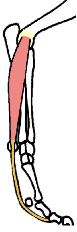
How does the SDFT muscle of the canine differ from that of the HORSE?

In the horse:

  • SDFT is only ONE branch

  • Receives an Accessory Check Ligament (ACL) from the radius

    • Found PROXIMAL to carpus

    • Limits the length of the tendon, and protects the muscle belly from overstretching 

<p>In the horse:</p><ul><li><p>SDFT is only ONE branch</p></li><li><p>Receives an<strong>&nbsp;Accessory Check Ligament (ACL)</strong> from the radius</p><ul><li><p>Found PROXIMAL to carpus</p></li><li><p>Limits the length of the tendon, and protects the muscle belly from overstretching&nbsp;</p></li></ul></li></ul><p></p>
8
New cards

How are the SDFT of the canine and horse SIMILAR? Think of 3 things

Both are VERY close to the surface; are almost palpable

Both split distally to allow passage of the deep digital flexor tendon 

Both insert at the middle phalanx 

9
New cards

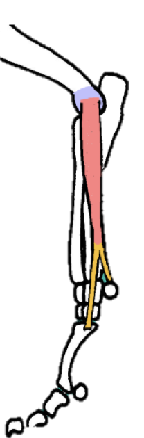
Describe the deep digital flexor tendon muscle… origin, insertion, location, function, nerve supply, and any unique characteristics for identification

Origin - HAS 3 ORIGINS

  • Medial epicondyle 

  • Radius 

  • Ulna

Insertion - ALL digits (palmar aspect of DISTAL phalanx; this is why SDFT has to split)

Location - caudal aspect of limb; crosses PALMAR aspect of CARPUS and all other joints

  • In the HORSE: runs over distal sesamoid (navicular bone)

Function - Carpal AND digital FLEXOR

Nerve supply - Median + Ulnar nerves

<p><strong>Origin -</strong> HAS 3 ORIGINS</p><ul><li><p>Medial epicondyle&nbsp;</p></li><li><p>Radius&nbsp;</p></li><li><p>Ulna</p></li></ul><p></p><p><strong>Insertion -</strong> ALL digits (palmar aspect of DISTAL phalanx; this is why SDFT has to split)</p><p></p><p><strong>Location - </strong>caudal aspect of limb; crosses PALMAR aspect of CARPUS and all other joints</p><ul><li><p>In the HORSE: <u>runs over distal sesamoid</u> (navicular bone)</p></li></ul><p></p><p><strong>Function - </strong>Carpal AND digital <strong>FLEXOR</strong></p><p></p><p><strong>Nerve supply - </strong>Median + Ulnar nerves </p><p></p>
10
New cards

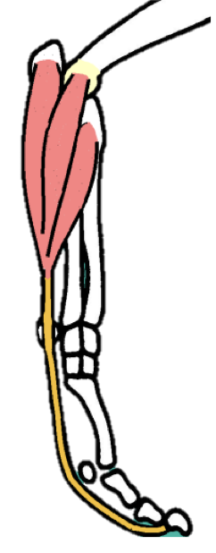
How is the DDFT SIMILAR to the SDFT in the horse?

Is 1 branch as well

ALSO HAS AN ACCESSORY CHECK LIGAMENT

  • This one comes from the DISTAL portion of the carpus… is an extension of the carpal joint capsule (see images)

  • Has the same function as the proximal ACL for the SDFT

11
New cards

Summary slide

knowt flashcard image
12
New cards

TIME FOR EQUINE VARIATIONS

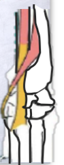
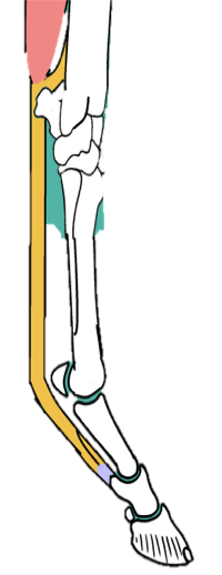
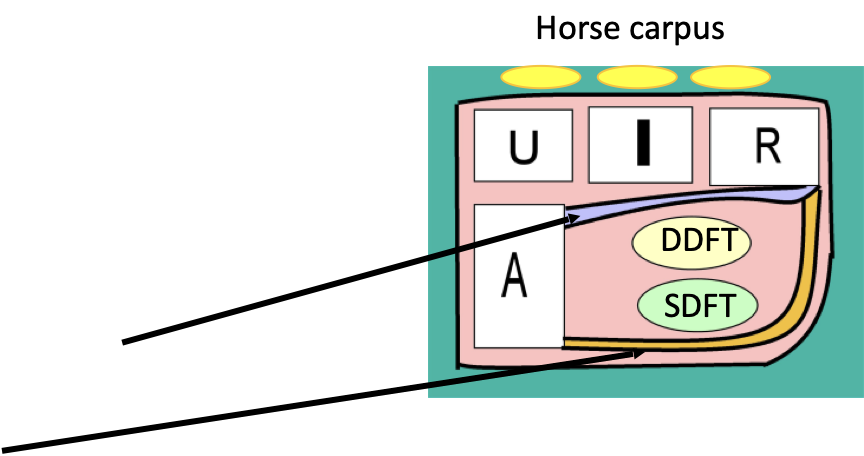
What is the carpal canal? What are its contents, and what makes up its boundaries?

The carpal canal, or “carpal tunnel”, is a passageway on the PALMAR aspect of the carpus…

  • The SDFT and DDFT run through here, along with blood vessels and nerves 

Boundaries:

  • Dorsally - palmar aspect of carpal joint capsule (purple line)

  • Laterally - the accessory carpal bone

  • Palmar - the flexor retinaculum

    • In orange; is a sleeve of fibrous tissue that encases the limb, like a compression sock 

In the dog, the SDFT runs OUTSIDE of this 

<p>The carpal canal, or “carpal tunnel”, is a passageway on the PALMAR aspect of the carpus…</p><ul><li><p>The SDFT and DDFT run through here, along with blood vessels and nerves&nbsp;</p></li></ul><p></p><p><strong>Boundaries:</strong></p><ul><li><p>Dorsally - palmar aspect of<u> carpal joint capsule</u>&nbsp;(purple line)</p></li><li><p>Laterally - the<u> accessory carpal bone</u></p></li><li><p>Palmar - the<u> flexor retinaculum</u></p><ul><li><p>In orange; is a sleeve of fibrous tissue that encases the limb, like a compression sock&nbsp;</p></li></ul></li></ul><p></p><p>In the dog, the SDFT runs OUTSIDE of this&nbsp;</p><p></p>
13
New cards

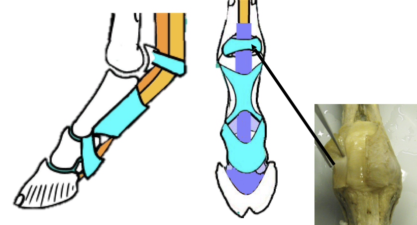
What is a “tendon sheath”, and what is its function?

Is a fluid filled tissue that surrounds the tendon, protecting it where it passes through confined spaces

  • Doesn’t completely wrap around, more like absorbs it (see images)

<p>Is a fluid filled tissue that surrounds the tendon, protecting it where it passes through confined spaces</p><ul><li><p>Doesn’t completely wrap around, more like absorbs it (see images)</p></li></ul><p></p>
14
New cards

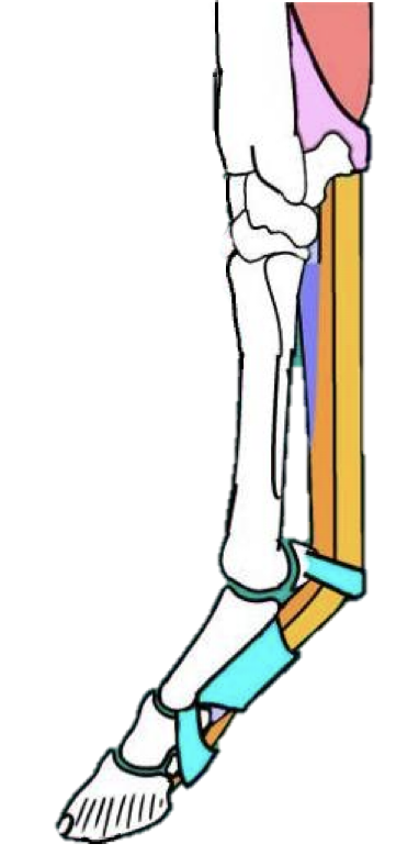
Describe how the tendons on BOTH the dorsal aspect and palmar aspect of the equine distal limb are stabilized… what particular ligaments can be found on the palmar aspect?

Dorsally - 

  • Only CDE is found here… held in place by retinaculum

Palmar - 

  • SDFT and DDFT are held in place by:

    • Carpal canal

    • ANNULAR LIGAMENTS (in blue)

      • Found in the fetlock and pastern regions

<p><strong>Dorsally -&nbsp;</strong></p><ul><li><p>Only CDE is found here… held in place by<u> retinaculum</u></p></li></ul><p></p><p><strong>Palmar -&nbsp;</strong></p><ul><li><p>SDFT and DDFT&nbsp;are held in place by:</p><ul><li><p>Carpal canal </p></li><li><p><strong><u>ANNULAR LIGAMENTS</u></strong> (in blue)</p><ul><li><p>Found in the fetlock and pastern regions </p></li></ul></li></ul></li></ul><p></p>
15
New cards

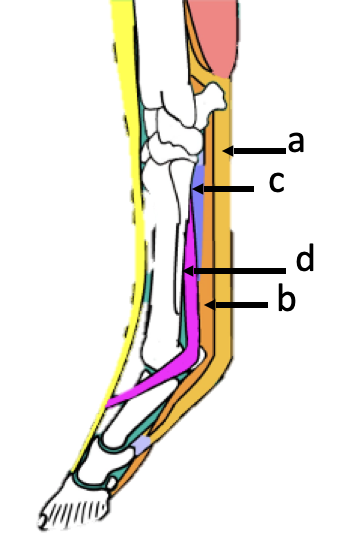
What is the order of tendons on the palmar aspect of the distal equine limb, starting from the skin:

Skin -

a) SDFT

b) DDFT
c) ACL / Check ligament (fused with DDFT)

d) Suspensory ligament (branches in 2)

<p>Skin -</p><p>a) SDFT</p><p>b) DDFT<br>c) ACL / Check ligament (fused with DDFT)</p><p>d) Suspensory ligament (branches in 2)</p>
16
New cards

Describe the concept of the “equine stay apparatus”… what needs to be done in order for this to work?

Most of the horse’s weight is borne on the forelimb… so a mechanism is needed to allow for passive weight-bearing, which lets a horse sleep standing up.

This requires MAINTENANCE OF EXTENSION; this is done by:

  • Proximal limb joints → prevention of flexion

  • Carpus → prevention of flexion and hyperextension 

  • Distal limb joints → prevention of hyperextension 

17
New cards

What muscles and ligaments of the proximal limb joints help to prevent flexion in the equine stay apparatus?

Scapula → Serratus ventralis muscle 

  • Suspends weight of the body between the forelimbs

Prevention of flexion:

  • Shoulder → biceps brachii

  • Elbow → caudal collateral ligaments + the alignment of the bones 

  • Carpus → lacertus fibrosis (fibrous tendon that attaches biceps brachii to metacarpals)

<p>Scapula →<strong> Serratus ventralis muscle&nbsp;</strong></p><ul><li><p>Suspends weight of the body between the forelimbs</p></li></ul><p></p><p>Prevention of flexion:</p><ul><li><p>Shoulder →<strong> biceps brachii</strong></p></li><li><p>Elbow → caudal collateral ligaments + the alignment of the bones&nbsp;</p></li><li><p>Carpus → lacertus fibrosis (fibrous tendon that attaches biceps brachii to metacarpals)</p></li></ul><p></p>
18
New cards

What muscles and ligaments of the carpus and distal limb joints help to prevent hyperextension in the equine stay apparatus?

Carpus: 

  • Palmar fibrocartilage joint reinforcement (in blue)

  • SDFT and check ligament

  • Retinaculum 

MCP / Fetlock joint : 

  • Suspensory ligament

  • Common digital extensor

  • Proximal sesamoids 

  • Distal sesamoidean ligaments

    • (short, cruciate, oblique & straight)

MCP, PIP, and DIP joints

  • DDFT & SDFT

    • Check ligaments

    • Annular ligaments

<p>Carpus:&nbsp;</p><ul><li><p>Palmar fibrocartilage joint reinforcement (in blue)</p></li><li><p>SDFT and check ligament</p></li><li><p>Retinaculum&nbsp; </p></li></ul><p></p><p>MCP / Fetlock joint :&nbsp;</p><ul><li><p>Suspensory ligament</p></li><li><p>Common digital extensor</p></li><li><p>Proximal sesamoids&nbsp;</p></li><li><p>Distal sesamoidean ligaments</p><ul><li><p>(short, cruciate, oblique &amp; straight)</p></li></ul></li></ul><p></p><p>MCP, PIP, and DIP joints</p><ul><li><p>DDFT &amp; SDFT</p><ul><li><p>Check ligaments</p></li><li><p>Annular ligaments</p></li></ul></li></ul><p></p>