C2.1 Chemical Signalling
1/42
There's no tags or description
Looks like no tags are added yet.
Name | Mastery | Learn | Test | Matching | Spaced |
|---|
No study sessions yet.
43 Terms
What are ligands?
Signalling molecules
What are examples of ligands?
Proteins + AA’s
Nucleotides
Steroids
Amines
How do ligands work?
- Ligands are secreted from a cell into the extracellular space
- Ligands are transported to a target cell
- Ligands bind to surface receptors on the target cell
Receptors may be proteins with binding sites
Chemical messengers inside the cell trigger a response
What is Quorom sensing?
How bacteria communicate with each other by releasing chemical signals (ligands).
How does Quorom sensing work?
Increase in bacteria population= more ligands released
Enough ligands bind to receptors on other bacteria —>triggers a change in gene expression.
Bacteria colony acts together switching genes off/on when quorum is reached.
What is Vibrio fischeri?
Live in a squid’s light organ
Help it camouflage using bioluminescence (light production).
How do Vibrio fischeri work?
Bacteria releases ligand called an autoinducer.
More bacteria growth= autoinducer concentration increases.
Autoinducer binds to a receptor protein (LuxR) inside other bacteria.
Enough autoinducer–LuxR complexes form—> triggers the production of an enzyme called luciferase.
Luciferase causes the bacteria to glow (bioluminesce).
What are cytokines?
Proteins released by nearly all cells in the body
- Help cells communicate + influence each other’s activity
Cytokines —> bind to receptors on surface other cells —> changes gene expression + behavior
2 main functions of Cytokines?
- Coordinate the immune response (especially between white blood cells).
- Regulate cell division and growth, like during embryo development.
3 main functions of calcium ions?
- Muscle contraction – Ca²⁺ enters muscle cells and allows protein fibers to slide and contract.
- Nerve transmission (synapse) – Ca²⁺ enters the presynaptic neuron and triggers the release of neurotransmitters.
- Can act as a second messenger inside cells after an external signal is received.
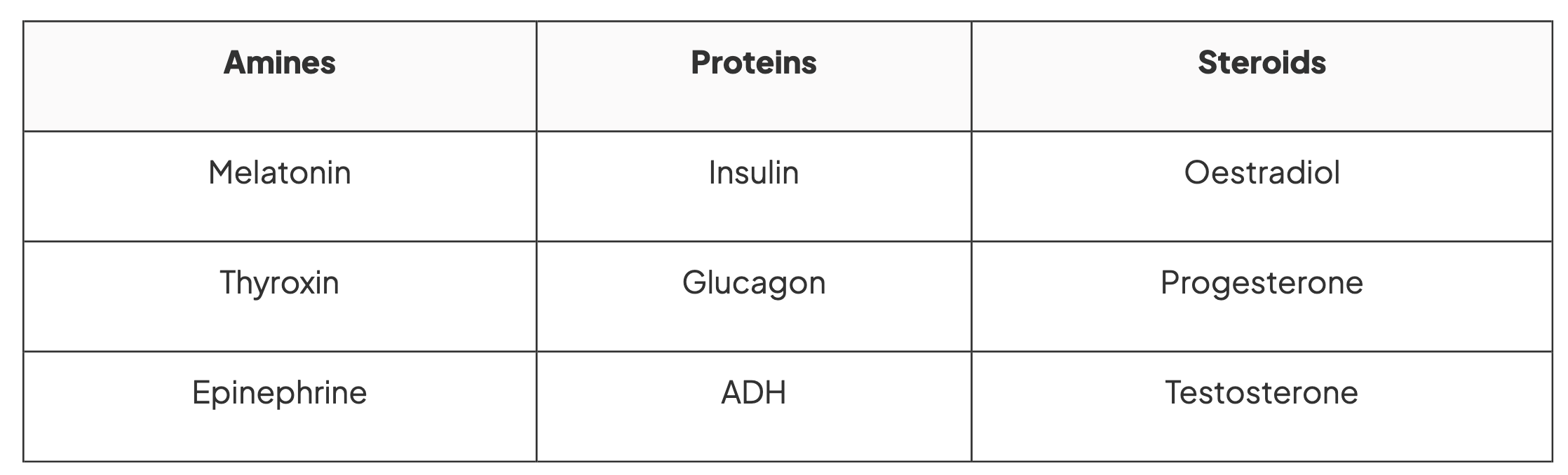
What are the 3 different categories of hormones?
Amines + proteins—> hydrophilic (cannot cross cross bilayers)
Steroid hormones—> hydrophobic (can cross cell membranes + bind to receptors in cell)
What are the examples of hormones in each category?
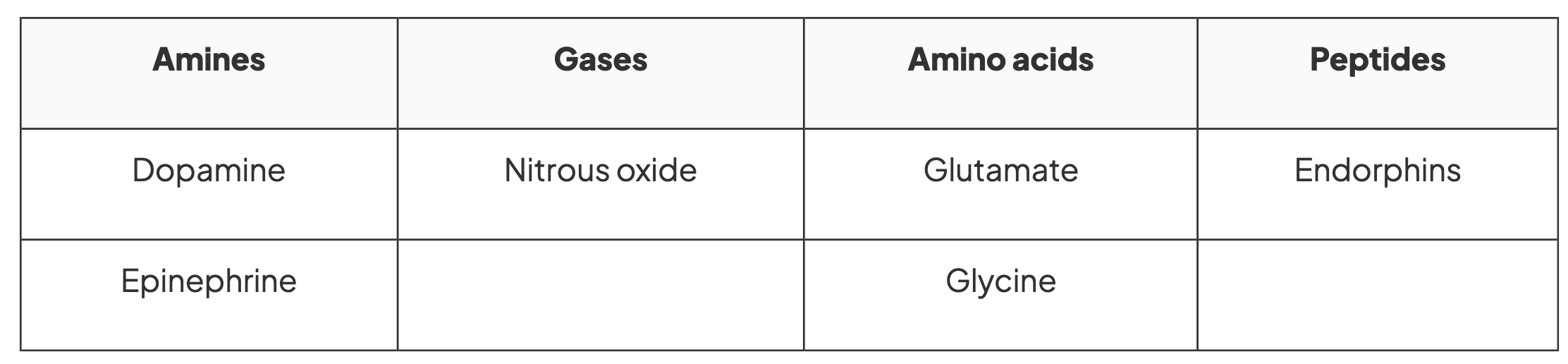
What are the 4 categories of neurotransmitters?
Amines, gases, amino acids and peptides
Most neurotransmitters are hydrophilic (can bind to receptors on cell membrane)
What are the examples of neurotransmitters in each category?
Transmembrane receptor proteins
Located in the cell membrane.
Span the entire width of the membrane
What is the structure of a transmembrane protein?
Ampiphatic
Hydrophilic regions on both ends (touching water inside and outside the cell).
A hydrophobic middle (interacts with the membrane’s fatty acid tails).
Function of transmembrane protein
Ligands (signalling molecules) bind to the outside part of the receptor.
This triggers a change inside the cell without the ligand entering.
Intracellular receptors
Found inside the cytoplasm or on the DNA in the nucleus.
Only non-polar, hydrophobic ligands (like steroid hormones) can pass through the cell membrane to reach these receptors.
What is a signal transduction pathway?
Different ligands and different receptors trigger different signal transduction pathways
Signal transduction pathway for transmembrane protein
Ligand bind to extracellular region of the transmembrane receptor protein
- Causes a change in shape of the internal region of the protein
Leads to phosphorylation events + second messenger
Signal transduction pathway for intracellular protein
A ligand binds to an intracellular receptor, forming a ligand-receptor complex
Intracellular ligand-receptor complexes are activated
Examples of signal transduction pathway responses
Regulation of gene expression through control of transcription or translation
Change in metabolic activity
Regulation of enzyme activity
Cell death
Rearrangement of the cytoplasm of the cell
Regulation of proteins
What is a G-protein-coupled-receptor (GPCR)?
Transmembrane receptor protein
Activates of G protein leading to changes inside the cell
What are G proteins?
Specialised proteins that bind to GTP or GDP
Act as a switch being activated or deactivated by signals
GTP
guanine triphosphate (active G proteins)
GDP
guanosine diphosphate (inactive G-proteins)
How are G-proteins are activated?
At rest:
- G-proteins are attached to the inside of the membrane, near a GPCR.
- They are inactive when bound to GDP.
Signal binds:
- A non-steroid ligand binds to the outside of the GPCR.
- This causes a shape change (conformational change) in the GPCR.
Activation:
- The shape change activates the G-protein.
- GDP is replaced by GTP and splits into a bound alpha subunit and a beta-gamma dimer
These subunits move along the membrane and activate other proteins, such as:
- Enzymes, Ion channels
This leads to the release of second messengers which start signal transduction inside the cell.
Switching off the signal:
GTP is hydrolysed (broken down) to GDP, inactivating the G-protein.
The subunits rejoin and attach back to the GPCR.
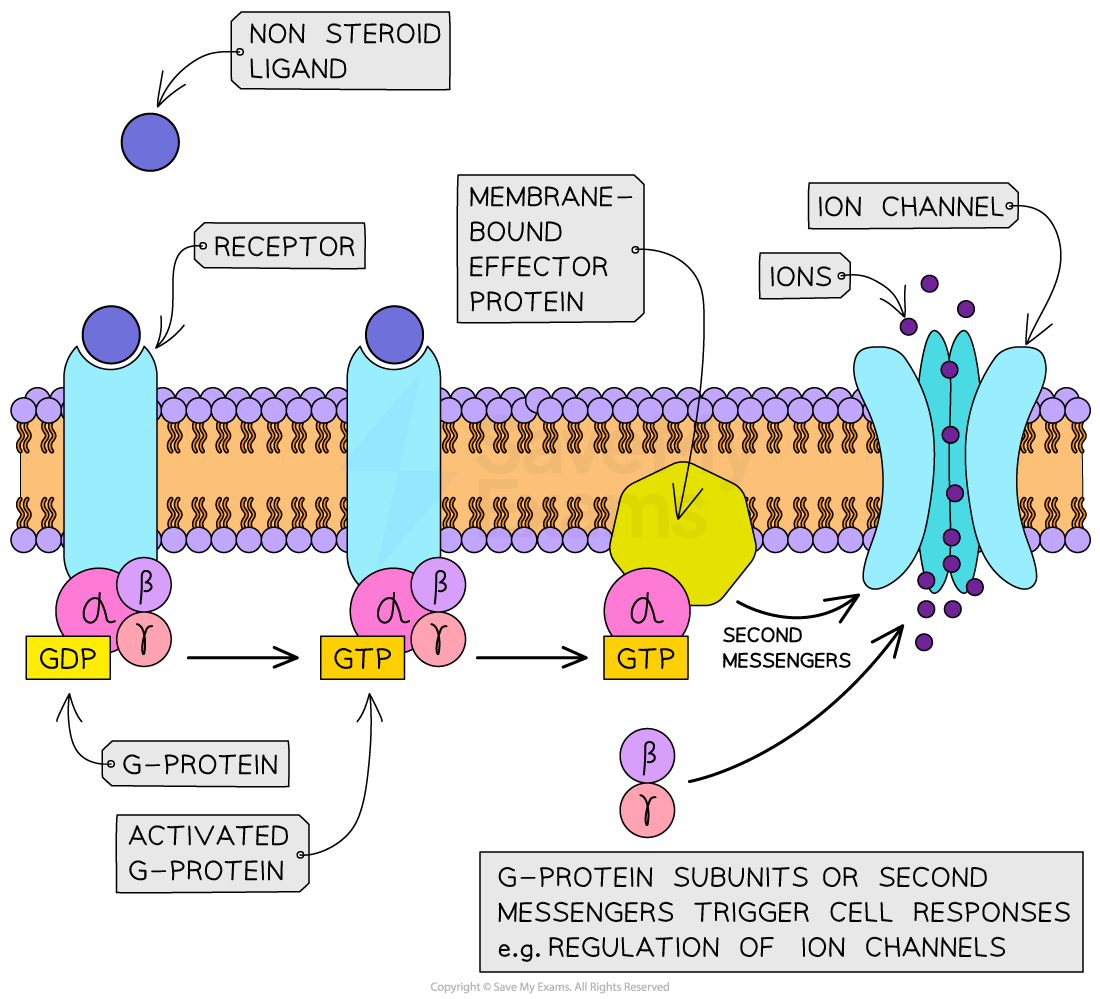
What does the G-protein splits into:
- A GTP-bound alpha subunit
- A beta-gamma dimer
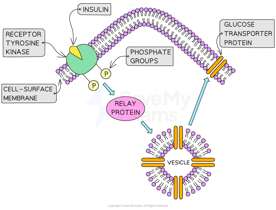
RTK’s
Receptor tyrosine kinases
- Transmembrane proteins responsible for different signal transduction pathways + responses
How do RTK’s work?
Ligand binds to extraceulluar side of RTK
A ligand (e.g. insulin) binds to the extracellular site of the RTK.
Activation of receptor’s intracellular part —> becomes phosphorylated
Phosphorylated tyrosines activate relay proteins —> start signal transduction pathways inside the cell.
One RTK can activate multiple pathways at once.
Insulin in RTK
Insulin binds to RTKs on target cells
RTK becomes phosphorylated, activating relay proteins.
Relay proteins trigger vesicles with glucose transporter proteins to move to the cell membrane.
Transporters increase glucose uptake by facilitated diffusion.
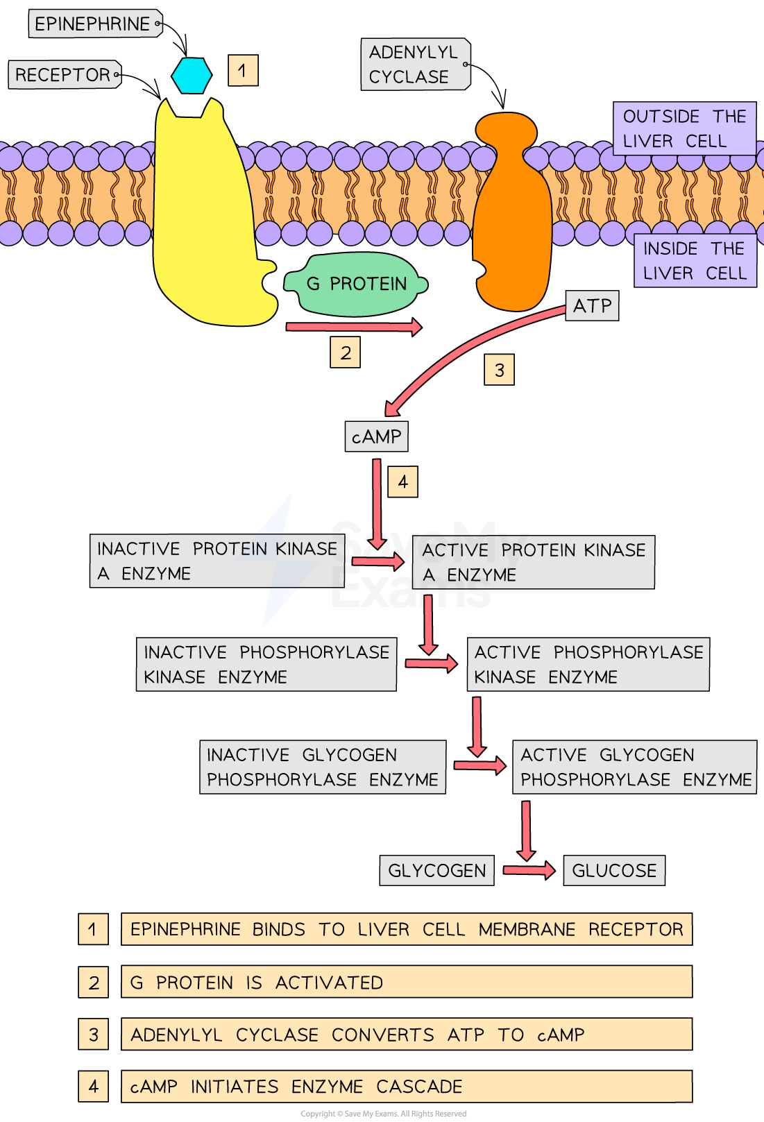
What is Ephinephrine?
Hormone (adrenaline)
Helps body respond to stress by increasing blood glucose levels.
How does epinephrine work (second messenger model)?
Epinephrine binds to receptors on the cell membrane of liver cells.
Activates enzyme Adenylyl Cyclase.
Adenylyl cyclase converts ATP to cAMP (cyclic AMP)- the second messenger.
cAMP activates protein kinase A.
Protein kinase A activates phosphorylase kinase by phosphorylation.
Phosphorylase kinase activates glycogen phosphorylase.
Glycogen phosphorylase breaks down glycogen into glucose (glycogenolysis).
Glucose is released into the blood → blood glucose concentration increases.
How are genes expressed?
Transcription factors are proteins that bind to specific DNA regions to control gene transcription.
If a gene is transcribed and translated, it is expressed.
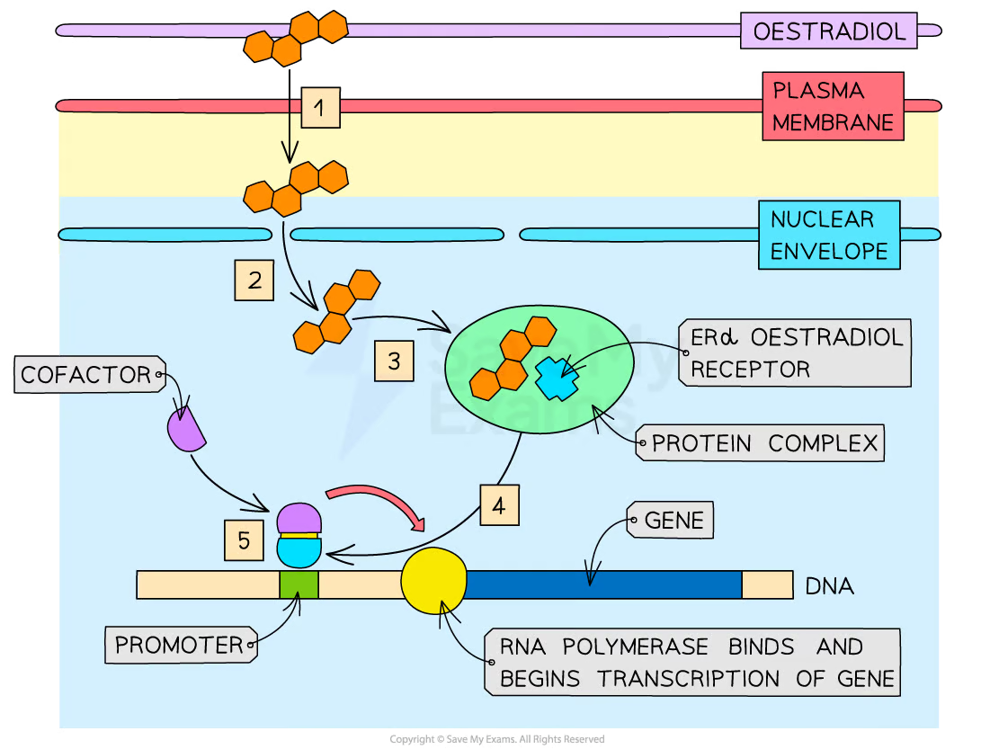
What are steroid hormones?
Small, hydrophobic, lipid-based hormones that can pass through nuclear pore bind to the intracellular receptor
What are steroid hormones responsible for?
Ligands responsible for the expression of many genes within a cell
What is the role of oestradiol hormone?
Involved in controlling the female fertility cycle and is also responsible for stimulating sperm production in males
Where is oestradiol produced?
Produced by the ovaries, placenta, and testes.
What is the oestradiol stimulation pathway?
The process by which oestradiol activates gene transcription through its receptor in the nucleus after diffusing through the cell membrane.
What is process of the oestradiol stimulation pathway?
Oestradiol diffuses through the cell membrane into the cytoplasm.
It enters the nucleus through a nuclear pore.
Inside the nucleus, oestradiol binds to an ERα oestradiol receptor held within a protein complex.
Binding causes a conformational change in the receptor.
The activated receptor detaches from the complex and moves to the target gene.
The receptor binds to a cofactor, allowing it to attach to the promoter region of the gene.
This stimulates RNA polymerase binding and initiates transcription.
Where is oestradiol controlled?
Controlled by hormones from the hypothalamus
- Gonadotropin-releasing hormone (GnRH)
What does Gonadotropin-releasing hormone (GnRH) stimulate?
Luteinising hormone (LH)
Follicle-stimulating hormone (FSH) from the pituitary gland
What is the function of