Topic 2: Organising animals and plants
 Call Kai
 Learn
Learn Practice Test
 Spaced Repetition
 Match
Match Flashcards
 Knowt Play
1/47

There's no tags or description
Looks like no tags are added yet.
Name | Mastery | Learn | Test | Matching | Spaced | Call with Kai | Chat |
|---|
No analytics yet
Send a link to your students to track their progress
48 Terms
The circulatory system
The heart
Blood vessels
Blood
What does blood carry
Oxygen
Glucose
Water
Hormones
Carbon Dioxide
Urea - Waste
Components of Blood
Red Blood cells
White Blood cells
Platelets
Plasma
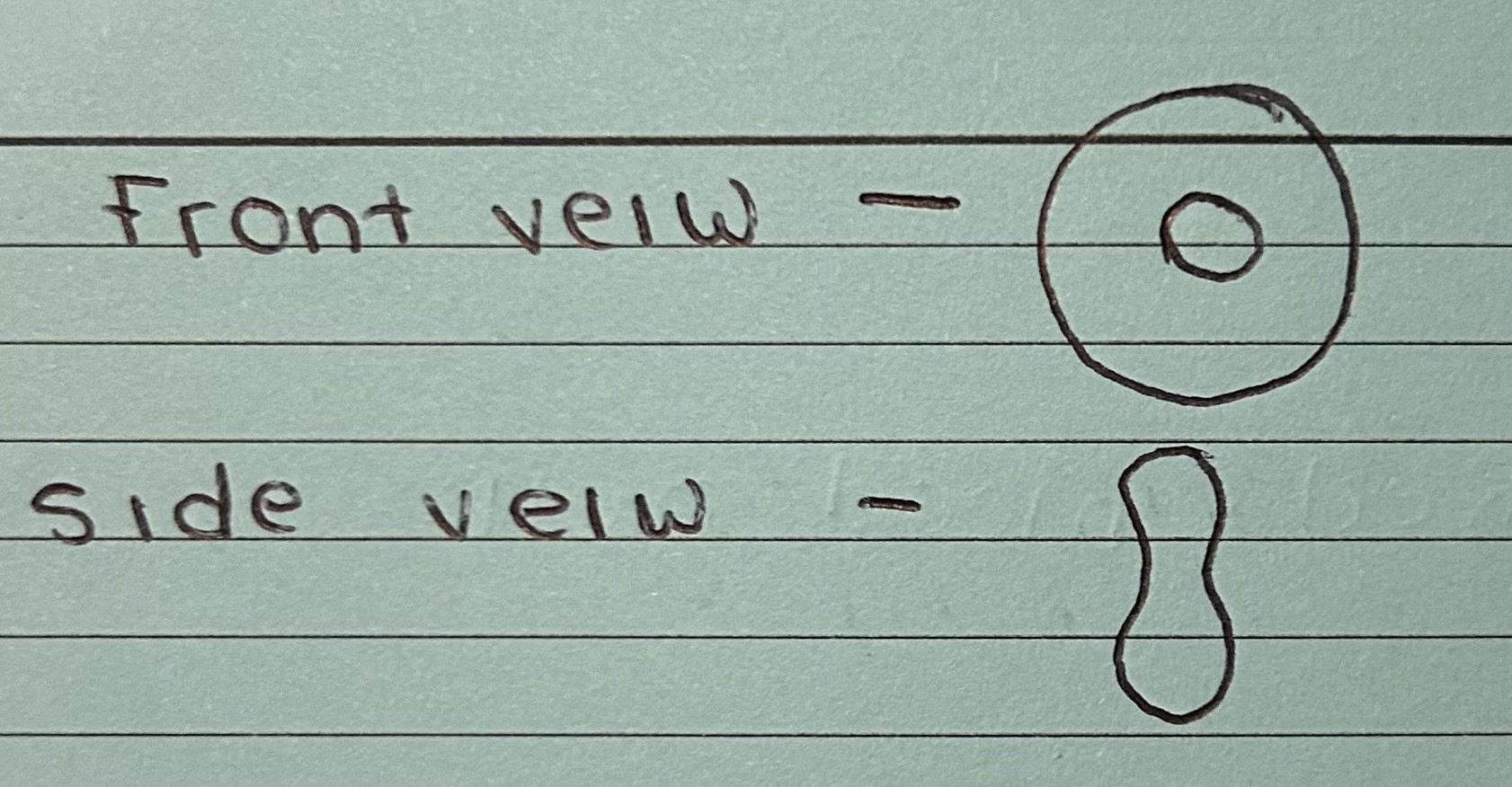
Red Blood Cells
Red blood cells contain haemoglobin to carry oxygen
They have a large surface area for absorbing oxygen
No nucleus
White Blood Cells
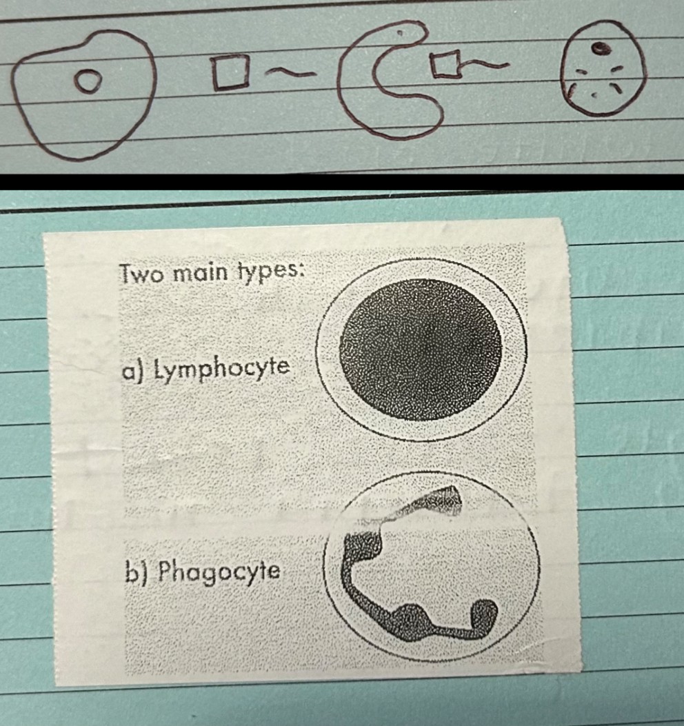
White blood cells helps your body defend against pathogens
Lymphocyte - make antibodies
Phagocyte - goes round eating bacteria in the body
Have a nucleus
Platelets
Platelets help your blood clot and help the body form scabs to help wounds heal
Plasma
Fluid of blood, and helps with blood clotting
Plasma carries 4 substances: water, glucose, hormones, CO2
The process of blood clotting
Blood clotting is the process that stops bleeding when you get hurt
First, blood vessels tighten to reduce blood flow
Then tiny blood cells called platelets, stick to the injury + form a plug
Then a series of proteins in the blood work together to create fibrin, which forms a net that traps blood cells + helps the clot get stronger
This clot helps close the wound, and once the vessel is healed the clot breaks down naturally
blood under a light microscope and how to recognise components
Red blood cells - pink
Platelets - pale pink
White blood cells cytoplasm - pale blue
White blood cells nuclei - magenta
Blood Vessels
Vein:
Transports blood from the body’s organs back to the heart
They have thin walls + contain valves that stop the blood from flowing backwards
Blue, Deoxygenated blood
Capillary:
Allows substances to diffuse to/from blood/tissues
Connect the arteries to the vein
Artery:
Transports blood away from the heart at high pressure
They have thick walls to handle the high pressure from the heart’s pumping action
Red, Oxygenated blood
The heart diagram
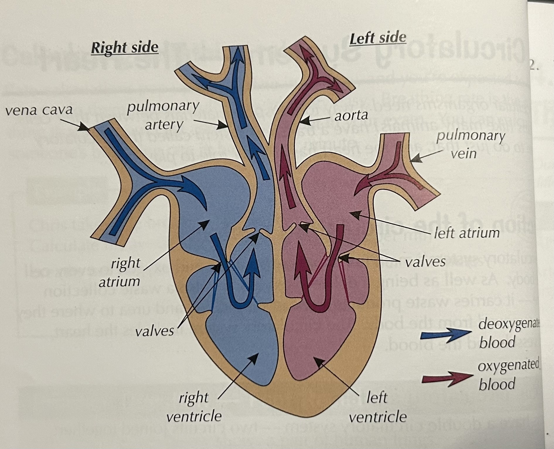
Vena Cava Function
Deoxygenated
This vein carries blood into the heart from the body
Aorta Function
Oxygenated
This artery carries blood around the body
Pulmonary vein and Pulmonary artery function
Pulmonary vein -
oxygenated
This vein carries blood into the heart from the lungs
Pulmonary artery -
Deoxygenated
This artery carries blood to the lungs
Left atrium and Right atrium function
Left atrium -
Oxygenated
Pumps blood from the lungs to the left ventricle (has thin walls)
Right atrium -
Deoxygenated
Pumps oxygen to the right ventricle (has thin walls)
Valves Function
Oxygenated and Deoxygenated
Stop blood going backwards
Left ventricle and Right ventricle Function
Left ventricle -
Oxygenated
Pumps blood around the body (has thick walls)
Right ventricle -
Deoxygenated
Pumps blood to the lungs, where gas exchange takes place (has thick walls)
What does the heart do
The heart is an organ that pumps blood around the body in a double circulatory system.
How does the heart uses its four chambers to pump blood around the body
Blood flows into the atria from the vena cava and the pulmonary vein
The atria contract, pushing the blood into the ventricles
The ventricles contract, forcing the blood into the pulmonary artery and the aorta, and out of the heart
The blood then flows to the organs through arteries, and returns through veins
The atria fill, and the whole cycle starts again over
Explain why an irregular heartbeat is detrimental to health
An irregular heart can make the heart less effective at pumping blood, which means the body doesn’t get enough oxygen. Which can lead to problems like stroke, heart failure, dizziness + fatigue. In severe cases, it can cause sudden heart failure. Treatment is important.
Different treatments for heart problems + their advantages and disadvantages
Lifestyle Change:
No surgery or medicine needed
Helps prevent further heart problems
Improves overall health
Requires effort + consistency and takes time to show results
Medications:
Easy to take (pills or injections)
Quickly relieves symptoms
No surgery needed
May have side effects
Might be taken long-term
Coronary Artery Bypass Surgery:
Can solve serious artery blockages long-term
Improves quality of life
Long-term recovery
Risk of infection
Implantable devices (pacemakers, defibrillators):
Helps control heart rhythm
Can prevent heart failure
Requires surgery to implant
May need replacements or adjustments over time
Heart transplant:
Life saving
Improves long-term health
Donors are hard to find
Requires medication to prevent (ejections which can have side effects)
What happens in coronary heart disease
In coronary heart disease, layers of fatty material build up inside the coronary arteries, narrowing them. This reduces the flow of blood through the coronary arteries, resulting in a lack of oxygen for the heart muscle. Stents are used to keep the coronary arteries open. Statins are widely used to reduce blood cholesterol levels, which slows down the rate of fatty material deposit.
Stents and Statins
Stents:
Stents are small, tube-like dives used to keep passageways, like blood vessels, open. They are often used in cases of narrowed or blocked arteries to improve blood flow. (they can be made from metal)
Statins:
A Statin is a medicine that lowers bad cholesterol in your blood, it helps prevent problems like heart attacks + strokes by stopping your body from making too much cholesterol
Stents and statins advantages and disadvantages
advantages and disadvantages
Stents:
Restores blood flow, relieving symptoms like chest pain
Quick recovery
Long-term effectiveness
Prevents further blockage
Risk of complications (infection, bleeding)
Long-term medication
Re-narrowing of the artery
Not a cure
Statins:
lowers cholesterol + reduces heart disease risk
Well studied + cost-effective
Reduce inflammation
Risk of diabetes
Possible side effects (muscle pain, liver issues)
Lifelong treatment
Does not directly treat blockages
Why might people have objections to heart transplants
Religion + cultural
Cost
Fear of rejection
Medical risks
Can place too much of a strain on your organs
Artifical and Natural pasemakers
Natural:
The heart has a natural pacemakers called the SA node, located in the right atrium. It sends out electrical signals to start each heartbeat making sure the heart contracts reguarly. This node keeps a normal resting heart of 60-100 bpm which is important for good blood flow
Artificial:
An artificial pacemaker is a small device that helps your heart beat regularly. It uses a battery + wires to send electrical signals to your heart. This makes sure your heart beats at the right speed especially when your natural pacemaker isn’t working well
What are antitoxins
antibiotics
Gas Exchange Diagram
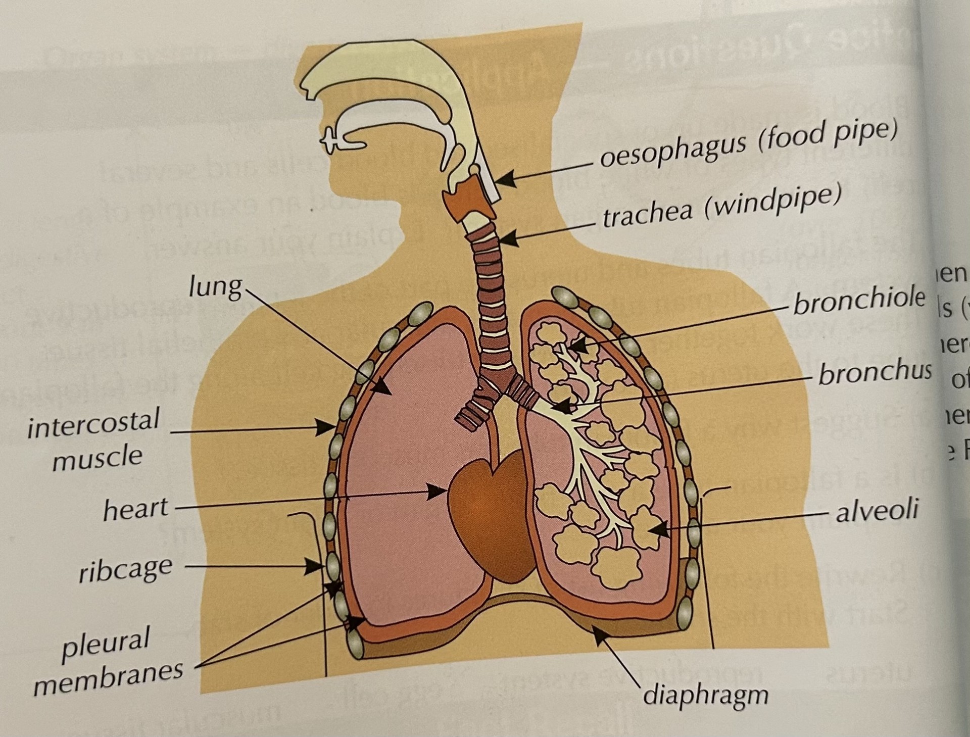
Functions of the labels to the gas exchange diagram
Nose - The nose filters, moistens, and warms the air before it enters the lungs
Trachea - The air travels from the nose down the trachea, which directs it to the lungs
Bronchi - The trachea splits into two bronchi, one for each lung. They carry air into smaller tubes
Bronchioles - These are smaller branches of the bronchi that lead to the tiny air sacs (alveoli) where gas exchange happens
Alveoli - Tiny sacs in the lungs where oxygen enters the blood and carbon dioxide is removed to be exhaled
Lungs - The lungs help us breathe by taking in oxygen + removing carbon dioxide from our body
Ribcage - Protects the lungs
Diaphragm - A muscle beneath the lungs that helps us inhale + exhale by moving up + down
What is the purpose of the lungs
You need to get oxygen from the air into your bloodstream so that it can get to your cells for respiration. You also need to get rid of the carbon dioxide in your blood. This exchange of gases all happens inside your lungs. Air is forced in and out of your lungs by the action of breathing
How are lungs adapted for gaseous exchange
Large surface area (many alveoli)
Thin walls for short diffusion distance
Moist lining to dissolve gases
Rich blood supply for quick gas exchange
Good ventilation (breathing in + out)
Process of gas exchange
Gas exchange happens in tiny air sacs called alveoli in the lungs. Oxygen from the air you breath in moves into the blood in the surrounding capillaries, while carbon dioxide from the blood moves into the alveoli to be breathed out.
Alveoli diagram

How are alveoli adapted
large surface area for gas exchange
thin walls one cell thick which allow gases to diffuse easily
moist surface, allows oxygen to dissolve
good blood supply, large surface area, maintain diffusion gradient
ventilation, maintain diffusion gradient
Stages of inhalation and exhalation in order
Inhalation:
Diagram contracts and flatens
Intercostal muscle contract
Ribs move upwards + outwards
Volume of the chest increases
Pressure inside the chest decreases
Air is drawn into the lungs
Exhalation:
Diagram relaxes and pushes upwards
Intercostal muscle relax
Ribs move downwards + in
Volume of the chest decreases
Pressure inside the chest increases
Air is forced out the lungs
Leaf diagram

Functions of the labels to the leaf diagram
Palisade cells - They have lots of chloroplasts for photosynthesis
Waxy Cuticle - Stops water loss
Guard cells - They open and close the stomata to control water loss (structure = cells that surround each stoma)
Stomata - Allow gases to move in + out of the leaf (Structure = small pores on the leaves)
Air spaces - For gas exchange
Spongy mesophyll - Helps gases like carbon dioxide + oxygen move around for photosynthesis + respiration (Structure = loosely arranged cells with air spaces)
Palisade mesophyll - Main area for photosynthesis where the leaf makes food from sunlight (Structure = tightly packed cells with many chloroplasts)
Epidermis - Protects the leaf + reduces water loss through waxy cuticle (Structure = a protective layer of cells covering the leaf)
Plant tissues
• epidermal tissues
• palisade mesophyll
• spongy mesophyll
• xylem and phloem
• meristem tissue found at the growing tips of shoots and roots.
Function of Meristem Tissue
Growth - responsible for plant growth by producing new cells through cell division
Differentiation - cells differentiate into various types of plant tissues
Repair - helps in the repair of damaged tissues
The Stomata
Which gases diffuse in + out
How they open + close
Factors that control the size
Out - Oxygen, In - carbon dioxide
Water enters guard cells, they swell open, water leaves them + they shrivel
Temp, humidity, wind, light
Explain why some of the cells are block shaped
to capture all light
Explain why the upper cells contain many chloroplasts
For photosynthesis
Describe the number of chloroplasts in the lower part of the leaf and why?
Less because there is less sunlight at the bottom
Why does the leaf have cells not tightly packed
Have a large surface area available for gas exchange
How plant organs are involved in the transport system
Roots:
They absorb water minerals from the soil, they bring up the plant + help in the uptake of essential nutrients, which are then transported upwards to the rest of the plant
Stems:
The stem acts as a support structure, holding a plant upright + connecting the roots to the leaves. It contains vascular tissue, including xylem + phloem, which transport water, minerals + sugars. The stem also transports water from the roots to the leaves through the xylem
Leaves:
The leaves are the main site for photosynthesis, where plants produce sugars. These sugars are then transported through the phloem to other parts of the plant for energy or storage with leaves.
Why transport in plants is important
Because it moves water, nutrients and sugars which are needed for growth, energy + survival. Without it the plant can’t grow, survive or reproduce
Explain why temperature, humidity, light intensity and the amount of air flow affect the rate of transpiration
Temperature + Light intensity:
Higher temperatures + light intensity increase transpiration by causing more water to evaporate + stomata to open
Humidity:
High humidity reduces transpiration by decreasing the water vapour gradient
Air flow:
Increased air flow speeds up transpiration by removing moisture around the leaf, helping more water evaporate
These factors control how much water the plant loses
Describe the differences between a moving bubble potometer and a mass photometer
Moving bubble potometer:
Measures water uptake by tracking the movement of a bubble in a tube. It gives an indirect measure of water absorption, but doesn’t directly measure transpiration
Mass photometer:
Measures water loss by tracking the change in mass of the plant or container. It provides a direct measure of water loss, but it is more complex