Tooth Morphology
1/37
There's no tags or description
Looks like no tags are added yet.
Name | Mastery | Learn | Test | Matching | Spaced |
|---|
No study sessions yet.
38 Terms
Primary/deciduous Dentition
20 teeth total (10 maxillary, 10 mandibular)
Includes incisors, canines, and molars (no premolars)
Permanent Dentition
32 teeth total (16 maxillary, 16 mandibular).
Includes incisors, canines, premolars, and molars.
Maxillary Arch
Upper arch; fixed to the skull
Mandibular Arch
Lower arch; movable via the temporomandibular joint
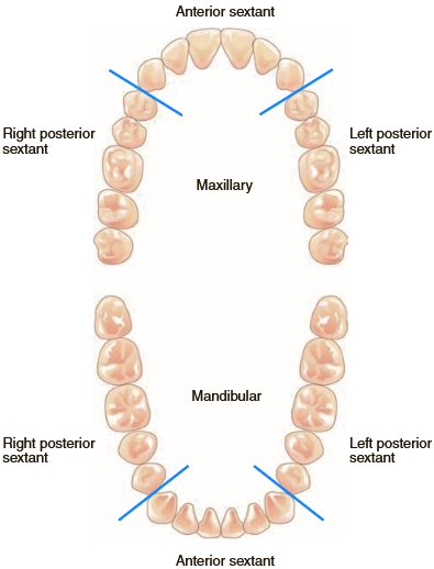
Divisions
Quadrants: Arches are divided into 2 halves by the midline (4 quadrants)
Sextants: Arches are divided into 6 sections (2 posterior, 1 anterior)
Incisors
Primary: 8 total (4 maxillary, 4 mandibular).
Permanent: 8 total (4 maxillary, 4 mandibular).
Function: Cutting food.
Canines
Primary: 4 total (2 maxillary, 2 mandibular).
Permanent: 4 total (2 maxillary, 2 mandibular).
Function: Tearing food.
Premolars (Bicuspids)
Primary: 0 (not present in primary dentition).
Permanent: 8 total (4 maxillary, 4 mandibular).
Function: Tearing and grinding food.
Molars
Primary: 8 total (4 maxillary, 4 mandibular).
Permanent: 12 total (6 maxillary, 6 mandibular).
Function: Grinding food
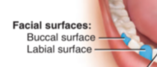
Facial
Toward the face (labial for anterior, buccal for posterior)
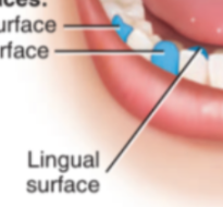
Lingual
Toward the tongue (palatal for maxillary)
Masticatory
Chewing surface (incisal for anterior, occlusal for posterior)
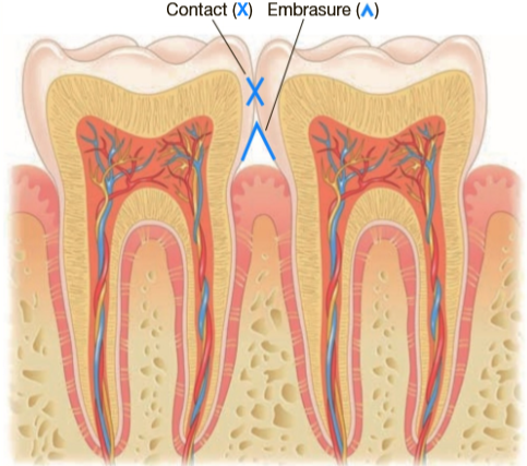
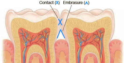
Embrasures
Triangular spaces between teeth near the gingiva

Contact Areas
Where adjacent teeth touch
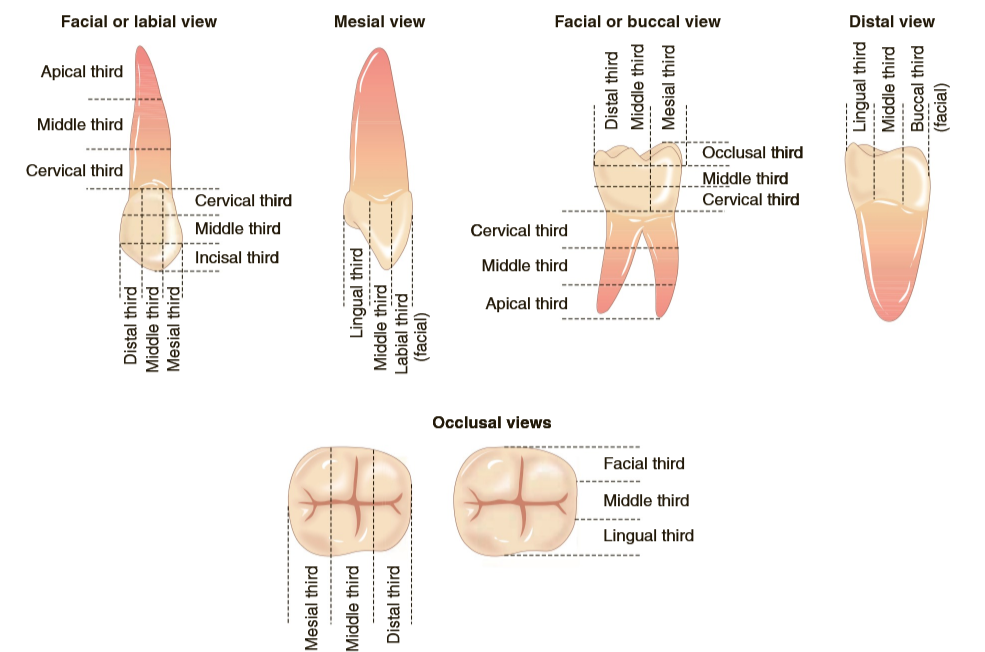
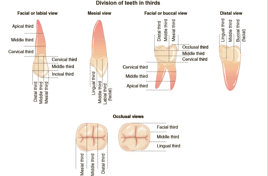
Divisions into Thirds
Crown and root divided into cervical, middle, and apical thirds
Occlusion
Relationship between maxillary and mandibular teeth when jaws are closed
Malocclusion
Abnormal tooth alignment (Class I, II, III based on Angle’s classification).
Class I: Normal relationship with misaligned teeth.
Class II: Mandibular arch distal to maxillary arch.
Class III: Mandibular arch mesial to maxillary arch.
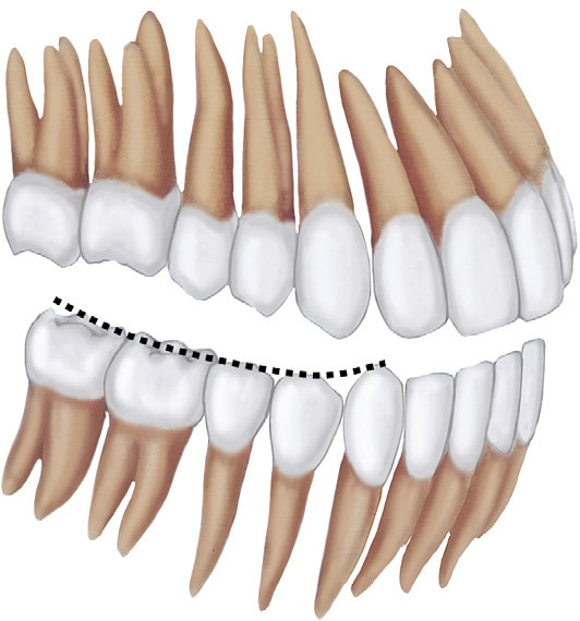
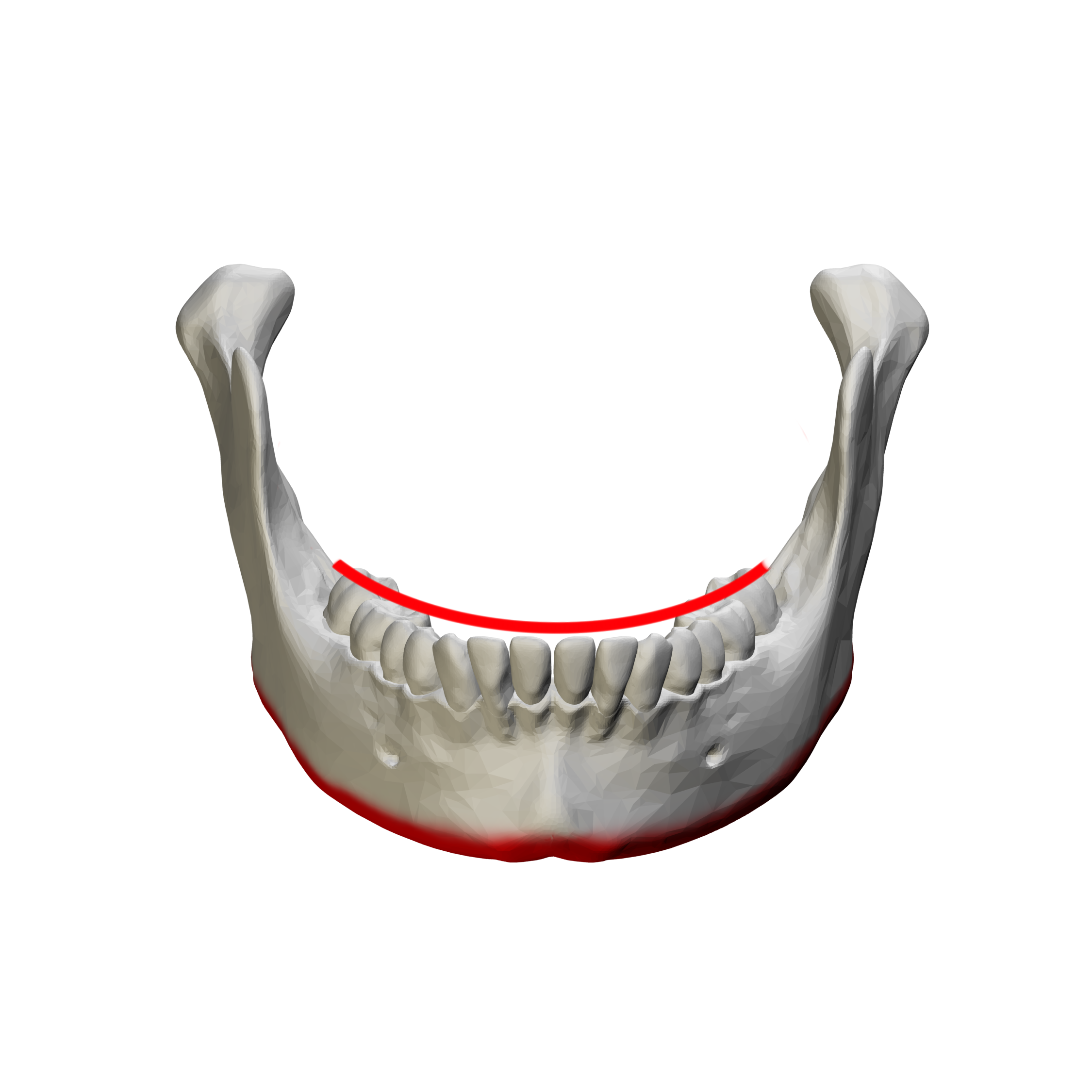
Curve of Spee
Curvature formed by the maxillary and mandibular arches in occlusion
Curve of Wilson
Cross-arch curvature of the posterior occlusal plane
Anterior Teeth
Support less occlusal force; posterior teeth bear most of the load
Maxillary Central Incisor
Longest crown, mamelons on incisal edge
Maxillary Canine
Longest root, cingulum, and marginal ridges
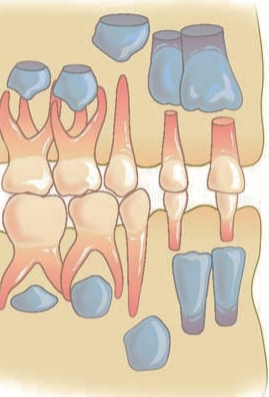
Maxillary First Molar
5 cusps (including cusp of Carabelli), trifurcated roots
Mandibular and maxillary Second Molar
4 cusps
mandibular: 2 roots
maxillary: 3 roots
Mandibular First Molar
5 cusps, bifurcated roots.
Third Molars (Wisdom Teeth)
Variable shape and size, often impacted
Mamelons
Bumps on the incisal edge of newly erupted incisors
Succedaneous Teeth
Permanent teeth that replace primary teeth (incisors, canines, premolars)
Nonsuccedaneous Teeth
Teeth that do not replace primary teeth
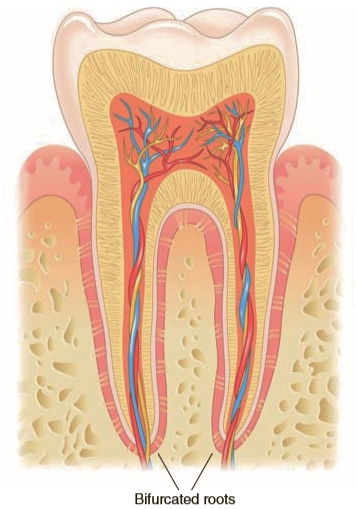
Bifurcated
2 roots on one tooth
Division of teeth in thirds
Embrasure
Triangular space in the gingival direction when two adjacent teeth are in contact
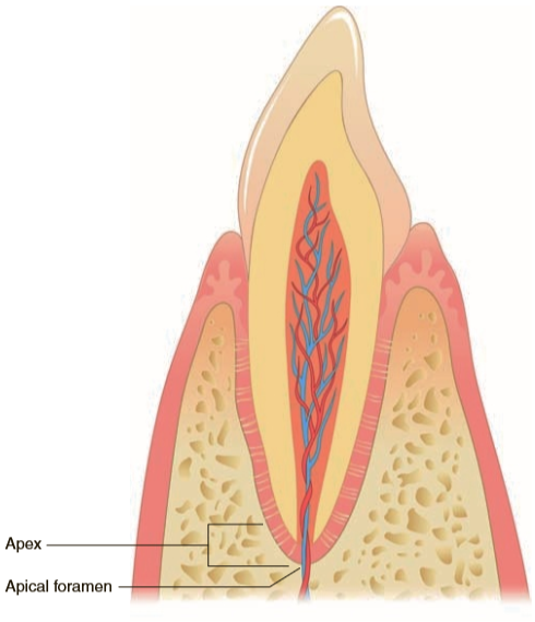
Apex: at or near the end of the root
Apical foramen: opening in the end of the tooth where nerve and blood vessels enter. There may be 1+ opening at the end of the root.
Cusp of Carabelli
Fifth cusp located on the mesial lingual surface of most maxillary first molars
Cingulum
Convex area on the lingual surface of the anterior teeth, near the gingiva
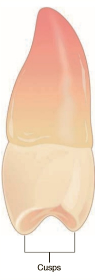
Cusp
Pointed or rounded mound on the crown of the tooth
Diastema
Space between two teeth, normally in reference to maxillary centrals
Groove
Buccal Groove:
Runs down the cheek (buccal) side to the biting (occlusal) surface.
The first molar has two buccal grooves, while the second and third molars have only one buccal groove
Central Groove:
Main groove on the biting (occlusal) surface of back teeth.
Developmental Groove:
Grooves formed when the tooth's crown is developing.
Marginal Groove:
Grooves at the edges that help food escape during chewing.
Supplemental Groove:
Smaller, branching grooves off the main (developmental) grooves, giving a wrinkled look.