APSLH CH 2 Anatomy of Respiration
1/54
There's no tags or description
Looks like no tags are added yet.
Name | Mastery | Learn | Test | Matching | Spaced | Call with Kai |
|---|
No analytics yet
Send a link to your students to track their progress
55 Terms
respiration
the exchange of gas b/w an organism and its environment involves inspiring oxygen and expiring carbon dioxide.
alveoli
tiny air sacs in the lungs where gas exchange occurs.
air pressure
the force exerted in the walls of a chamber by molecules of air
boyles law
states that the pressure of a gas increases as its volume decreases, provided the temperature remains constant and vice versa.
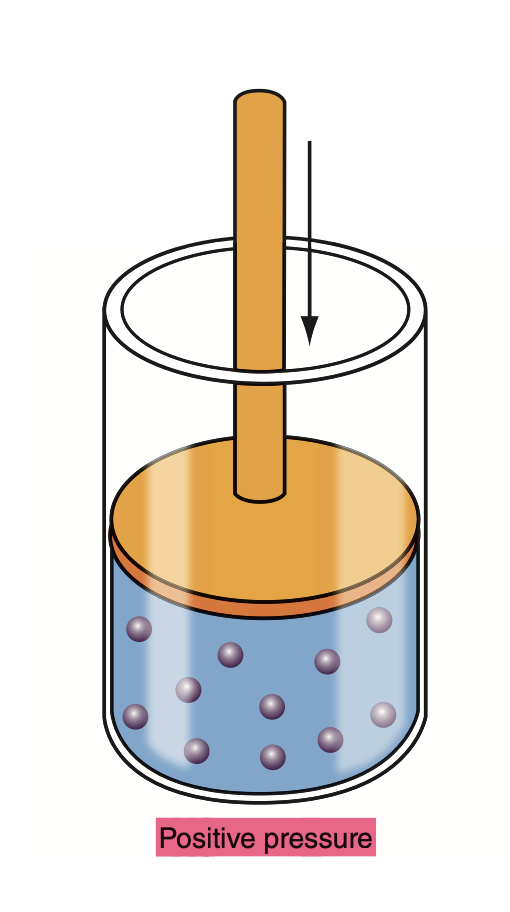
positive pressure
when the volume of a chamber has been reduced, leading to an increase in pressure relative to the surrounding atmosphere.
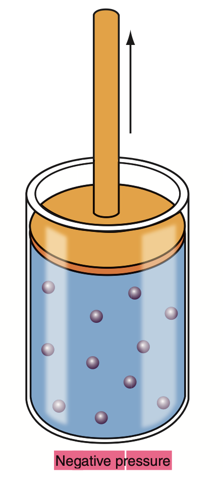
negative pressure
when the volume of a chamber increases leading to a decrease in pressure relative to the surrounding atmosphere.
divisions of the vertebral column
include cervical, thoracic, lumbar, sacral, and coccygeal regions.
rib types
include ribs 1-7 true, 8-10 false, and 11-12 floating .
rib cage mobility
refers to the ability of the rib cage to expand and contract during breathing via cartilage, allowing for lung inflation and deflation. increasing size of the thoracic cavity.
where do false ribs attach
False ribs attach to the sternum indirectly through the cartilage of the rib above them.
where do floating ribs attach
Floating ribs do not attach to the sternum or to other ribs, instead they attach to the vertebral column only.
components of the sternum
include the manubrium, body, and xiphoid process.
trachea
a tube that connects the throat to the lungs, allowing the passage of air. It is also known as the windpipe and is supported by 16-20 C-shaped hyaline cartilage rings.
types of bronchi
The main bronchi divide into the right and left primary bronchi, which further branch into secondary (lobar) bronchi and tertiary (segmental) bronchi. These bronchi are part of the airway system that directs air into the lungs.
pectus excavatum
A condition characterized by an abnormal indentation of the breastbone (sternum), which can affect chest shape and may lead to respiratory issues and heart compression.
pectus carinatum
A condition marked by a protrusion of the breastbone (sternum), often referred to as "pigeon chest," which can lead to cosmetic concerns and, in some cases, respiratory problems.
poland’s syndrome
A congenital condition characterized by the underdevelopment or absence of chest muscles on one side of the body, often leading to structural abnormalities of the breastbone and ribcage.
cleft sternum
A rare congenital defect where the sternum is not fully formed, leading to a split or failure to fuse properly.
ectopia cordis
A rare congenital condition where the heart is partially or totally outside of the thoracic cavity due to improper development of the chest wall.
trachea smooth muscle function
The smooth muscle in the trachea helps regulate airflow by constricting or dilating the airways, thus playing a crucial role in maintaining unobstructed breathing.
hyaline cartilage and fibroelastic membranefunction in trachea
Hyaline cartilage maintains tracheal structure and prevents collapse during breathing, while the fibroelastic membrane allows flexibility and elasticity, facilitating normal ventilation and movement of the trachea.
oesophagus collapsibility
The oesophagus is highly collapsible, allowing it to expand and contract during swallowing and accommodating various food sizes. This flexibility is essential for the efficient passage of food to the stomach.
bronchioles
The bronchioles are small airways within the lungs that branch from the bronchi, allowing airflow to the alveoli. They play a vital role in regulating airflow and gas exchange.
terminal bronchioles
The terminal bronchioles are the smallest branches of the bronchioles in the lungs, marking the transition from the conducting zone to the respiratory zone, where gas exchange begins.
right lung lobes
The right lung lobes consist of three distinct lobes: the upper, middle, and lower lobes. They are larger than the left lung lobes and facilitate airflow and gas exchange.
left lung lobes
The left lung lobes consist of two distinct lobes: the upper and lower lobes. They are smaller than the right lung lobes to make room for the heart and other structures in the thoracic cavity.
branching structure of bronchioles function
The branching structure of bronchioles is essential for distributing air throughout the lungs and optimising gas exchange by increasing the surface area available for diffusion.
gastroesophageal reflux
A condition where stomach acid flows back into the esophagus, causing symptoms like heartburn and regurgitation. This occurs due to a weak lower esophageal sphincter.
nissen fundoplication
A surgical procedure to treat gastroesophageal reflux disease (GERD) by wrapping the top of the stomach around the lower esophagus to strengthen the lower esophageal sphincter.
gastronomy
A surgical procedure to create an opening in the stomach for feeding or drainage.
jejunostomy
A surgical procedure that creates an opening in the jejunum for feeding or drainage.
alveolar ducts
A component of the respiratory system that connects the respiratory bronchioles to the alveolar sacs (containing alveolus), allowing for gas exchange.
alveolar sacs
Clusters of alveoli at the end of alveolar ducts where gas exchange occurs in the lungs.
emphysema
A chronic lung condition characterized by the destruction of alveoli, leading to reduced airflow and difficulty in breathing. It is often caused by long-term exposure to irritants such as cigarette smoke.
cells found in alveolar sacs that are responsible for gas exchange
type I and type II pneumonocytes. Type I cells facilitate gas exchange, while type II cells produce surfactant to reduce surface tension.
how do pneumonocytes regenerate
Pneumonocytes regenerate primarily through the proliferation of type II cells, which can differentiate into type I cells when needed, particularly after injury.
where does oxygen go from lungs
Oxygen from the lungs travels to the bloodstream, where it binds to hemoglobin in red blood cells for transport to tissues throughout the body.
aspiration
the inhalation of foreign material, such as food or liquid, into the lungs.
who is at high risk for aspiration
Individuals with swallowing difficulties, neurological disorders, or reduced consciousness.
what are the defences against pollution of the respiratory tract
several mechanisms including nostril hairs and mucous production which trap larger foreign particles
cilia along respiratory tract trap and move particles, and reflex actions like coughing and sneezing to expel irritants.
why does enphysema cause barrel chest
Emphysema leads to hyperinflation of the lungs, resulting in a characteristic barrel-shaped chest due to the increased air volume and loss of elasticity in the lung tissue.
vertical dimension of thoracic expansion
The vertical dimension of thoracic expansion refers to the increase in the height of the thoracic cavity during inhalation. This expansion facilitates lung inflation and enhances the efficiency of respiration.
transverse dimension of thoracic expansion
The transverse dimension of thoracic expansion refers to the increase in width of the thoracic cavity during inhalation, allowing for greater lung capacity and improved airflow.
upper respiratory tract
includes the nasal cavity, pharynx, and larynx, serving as a passage for air to enter the lower respiratory tract while actively conditioning the inhaled air.
lower respiratory tract
The lower respiratory tract consists of the trachea, bronchi, bronchioles, and lungs, responsible for conducting air to the alveoli where gas exchange occurs.
pleural lining function
The pleural lining functions to facilitate smooth movement of the lungs during breathing, provide cushioning, and create a pressure gradient that aids in lung expansion.
visceral pleura
The visceral pleura is a membrane that closely envelops the lungs, providing a protective layer and reducing friction during respiration.
parietal pleurae
The parietal pleurae are the membranes that line the thoracic cavity, providing a protective barrier and allowing smooth lung movement by reducing friction during breathing.
pleurisy
Pleurisy is an inflammation of the pleura, causing pain during breathing and often leading to difficulty in lung expansion.
diaphragm
The diaphragm is a dome-shaped muscle located beneath the lungs that plays a crucial role in the process of breathing by contracting to expand the thoracic cavity and facilitate inhalation.
cuboidal cells function
Cuboidal cells in the lungs secrete surfactant and help in the exchange of gases, contributing to the overall functionality of the alveoli.
what innervates diaphragm
The diaphragm is primarily innervated by the phrenic nerve, which originates in the cervical spinal cord.
what are the muscles of inspiration
The muscles of inspiration primarily include the diaphragm and the external intercostal muscles. These muscles work together to increase the volume of the thoracic cavity, facilitating air intake into the lungs.
what are the accessory muscles in inspiration
Accessory muscles in inspiration include the sternocleidomastoid, scalene muscles, and pectoralis minor. These muscles assist the diaphragm and external intercostals during heavy or labored breathing.
what are the muscles in forced expiration
The muscles involved in forced expiration include the internal intercostal muscles, abdominal muscles (such as the rectus abdominis and obliques), and transversus thoracis. These muscles help decrease the volume of the thoracic cavity to expel air from the lungs more forcefully.