Cranial Nerves, Eye, Muscles of Mastication, Facial Muscles
1/117
Earn XP
Description and Tags
TO BE ADDED (just for identify and justify for now)
Name | Mastery | Learn | Test | Matching | Spaced | Call with Kai |
|---|
No analytics yet
Send a link to your students to track their progress
118 Terms
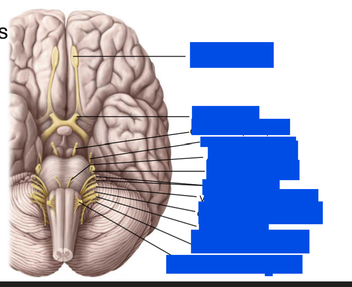
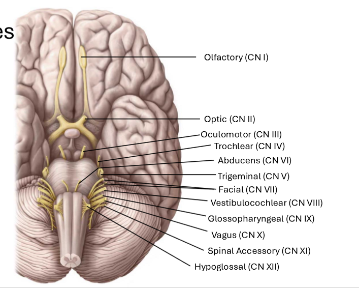
Label these nerves
What are some neumonics for memorizing the nerves?
Oh, Oh, Oh, They Traveled And Found Voledemort Guarding Very Secret Horocruxes
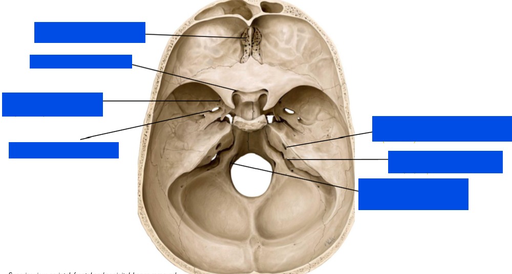
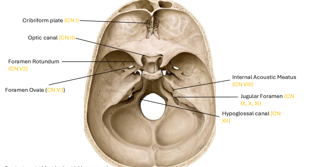
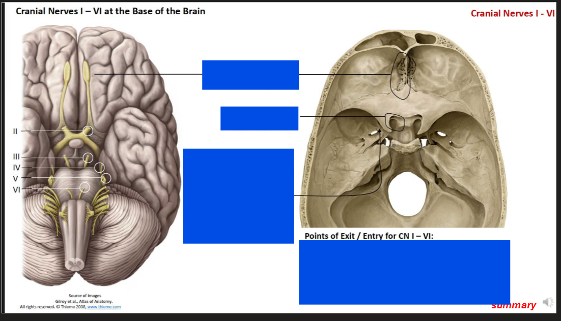
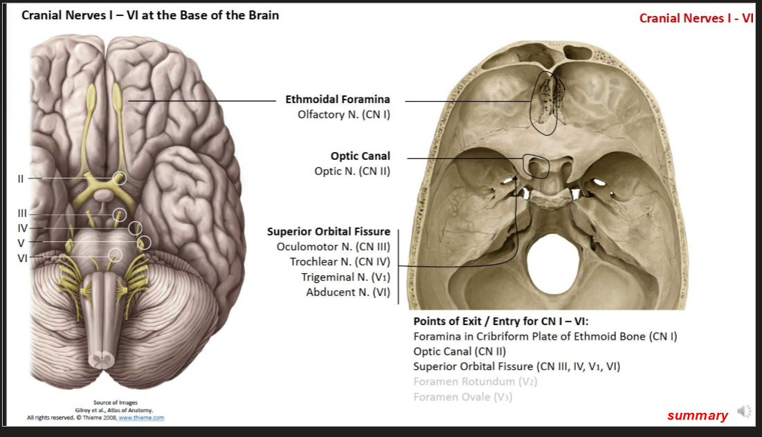
What nerve exits from the cribriform plate?
Olfactory Nerve (CN I )
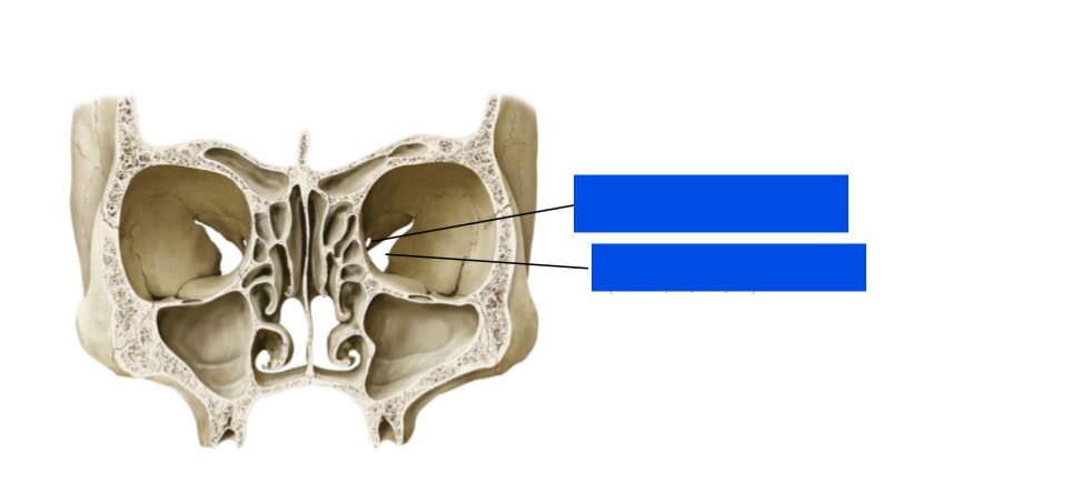
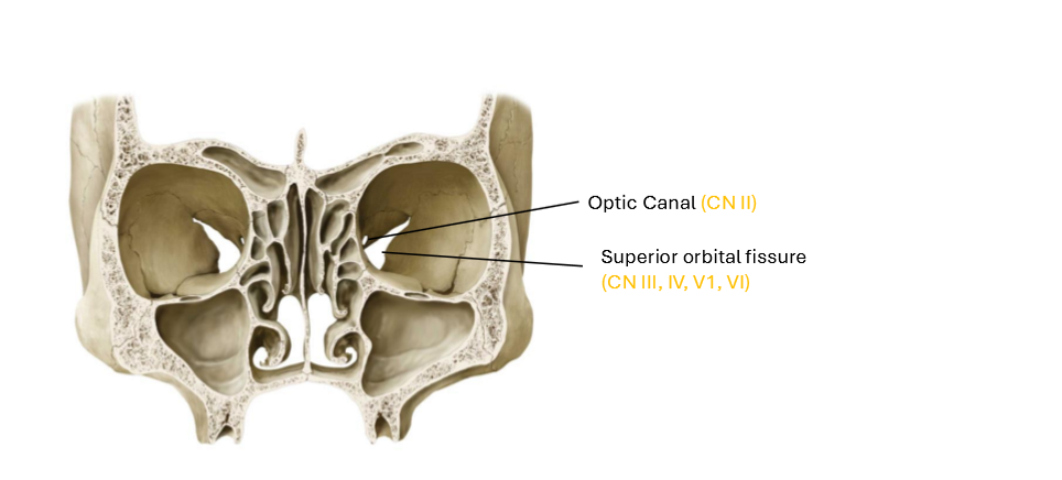
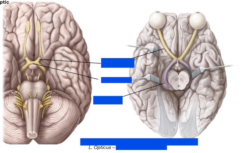
What nerve exits from optic canal?
Optic Nerve (CN II)
What nerve exits from Superior Orbital Fissure
Occulomotor, Trochlear, Trigeminal V1 (Opthalmic nerve), and Abducens nerve (CN III - VI)
What nerve exits from the forament rotundum
Trigeminal V2 (maxillary nerve)
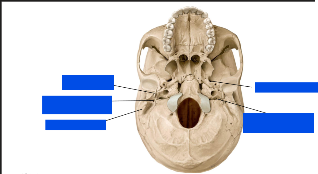
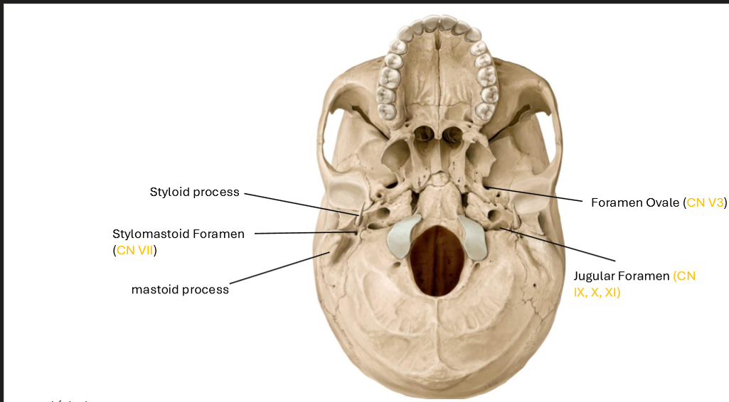
What nerve exits from the foramen ovale
trigeminal V3 (mandibular nerve)
What nerve exits from the sylomastoid foramen
Facial nerve (CN VIII)
What nerve exits internal acoustic meatus
Vestibulocochlear nerve (CN VIII)
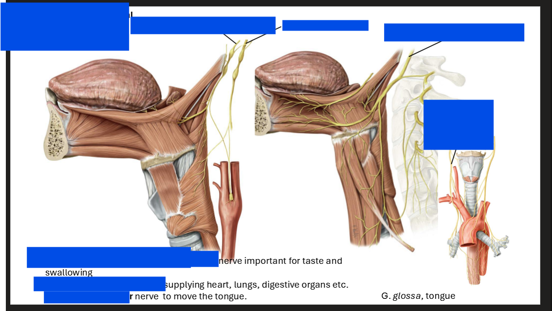
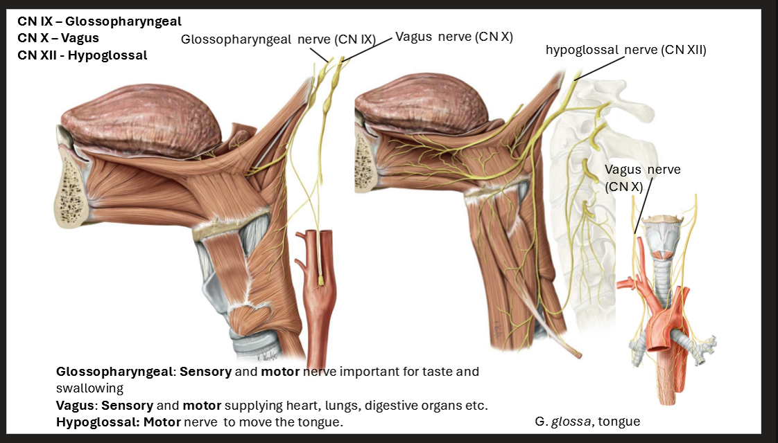
What nerve exits the jugular foramen
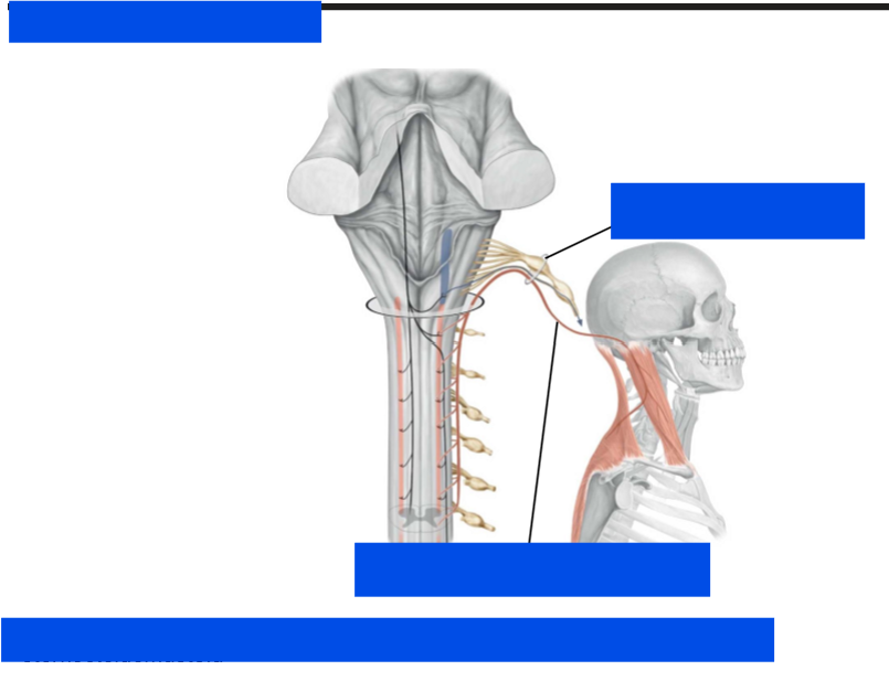
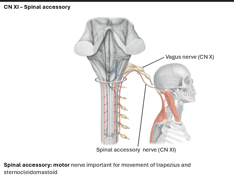
Glosspharyngeal, Vagus, and Spinal accessory nerve (CN IX - CN XI)
What nerbe exits from the hypoglossal canal
hypoglossal nerve (CN XII)
label (with the nerves)
label (with the nerves)
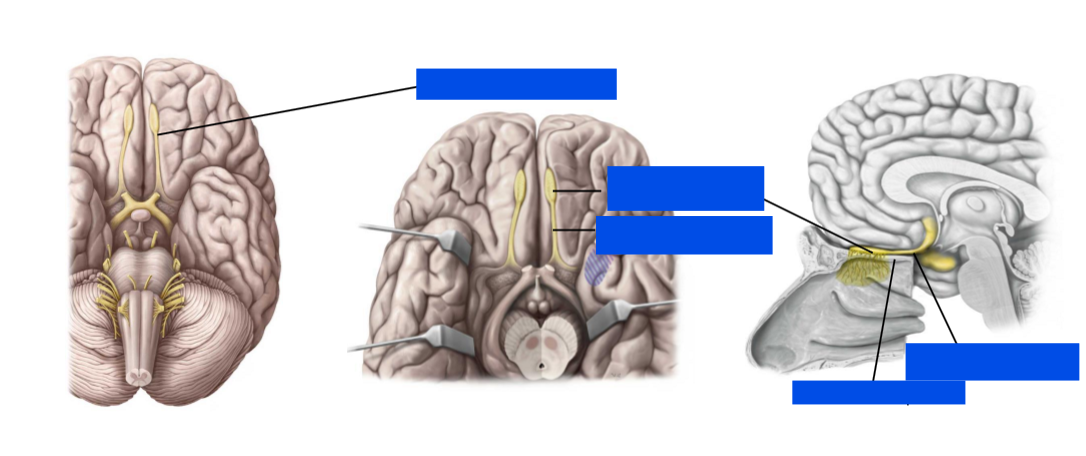
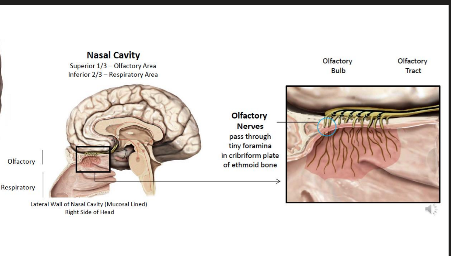
Important for SMELL
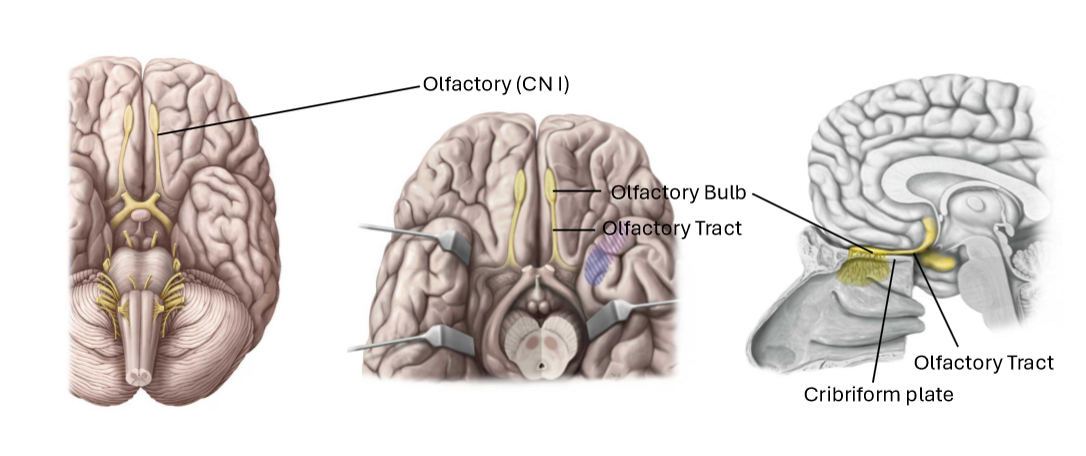
Describe the trajectory of the olfactory nerves
pass through the tiny foramina in the cribiform plate of the ethmoid bone
The olfactory area also includes the superior 1/3rd of the nasal cavity
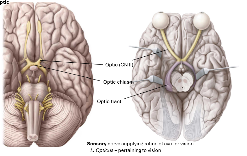
Sensory nerve, supplying the retina of the eye for vision
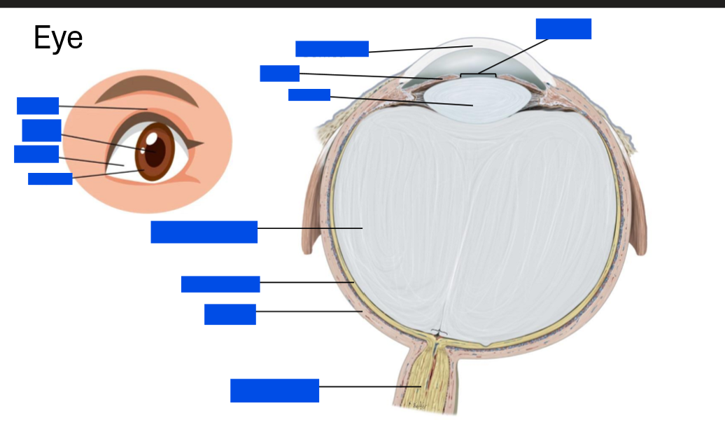
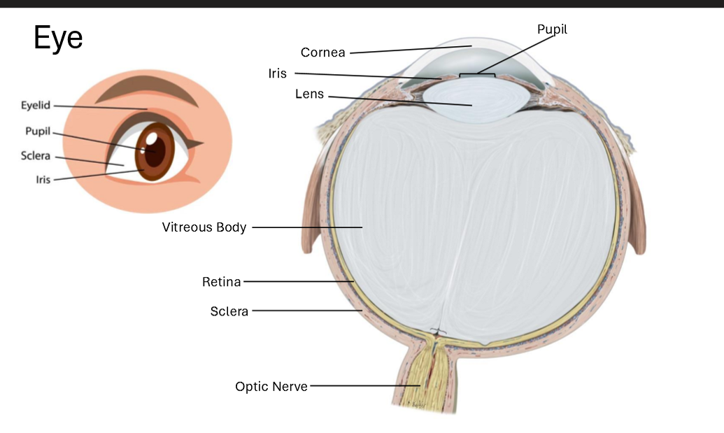
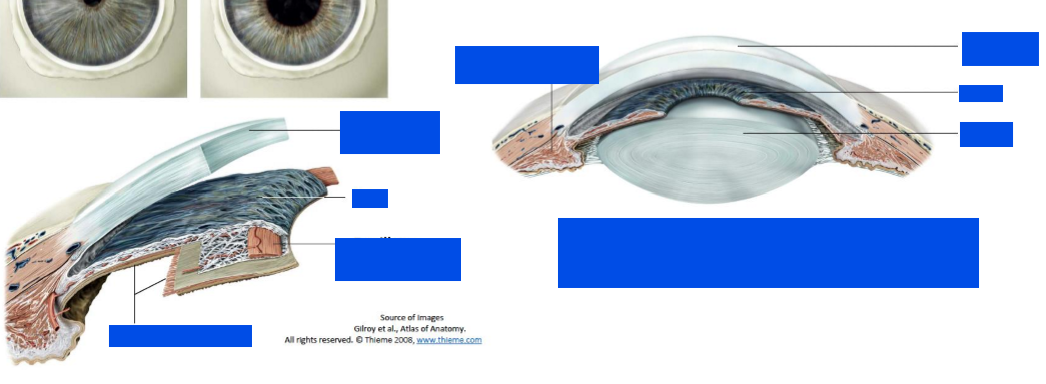
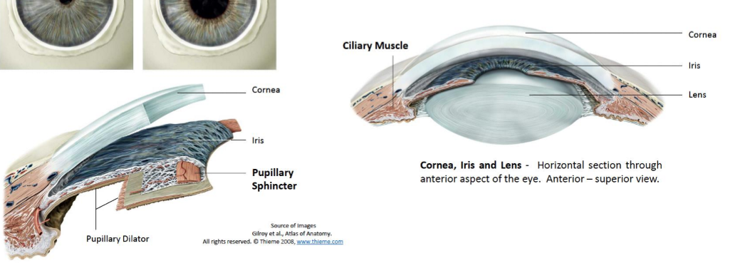
Label the eye
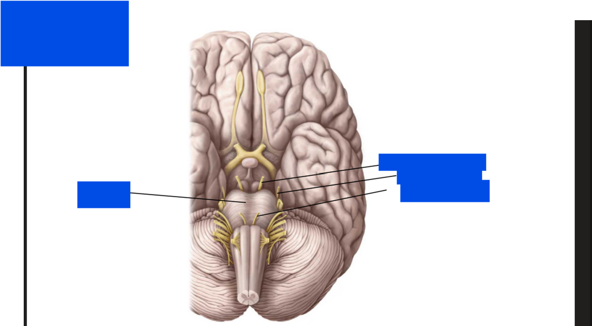
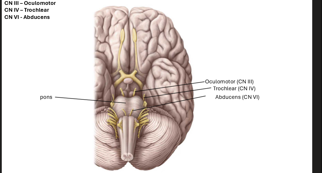
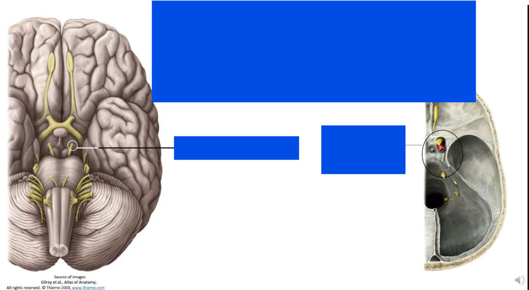
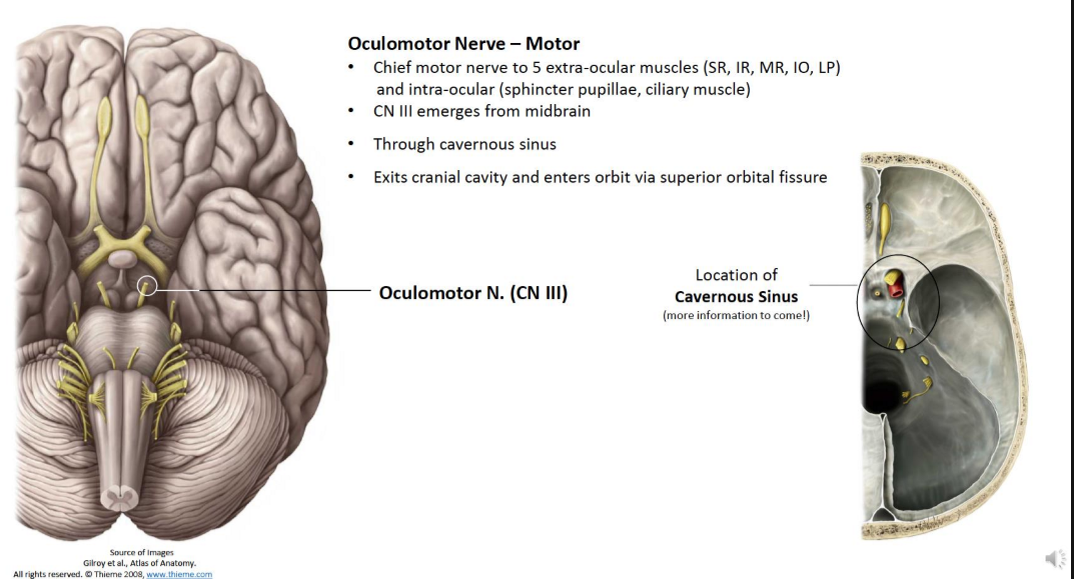
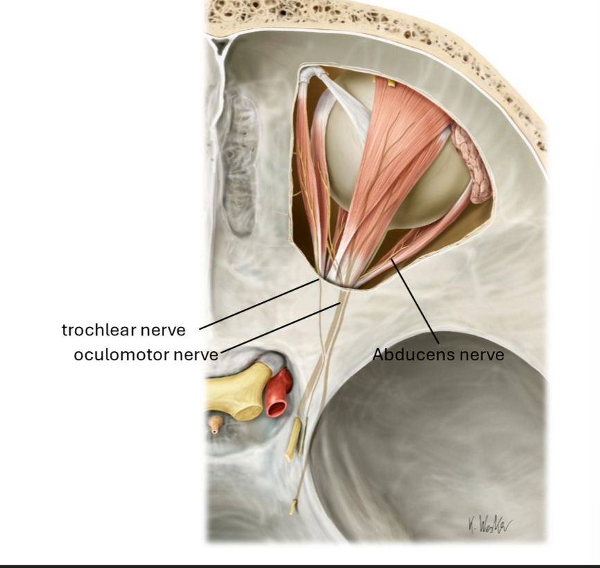
Describe the occulomotor nerve
Chief motor nerve to 5 extra-ocular muscles (SR, IR, MR, IO, LP) and intra-ocular (sphincter pupillae, cillary muscle)
CN III emerges from midbrain
through cavernous sinus
Exits cranial cavity and enters orbit via superior orbital fissure
What does the occulomotor nerves innervate?
The Sphincter Pupillae and Cillary Muscle
carries parasympathetic information
to the smooth muscle of the sphincter pupillae - causes constriction of pupil
to the cillary muscles - produces accommodation (lens rounded) for near vision
What are the two intraocular muscles that regulate pupil size
Pupillary Sphincter - narows the pupil (parasympathetic innervation)
Pupillary Dilator - enlarges the pupil (sympathetic innervation)
Describe the Trochlear Nerve
Motor nerve to one extra-ocular muscle (SO)
CN IV emerges from posterior surface of midbrain
passes anteriorly around brainstem, through cavernous sinus
exits cranial cavity and enters orbit via superior orbital fissure
Describe the Abducent Nerve
Motor nerve to one extra-ocular eye muscle (LR)
CN VI emerges from brainstem between pons and medulla
through the cavernous sinus
Exits cranial cavity and enters orbit via superior orbital fissure
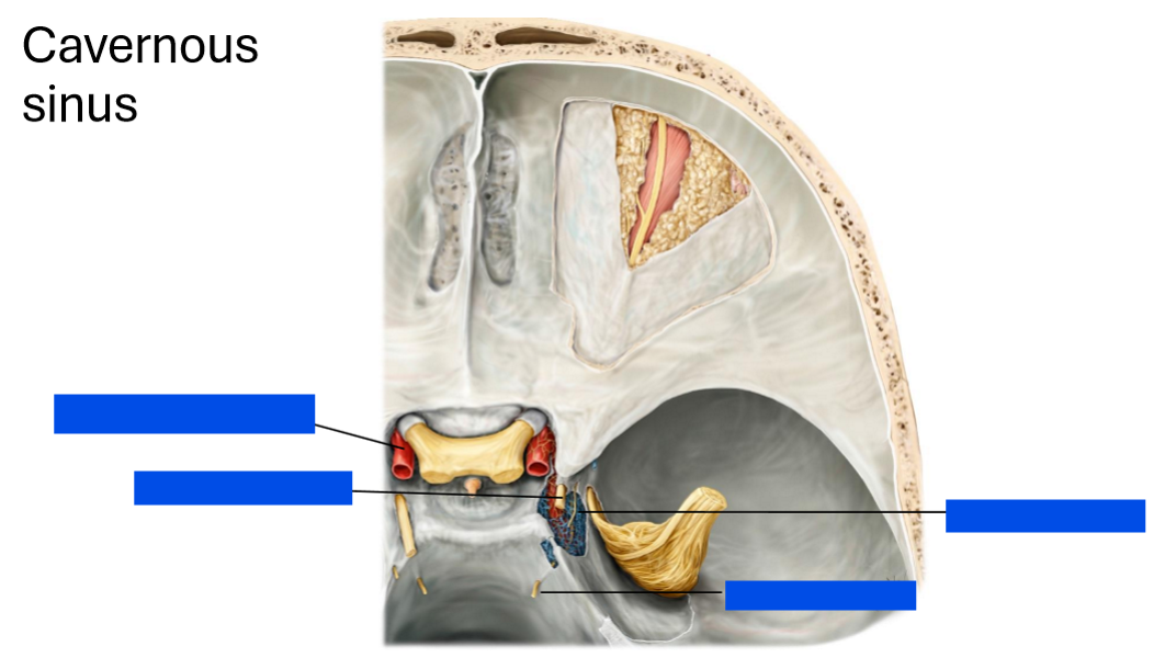
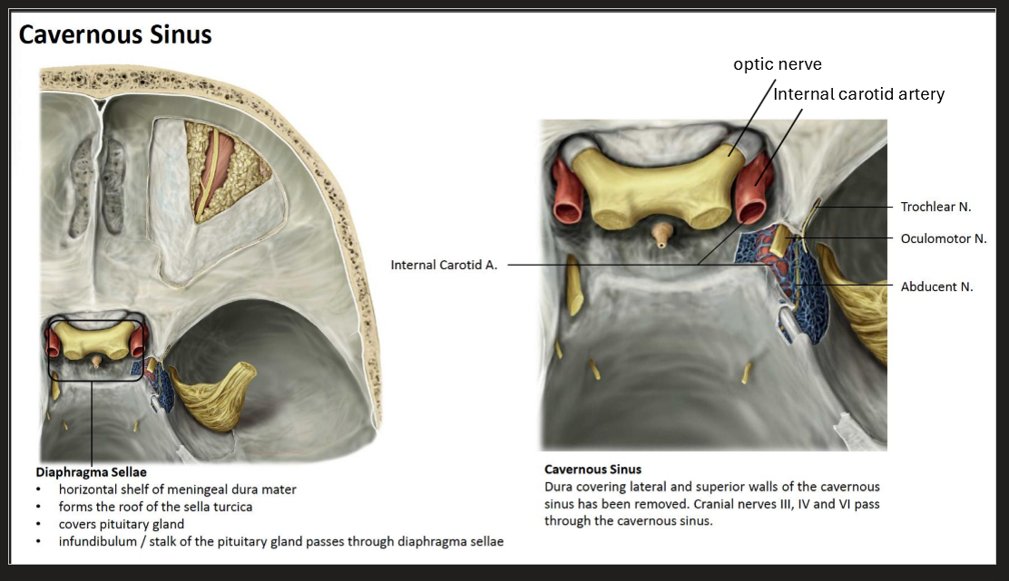
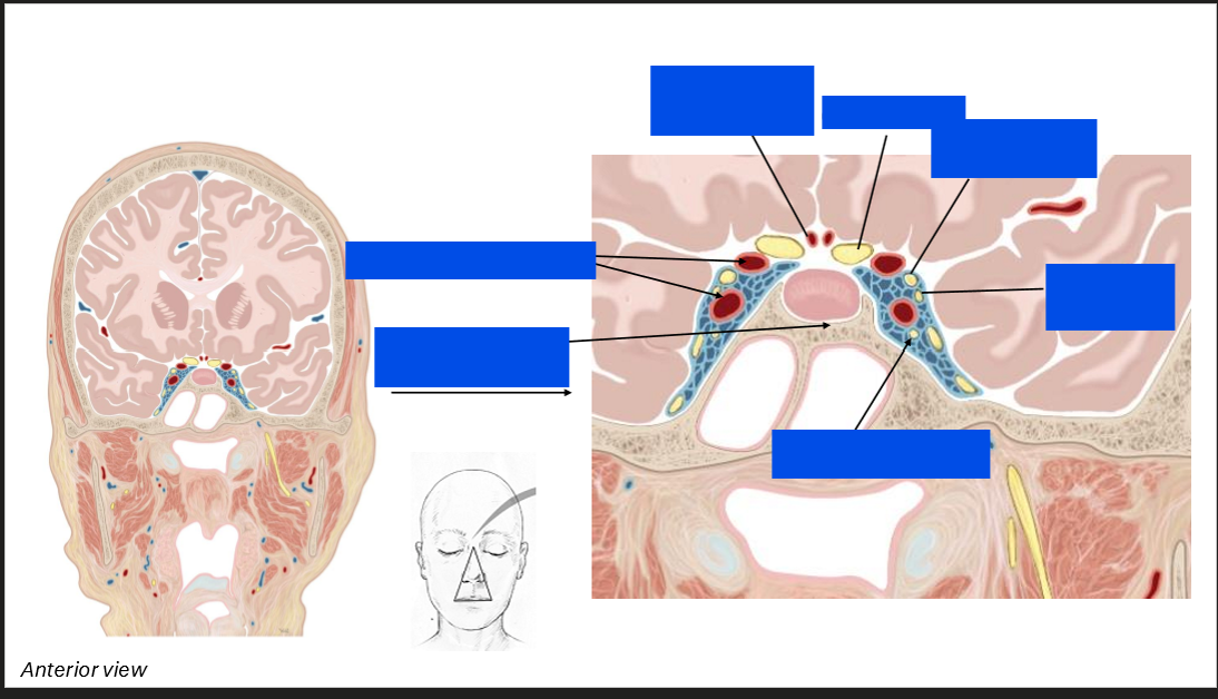
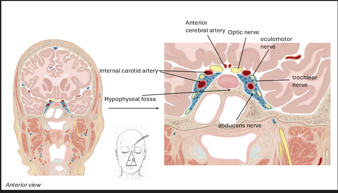
Label the cavernous sinus
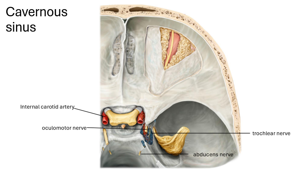
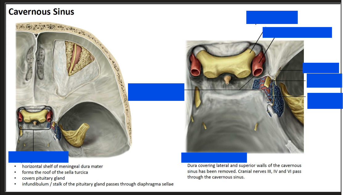
Label the cavernous sinus
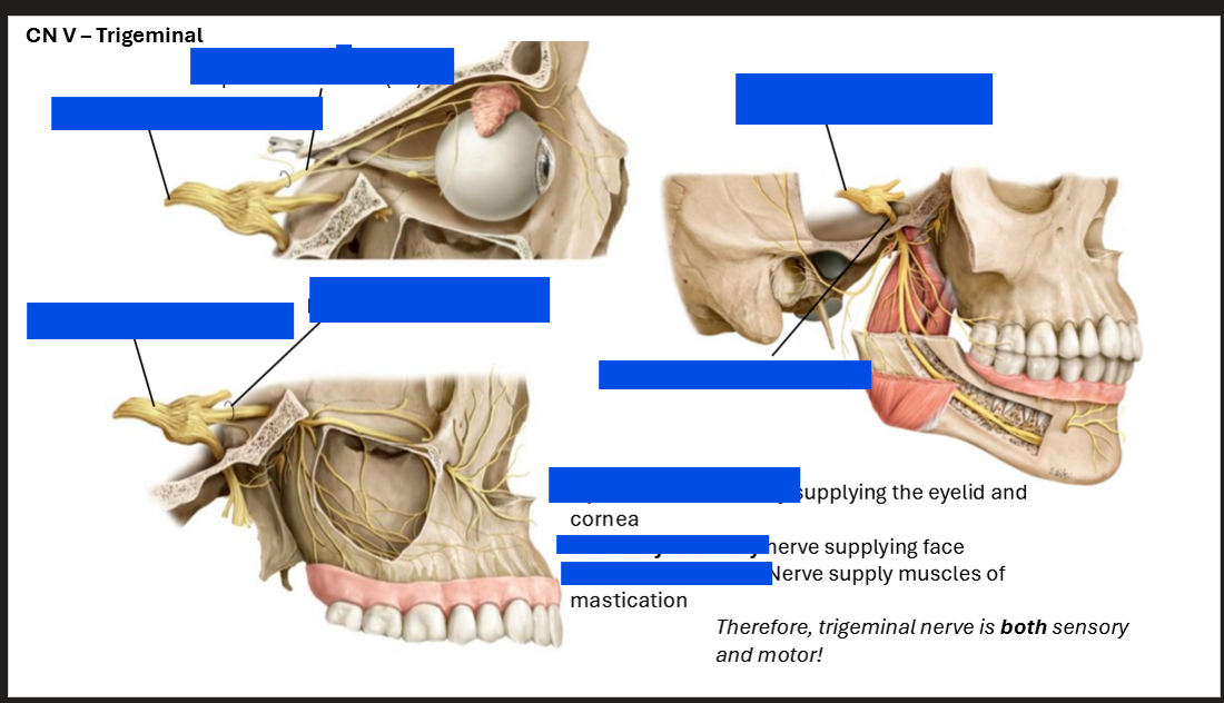
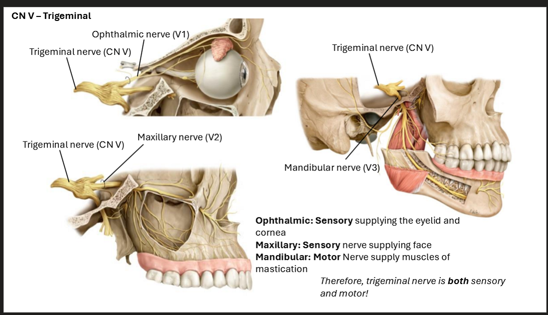
What are the branches of the trigeminal nerve
The trigeminal nerve iteslf emerges from the lateral aspect of pons - large sensory root; small motor root
Opthalmic nerve → exits superior orbital fissure
maxillary nerve → exits forament rotundum
mandibular nerve → exits foramen ovale
Sensory - face, oral, nasal, and sinus mucosa; teeth; anterior 2/3 tongue
Motor - muscles of mastication and 4 other small muscles
Label the exits of the brain and which nerve exits from them
Label the trigeminal nerve
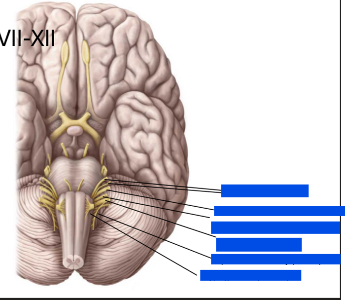
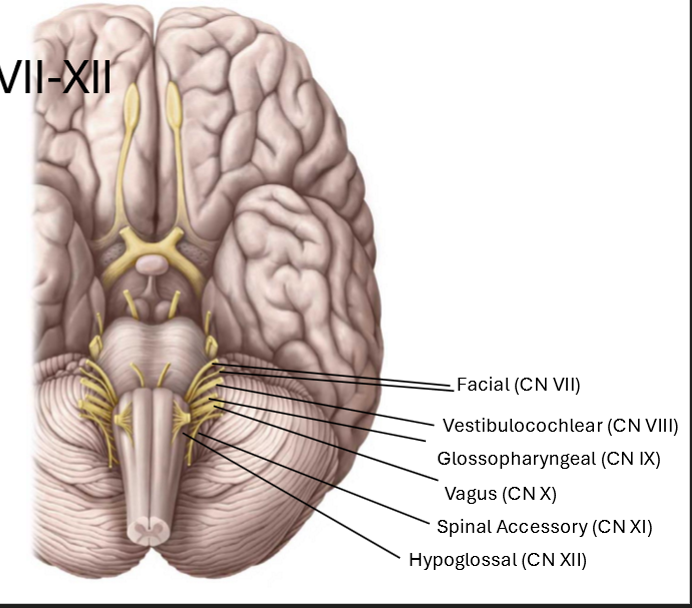
Describe the cranial nerves from CN VII-XII
CN VII and VIII - between pons and medulla (CN VI also in this region)
CN IX - XI are lateral to the medulla oblongota, behind the olive
CN XII more medial between pyramid and olive
Label
What are the neumotics for the facial nerves?
Temporal - To
Zygomatic - Zanzibar
Buccal - By
Mandibular - Motor
Cervical - Car
Sensory and motor nerve that supply nerves for taste, and muscles of facial expression
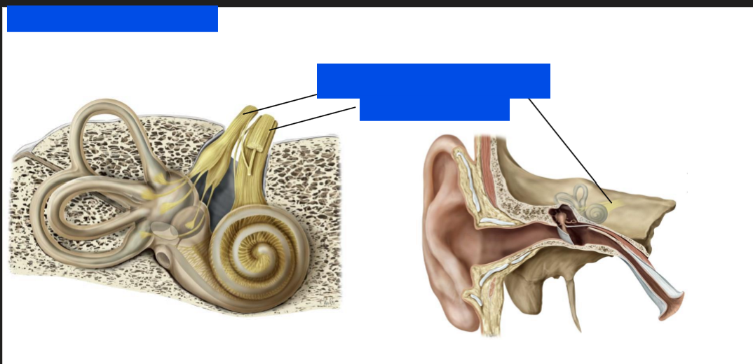
Label this nerve
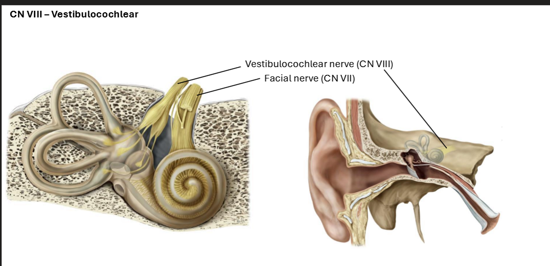
Label these nerves
Label this nerve
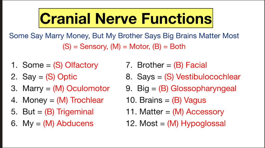
Whats a neumonic for cranial nerve functions?
Some Say Marry Money, But My Brother Says Big Brains Matter Most
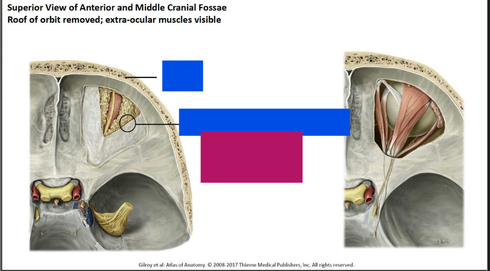
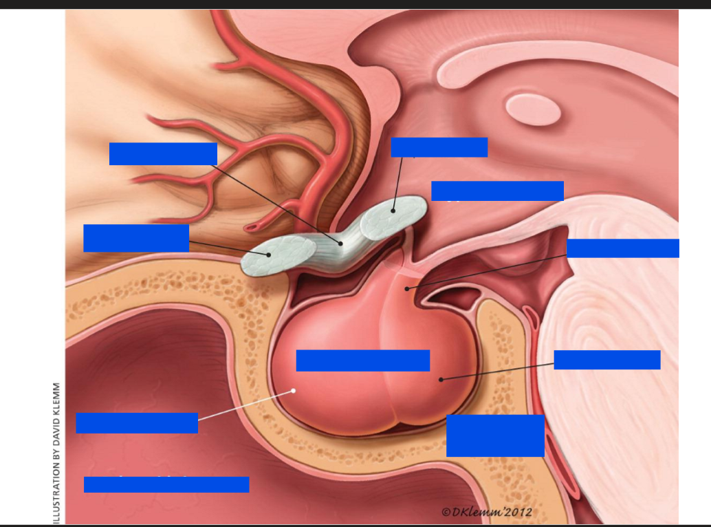
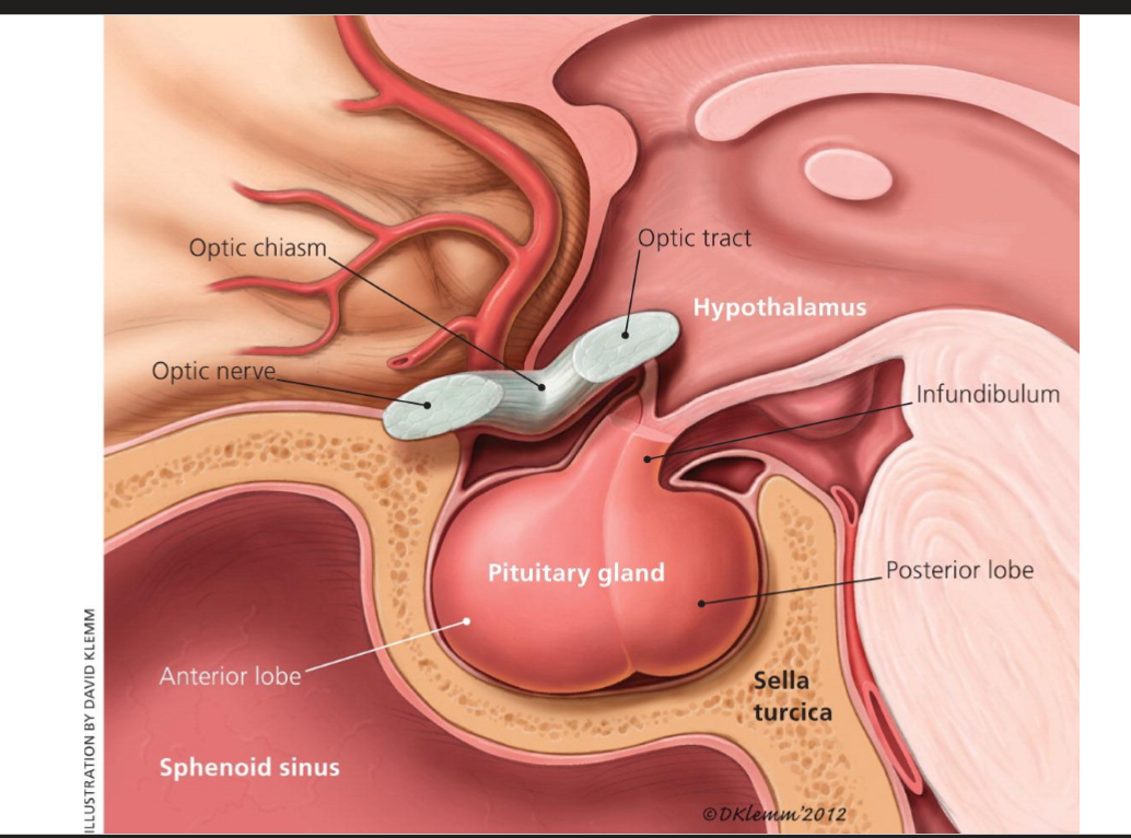
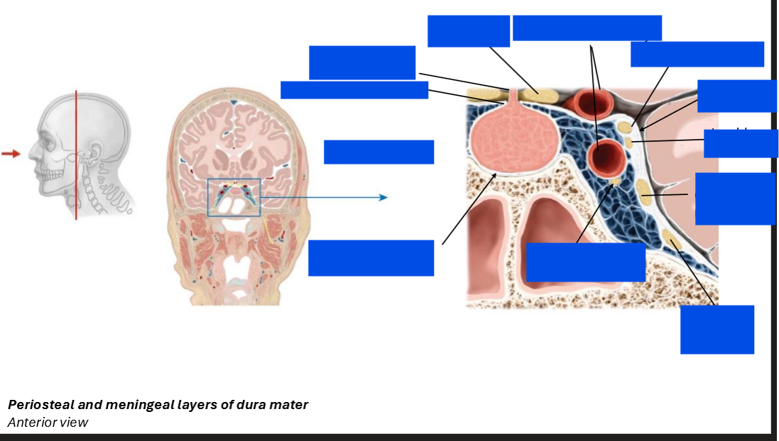
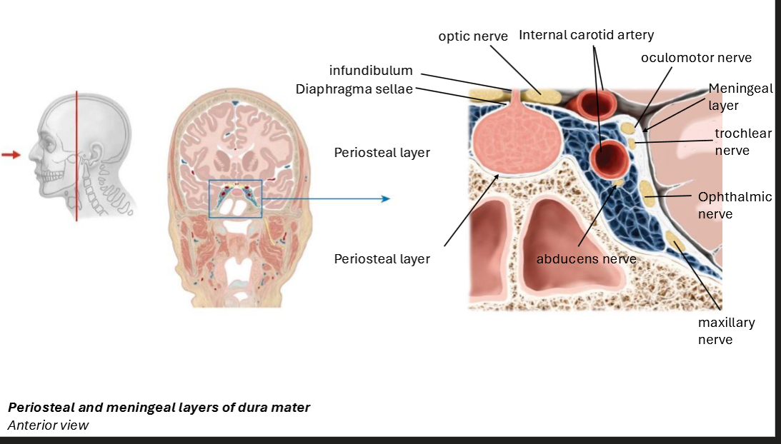
Label this diagram
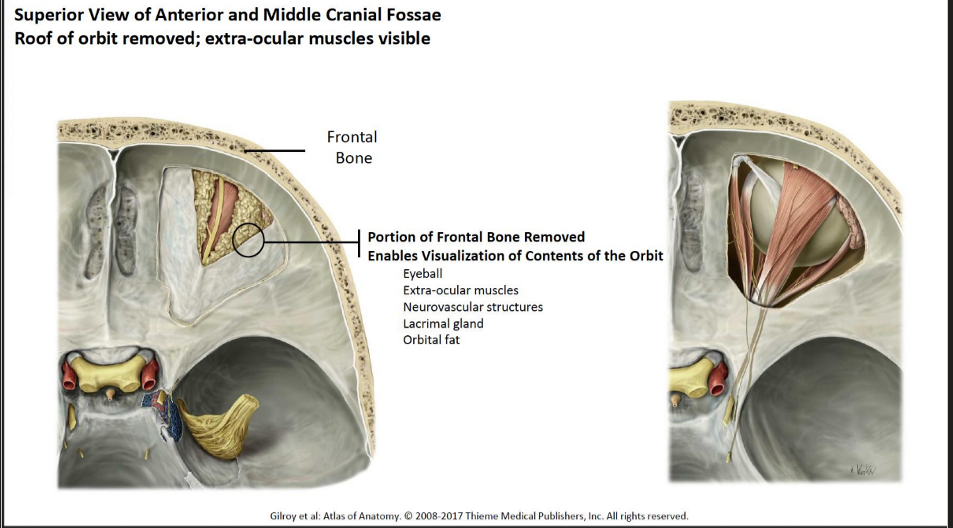
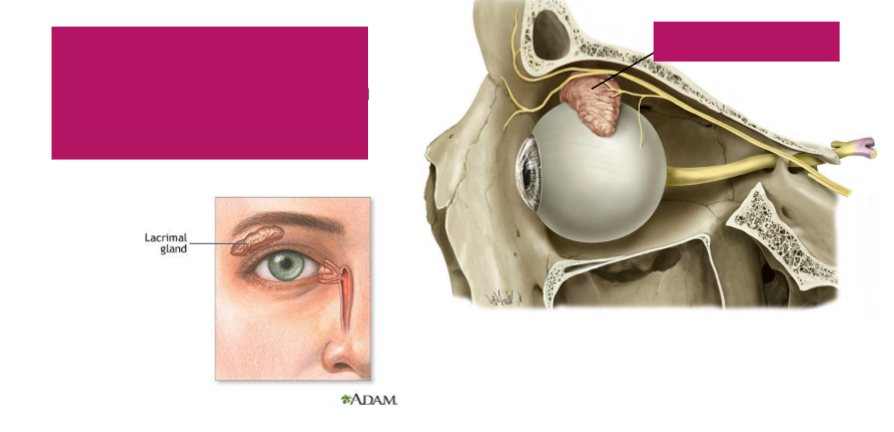
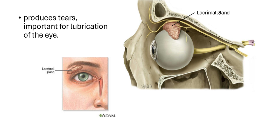
What is this structure and it’s function?
Produces tears important for lubrication of the eye
How many muscles are in the eye?
7 muscles of the orbit
6 involved in moving the eye, 1 involved in moving the eyelid
Describe the different movements of the eye
Intorsion: Internal rotation
Extorsion: external rotation
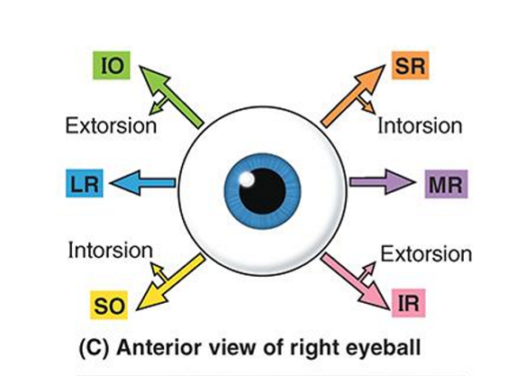
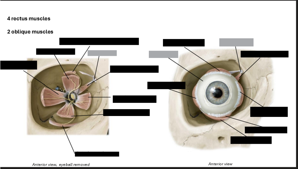
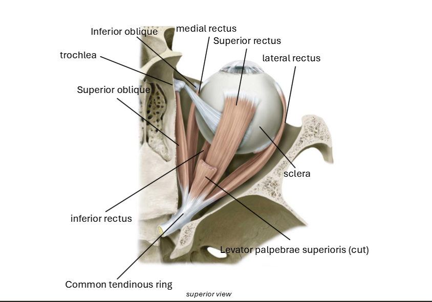
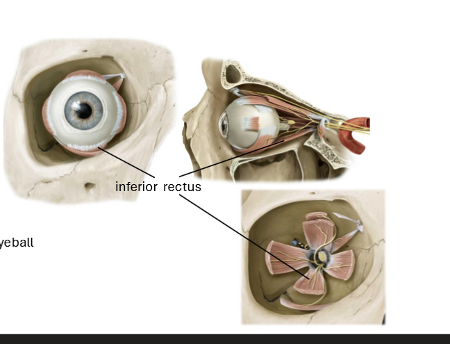
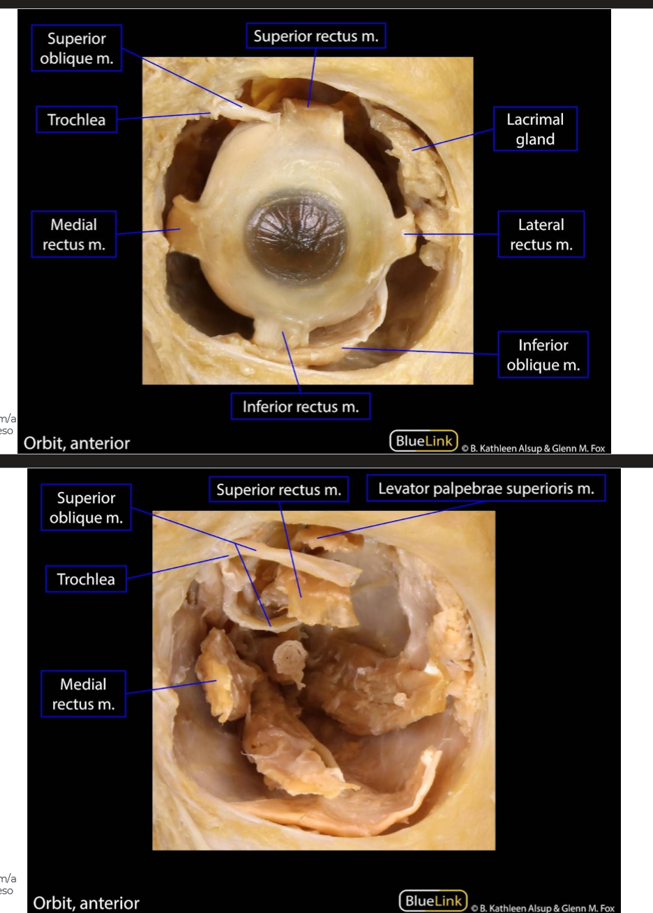
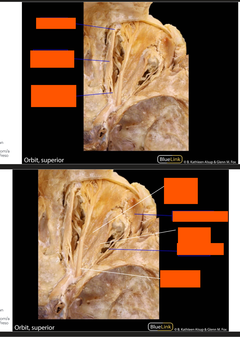
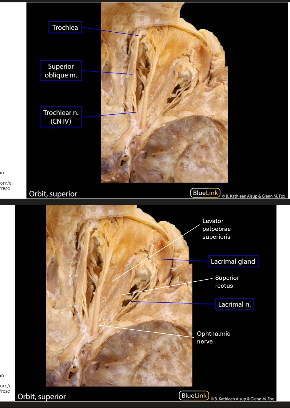
Label the eye muscles
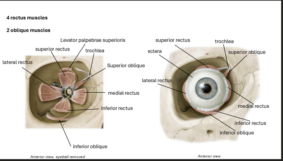
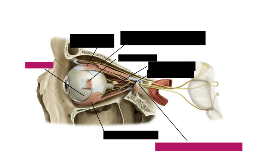
Label the eye muscles
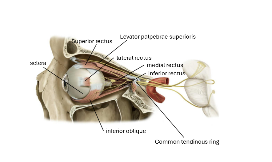
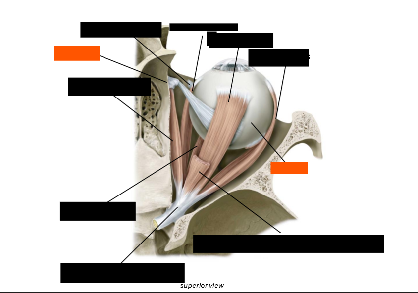
Label the eye muscles
Levator Palpebrae Superioris Origin
Sphenoid bone
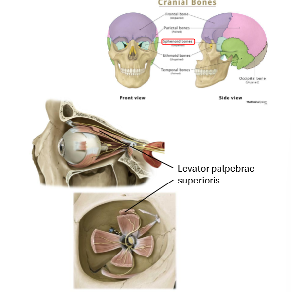
Levator Palpebrae Superioris Insertion
Superior Eyelid

Levator Palpebrae Superioris Innervation
Oculomotor nerve (CN III)

Levator Palpebrae Superioris Action
Elevation of the upper eye lid

Superior Oblique Origin
Sphenoid Bone
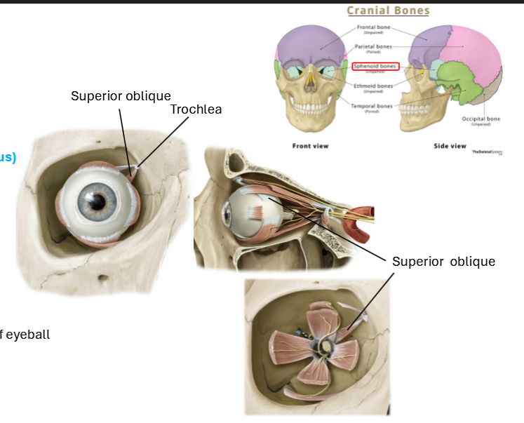
Superior Oblique Insertion
Sclera (deep to superior rectus)

Superior Oblique Innervation
Trochlear nerve (CN IV)

Superior Oblique action
Intorsion of the eyeball
Depression of eyeball
Abduction of eyeball

Inferior Oblique Origin
Floor of orbit

Inferior Oblique Insertion
Sclera (deep to lateral rectus)
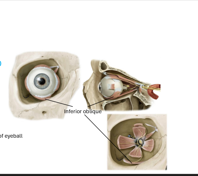
Inferior Oblique Innervation
Oculomotor nerve (CN III)

Inferior Oblique Action
Extorsion of eyeball
Elevation of eyeball
Abduction of eyeball

Superior Rectus Origin
Common Tendinous Ring
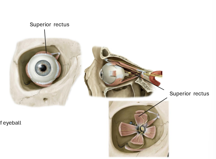
Superior Rectus Insertion
Sclera

Superior Rectus Innervation
Oculomotor nerve (CN III)

Superior Rectus Action
Intorsion of eyeball
Elevation of eyeball
Adduction of eyeball

Inferior Rectus Origin
Common Tendinous Ring

Inferior Rectus Insertion
Sclera

Inferior Rectus Innervation
Oculomotor nerve (CN III)

Inferior Rectus Action
Extorsion of eyeball
Depression of eyeball
Adduciton of eyeball
Medial Rectus Origin
Common Tendinous Ring
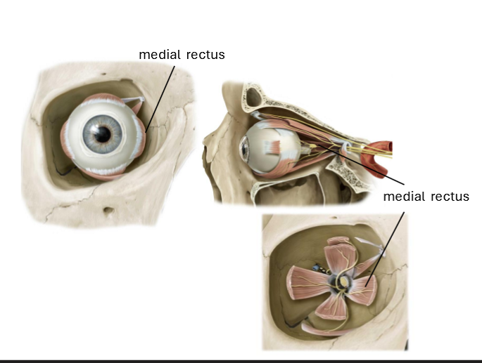
Medial Rectus Insertion
Sclera

Medial Rectus Innervation
Oculomotor nerve (CN III)

Medial Rectus Action
Adduction of eyeball

Lateral Rectus Origin
Common Tendinous Ring
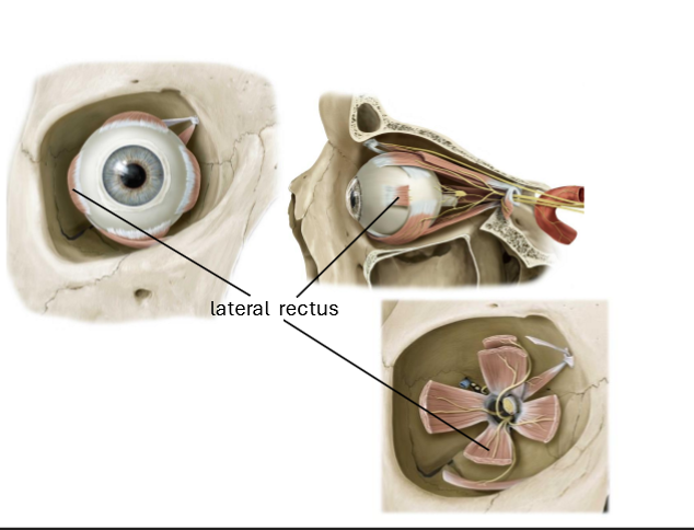
Lateral Rectus Insertion
Sclera

Lateral Rectus Innervation
Abducens nerve (CN VI)

Lateral Rectus Action
Abduction of eyeball

Describe the actions of each of the eye muscles
Obliques - abduction
Superior and inferior rectus - adduction
Superiors (oblique and rectus) - intorsion
Inferiors (oblique and rectus) - extorsion
(and then adduction and abduction for medial and lateral rectus)
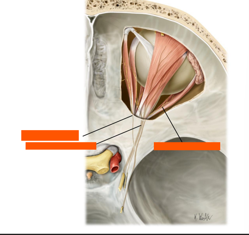
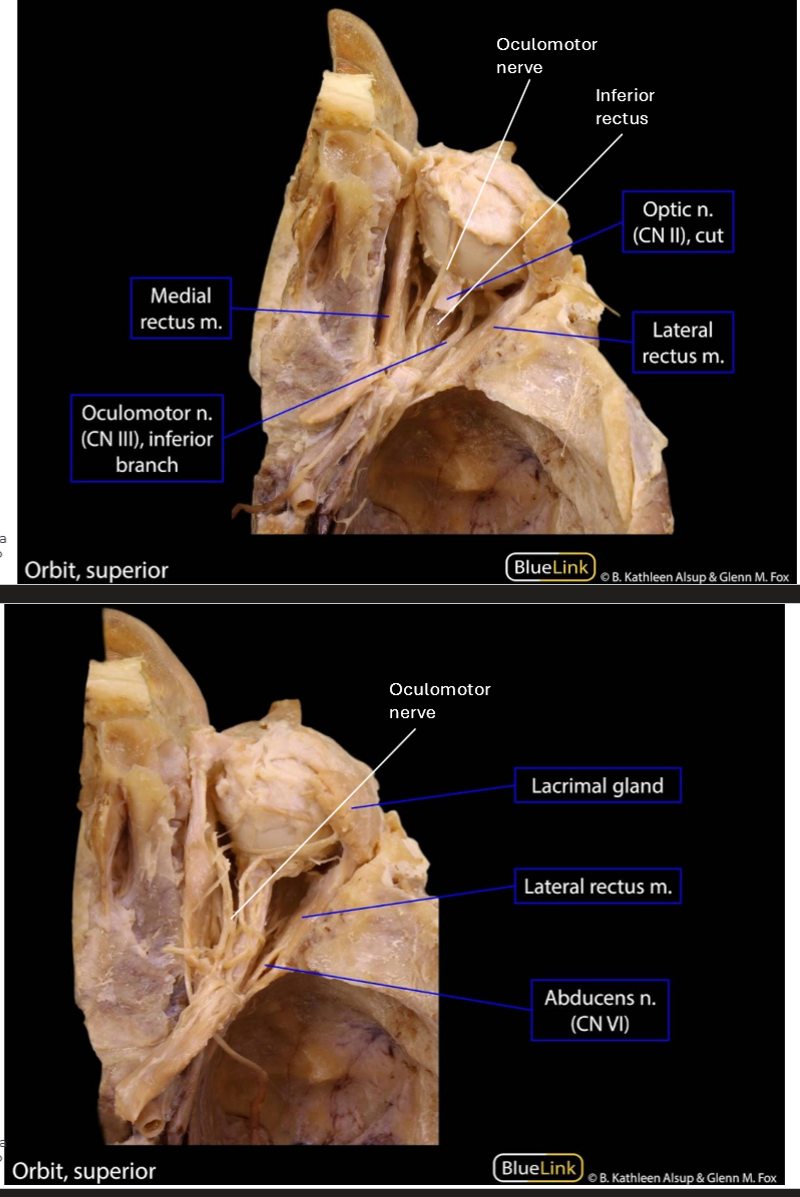
Label these nerves
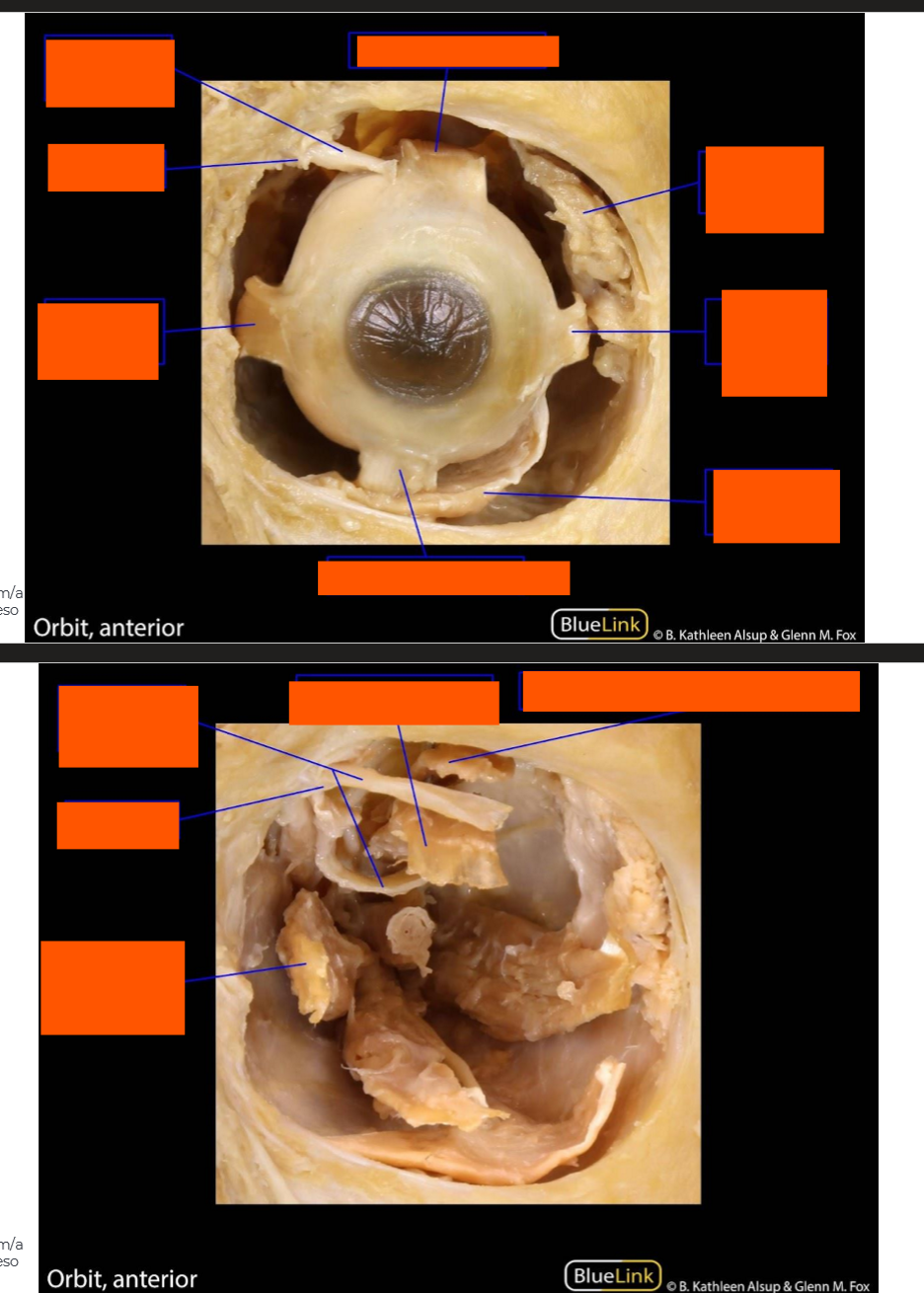
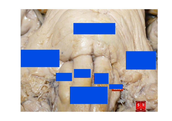
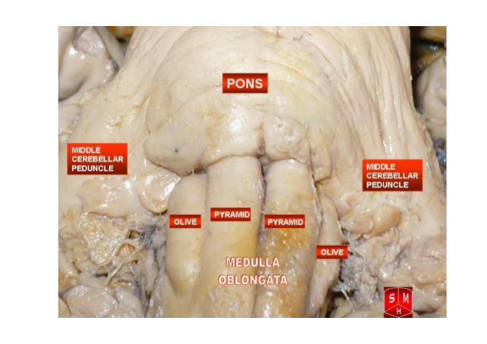
Label this prossection
Label this prosection
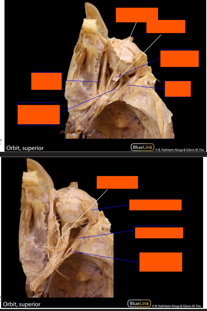
Label this prosection
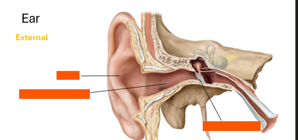
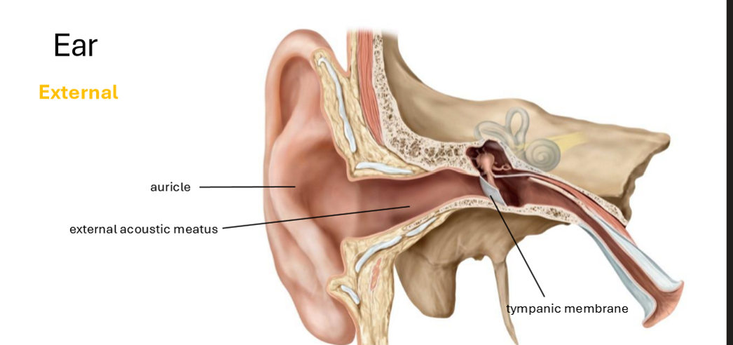
Label the external ear
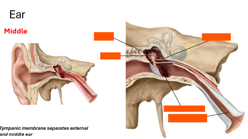
Label the middle ear
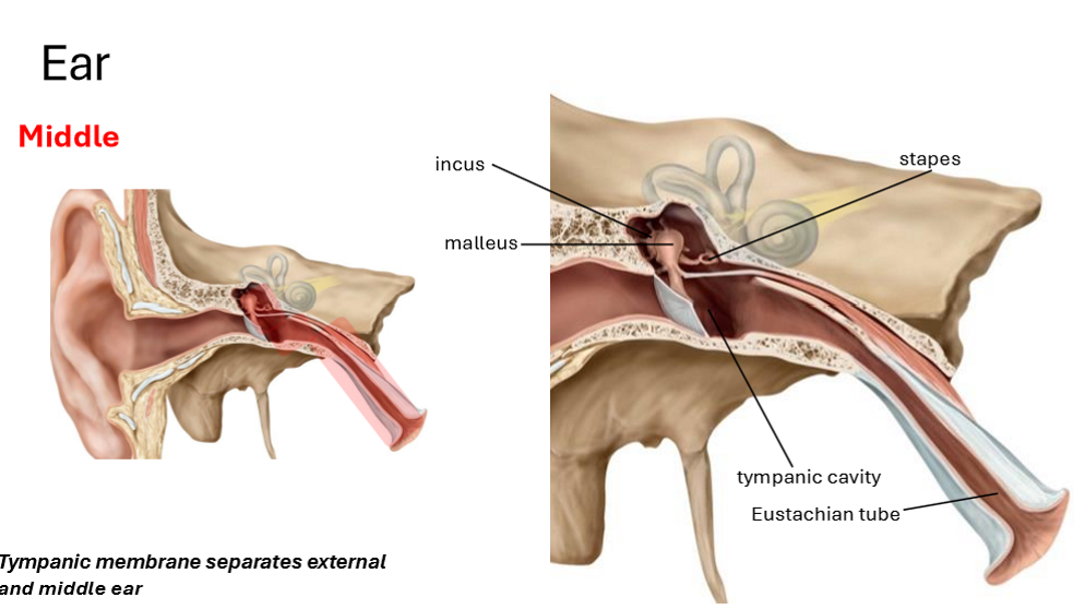
What separates the external and middle ear?
Tympanic membrane
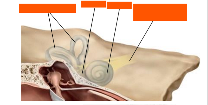
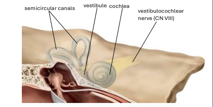
Label the internal ear
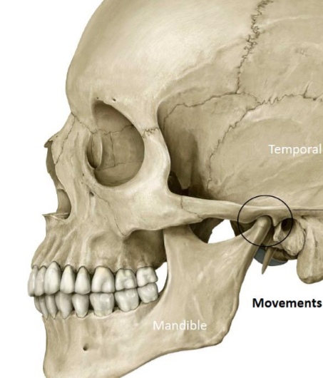
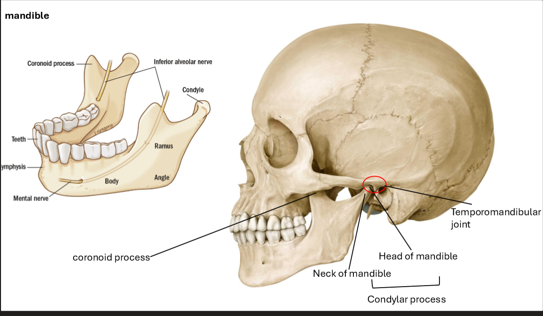
Describe the Temporomandibular joint
Articulation between mandibular fossa (temporal bone) and head of condyloid process of mandible
Movements include
Elevation (close mouth)
Depression (open mouth)
Protrusion (protrude chin)
Retrusion (retrude chin)
Lateral movements (grinding and chewing)
Describe the space of the infratemporal fossa
Medial/deep and inferior to zygomatic arch
Consider space between:
Ramus of Mandible
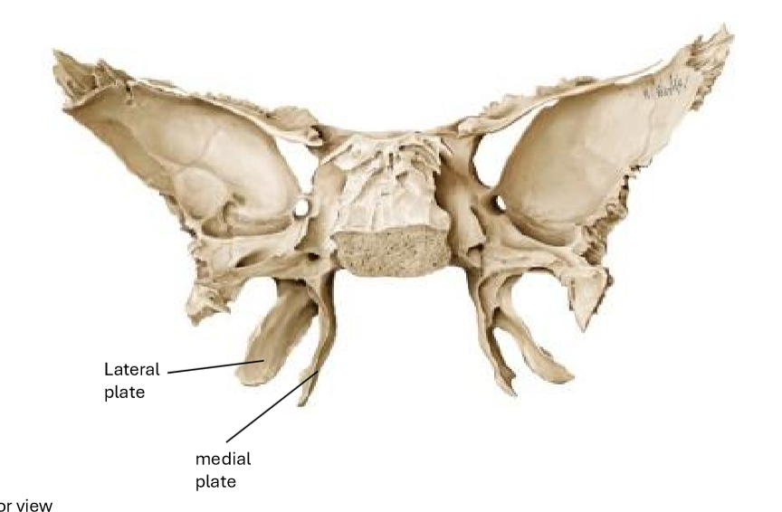
Lateral Pterygoid Plate
Maxila
Inferior Surface of Greater Wing of Sphenoid Mastoid and styloid processes of temporal bone
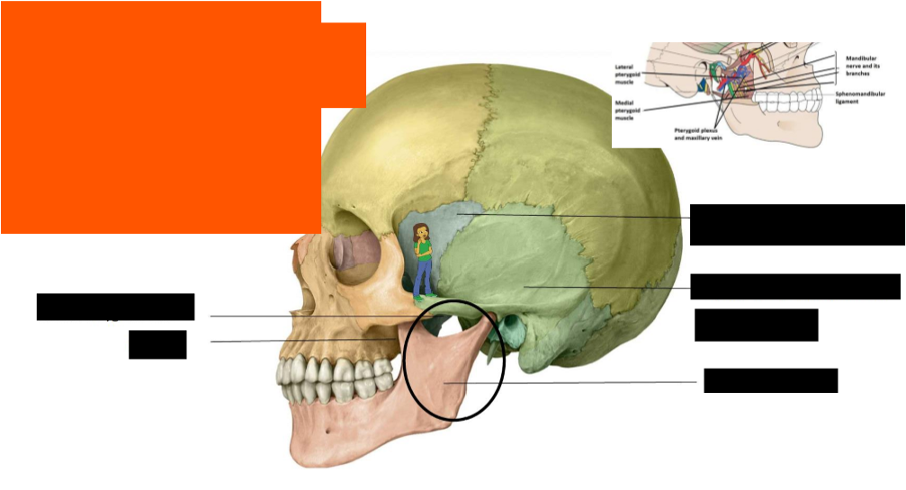
Label the structures in the infratemporal fossa
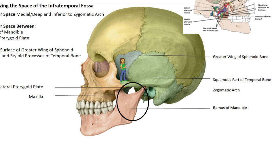
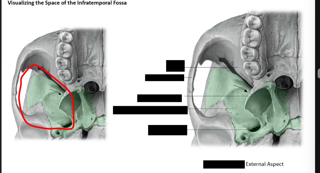
Label the infratemporal fossa
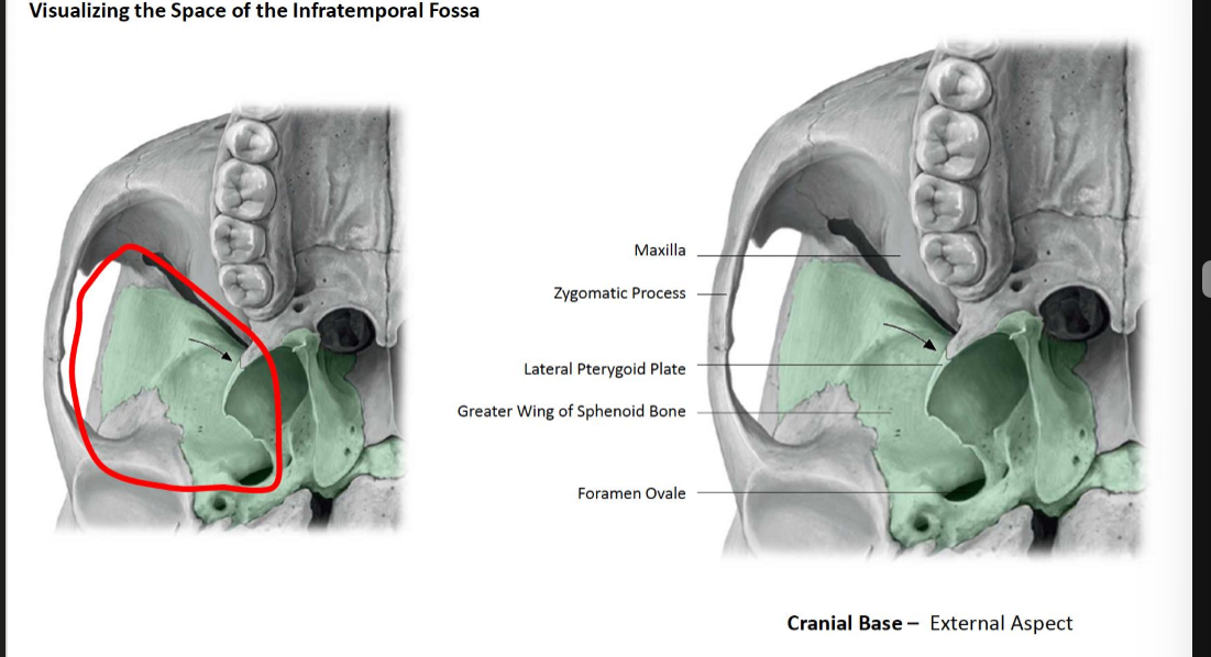
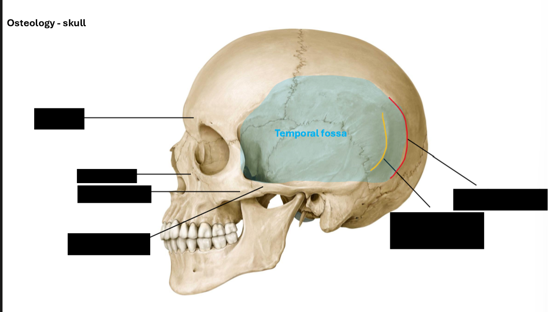
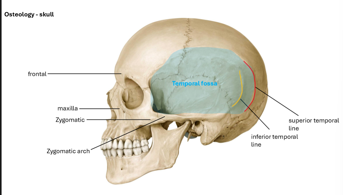
Label the osteology of the skull
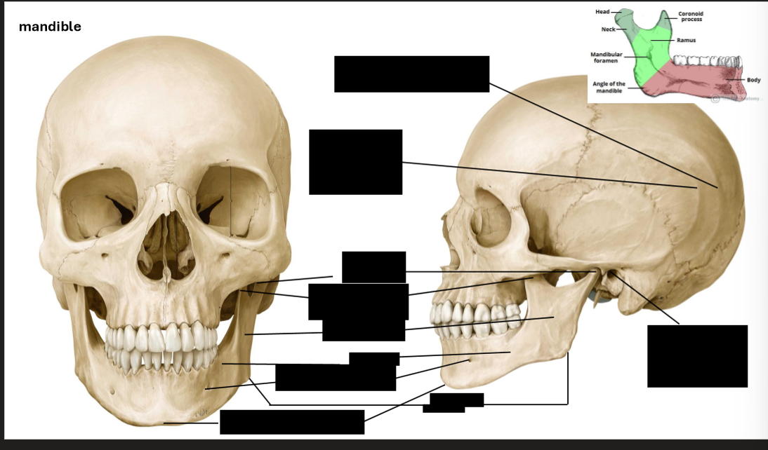
Label the mandible
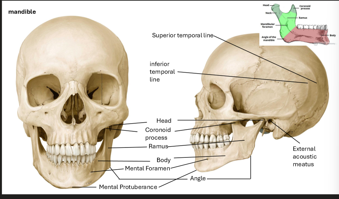
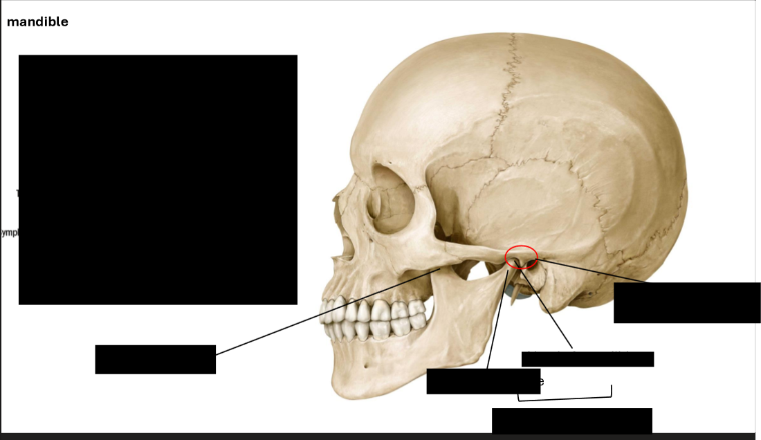
Label the Mandiblee
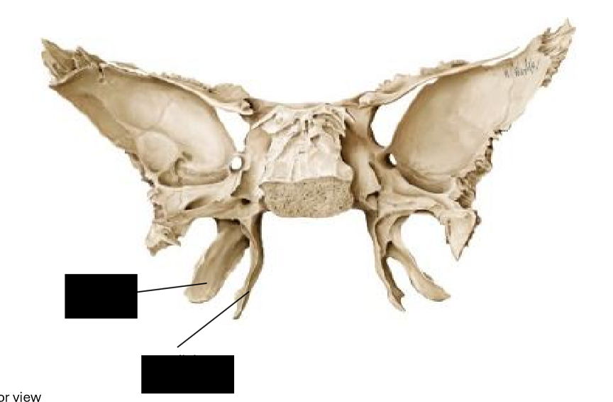
Label diagram
Of pterygoid process
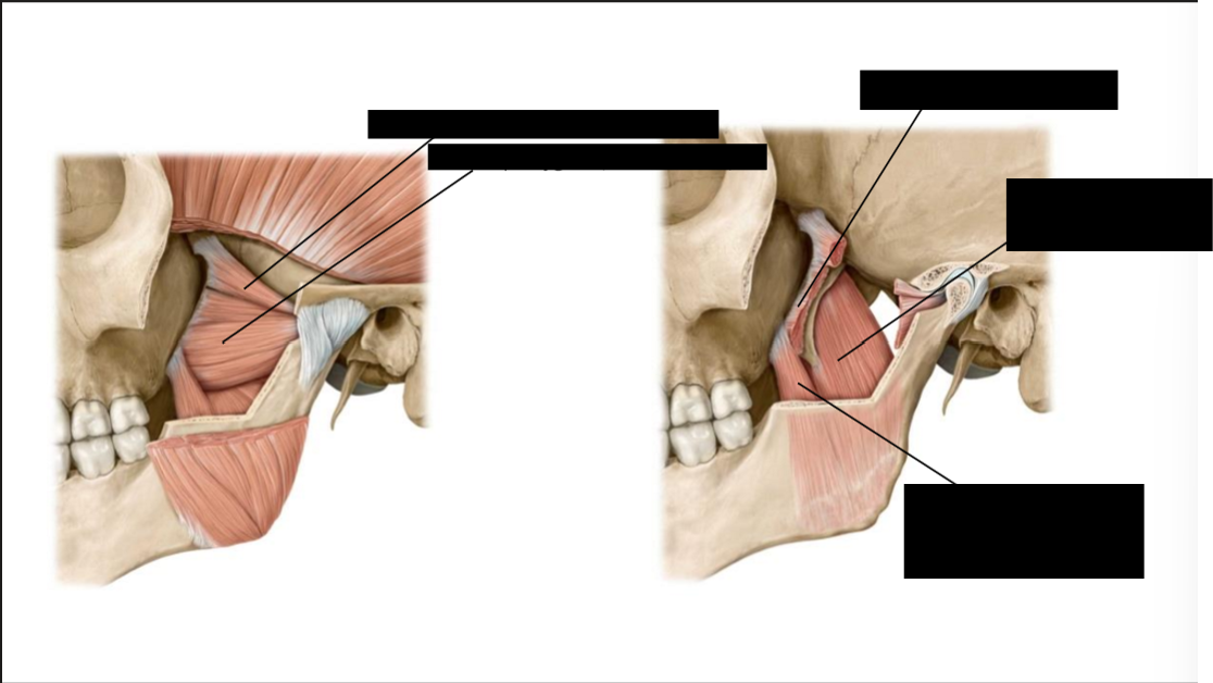
What are the muscles of mastication innervated by?
Trigeminal Nerve - Mandibular Division (V3)
What is the Infratemporal fossa bounded by?
Ramus of Mandible
Maxilla
Greater Wing of Sphenoid Bone
Lateral Pterygoid Plate (of sphenoid Bone)
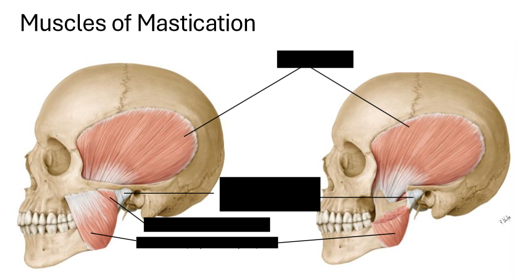
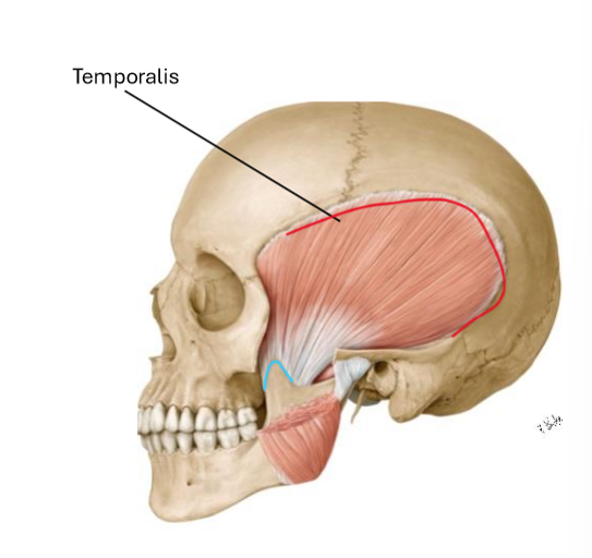
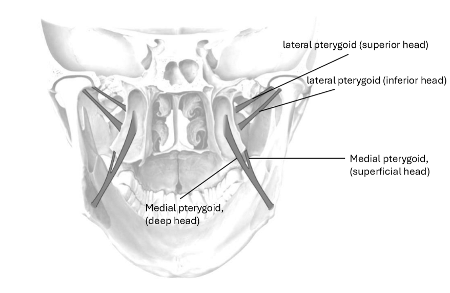
Label the Muscles of Mastication
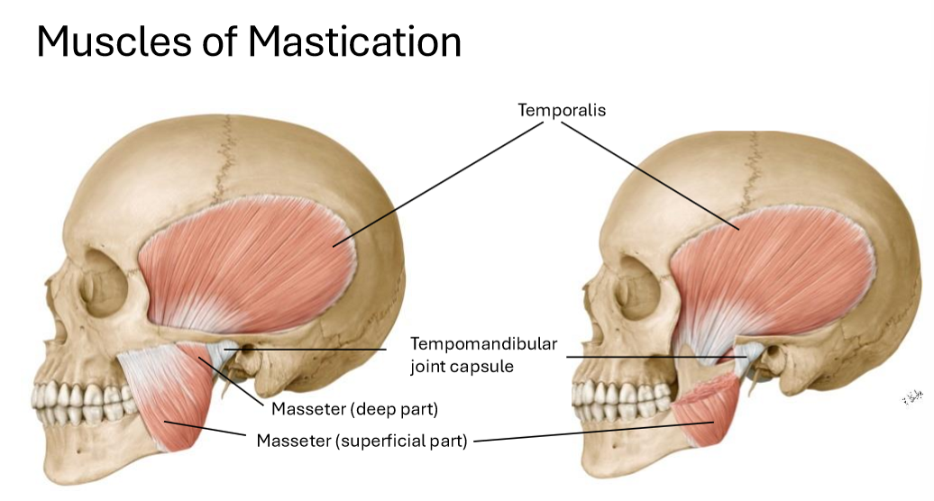
Label these muscles of mastication
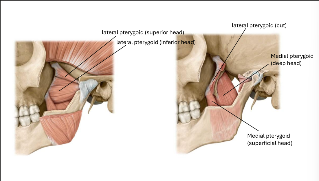
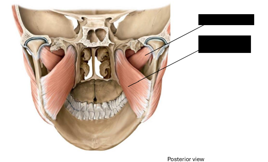
Label these muscles of mastication
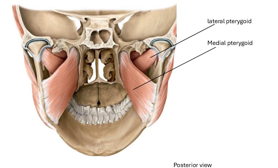
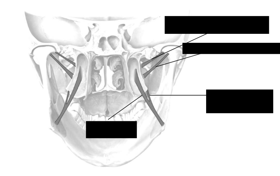
Label these muscles of mastication
Temporalis Origin
Temporalis Fossa

Temporalis Insertion
Coronoid Process of Mandible