chapter 4: a&p (tissues)
what is a tissue?
- Group of cells found together within a body that share an embryonic origin
- Histology: microscopic study of tissue appearance, organization, and function
- epithelial tissue
- connective tissue
- muscle tissue
- nervous tissue
how do we study tissues?
1. Samples are fixed: preserved in formalin or frozen
2. Samples are cut into sections
3. Samples are stained: dyed to enhance specific structures or contrasts
4. Samples are viewed
- Light microscopy can view many different tissues and dyes
- Transmission electron microscopy (TEM) views high magnification in grayscale
- Scanning electron microscopy (SEM) allows 3D views of tissue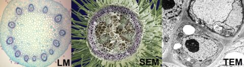
Tissue Types
- Epithelial: sheets that cover surfaces or line internal cavities
- Connective: bind cells and organs together, function in protection, support, and integration
- Muscle: excitable; response to stimuli and contract
- Nervous: excitable; propagate electrochemical signals to communicate across the body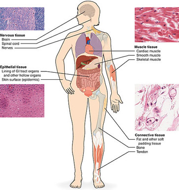
Epithelial Tissues
- Sheets of cells exposed to the environments
- Coverings and linings: skin, organ system cavities, organ walls
- Glandular epithelium
Characteristics:
- Polarity (Apex & Basal)
- Basal lamina: supporting glycoprotein sheet
- Tight Junctions & Desmosomes
- Highly regenerative
- Avascular (no blood vessels) & Innervated (nerves)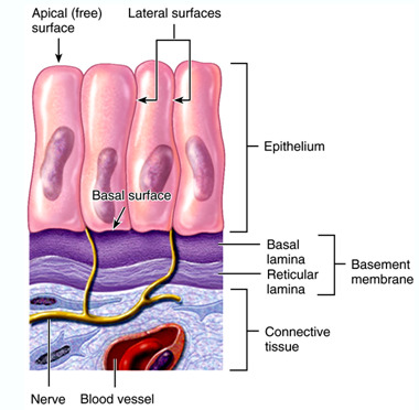
Epithelial Tissue Identification:
1. # of rows → stratified or simple?
2. shapes → squamous, cuboidal, or columnar?
Epithelial Types
- Simple Squamous: endothelium of vessels, mesothelium of serous membranes (ex: lungs, kidneys, capillary)
- Simple Cuboidal: secretion & absorption (ex: kidney, glands)
- Simple Columnar: absorption & secretion (ex: airway, gi tract)
- Stratified Squamous: most common (ex: skin, mouth)
- Stratified Cuboidal & Columnar: in some glands, but are both very rare
Other Epithelia
- Pseudostratified: single layer of cells attached to basal lamina, but apical surfaces reach different heights, nuclei at different heights (ex: testes, airway)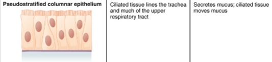
- Transitional: stratified in range of cuboidal and squamous shapes to enable stretch (ex: bladder)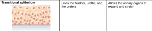
Glandular Epithelium
- Gland: one or more cells that produce and secrete a specific product
- Endocrine glands: ductless glands, secrete hormones directly into tissues
- (ex: thyroid hormone, insulin, epinephrine/adrenaline, testosterone, estrogen)
- Exocrine glands: ducts lead to the epithelial surface
- (ex: mucus, sweat, saliva, breast milk)
Glandular Structure
- Unicellular Glands: goblet cells
- Multicellular glands: (pockets (alveolar/acinar) or tubes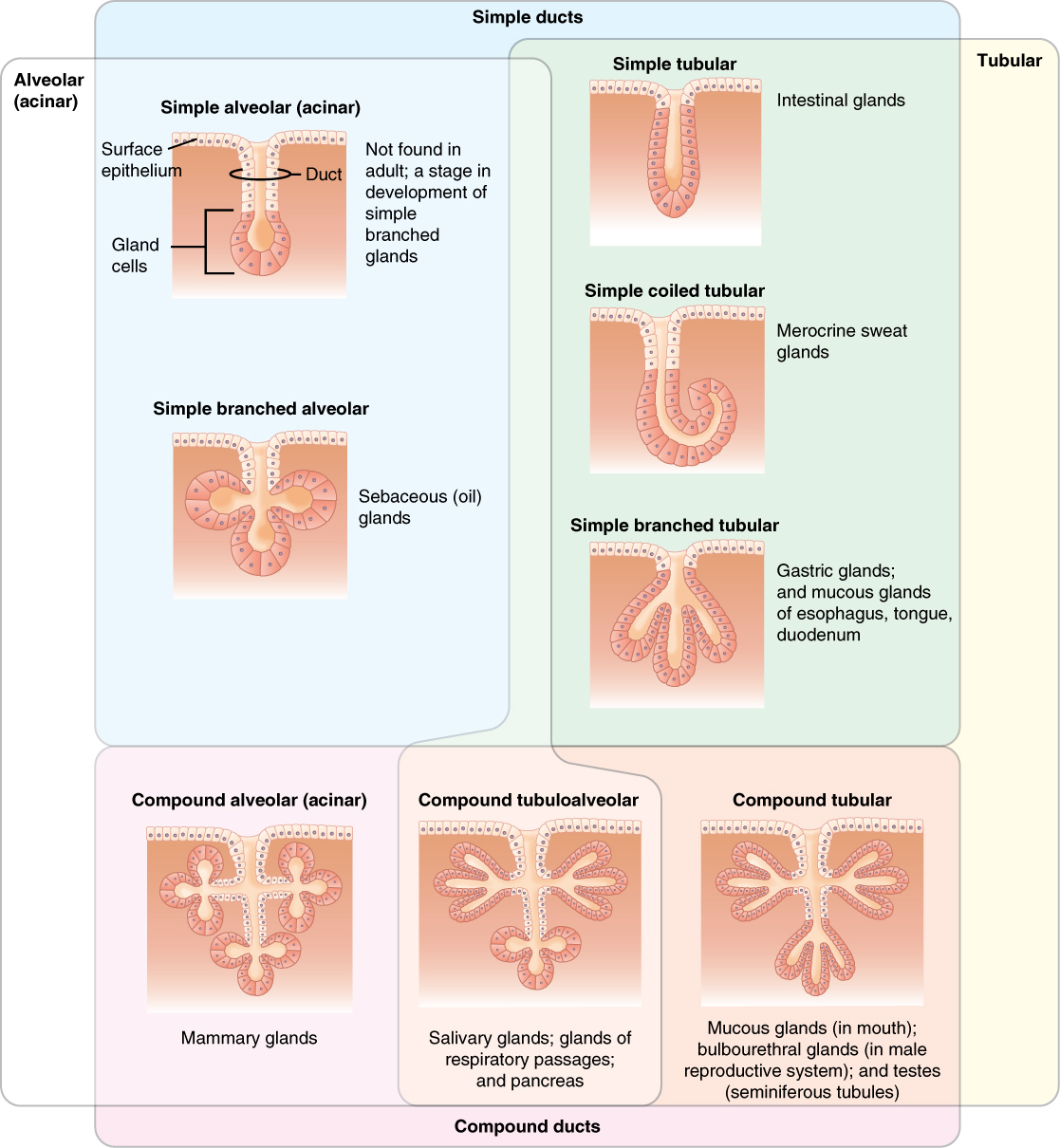
Secretion Methods
- Merocrine: vesicles emptied into extracellular space
- Apocrine: release portion of cell
- Holocrine: cell ruptures and is destroyed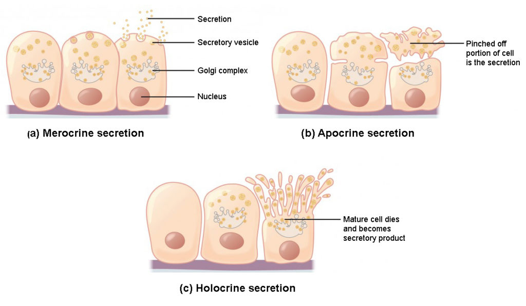
Connective Tissues
- loosely dispersed cells in a matrix with extensive extracellular material (ex: blood, cartilage, bone, etc)
- Ground Substance: material filling space between cells
1. interstitial fluid
2. adhesion proteins
3. proteoglycans
(glycoprotein)
- Protein Fibers throughout
- support & connect other tissues
- protection & immune defense
- transport
- storage
Tissue → Organs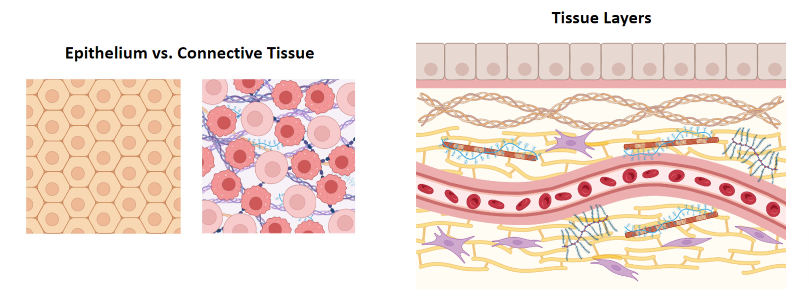
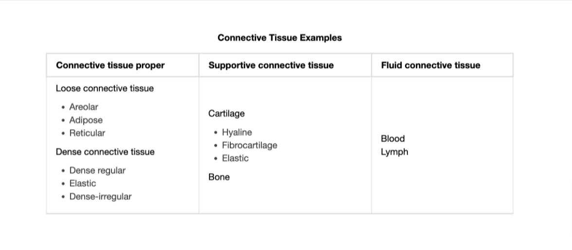
Connective Tissue: Proper
- Fixed Cells:
1. Fibrocytes (Fibroblasts)
- secrete protein, abundants
2. Adipocytes
- store adipose/fat
- can either be brown or white
3. Mesenchymal Cell
- adult STEM cells
- WBC & RBC develop
4. Macrophages
- immune cell & eat pathogens & signal immune system
5. Mast Cells
- immune cells & release histamine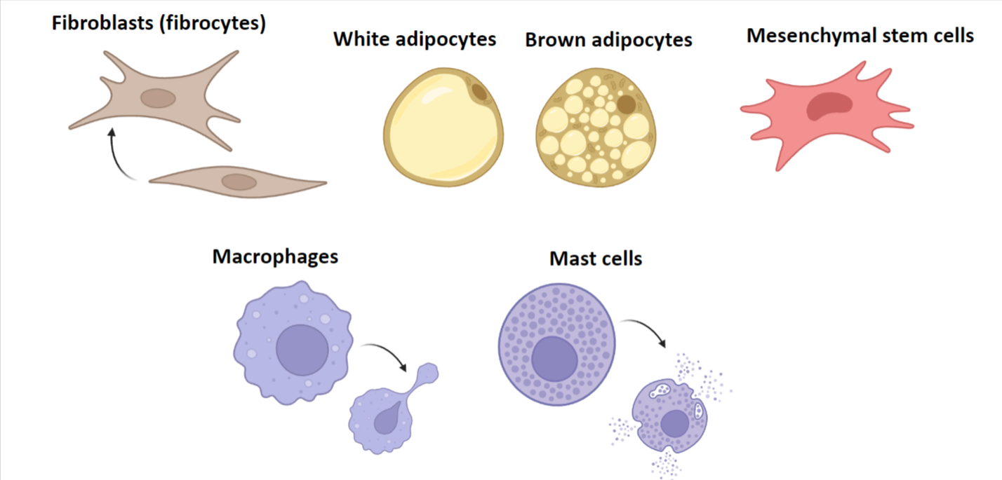
Extracellular Matrix
- Collagen:
resist, stretch, tendons & ligaments
- Elastic Fiber (Elastin):
stretch & compress → og shape; dermis
- Reticular Fibers:
organ walls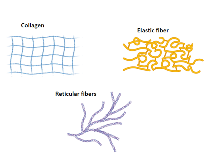
Loose Connective Tissue
- Adipose: metabolism, fast capillary
1. white: insulation, protection from injury
2. brown: “baby fat” ; thermogenic metabolism
- Reticular: support soft organs
- Areolar: cushion organs, mediates inflammation
- between muscles & vessels, under epithelia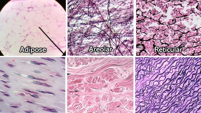
Dense Connective Tissue
- Dense Regular: attaches muscles and bones to each other
- parallel collagen, little elastin
- Dense Irregular: structural strength in multiple directions
ex: dermis, digestive tract
- Elastic: regular connective tissue with high levels of elastinallows recoil after stretching
ex: large arteries, bronchial tubes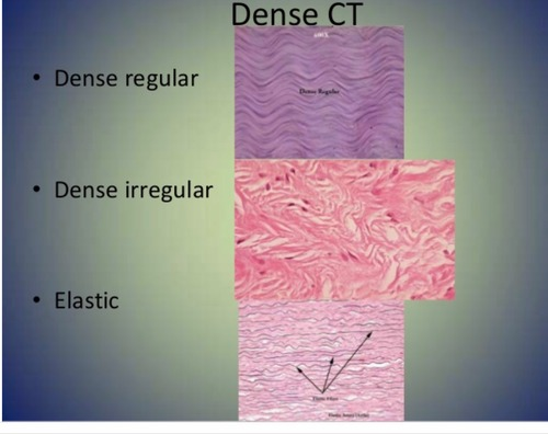
Supportive Connective TissueCartilage: chondrocytes in lacunae, ground substance full of proteoglycans
- Avascular & not innervated
1. Hyaline — strong, dense (ex: nose, ribcage, embryonic skeleton)
2. Fiber — thick bundle (ex: intervertebral discs)
3. Elastic — bendy (ex: ear)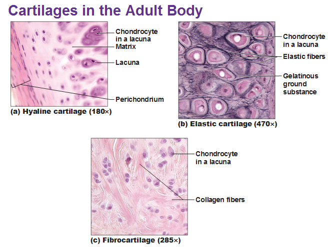
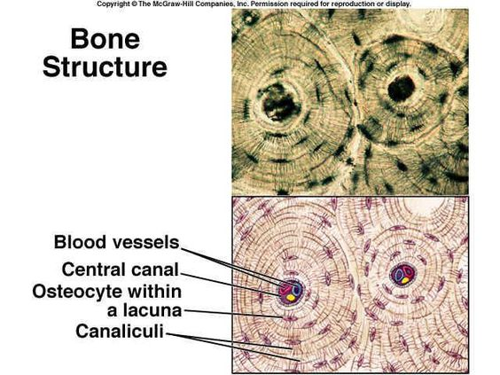
Bone: collagen fibers embedded in hydroxyapatite (calcium phosphate); highly vascular
Fluid Connective TissueBlood: erythrocytes transport oxygen, leukocytes fight pathogens, platelets promote blood clotting
Lymph: white blood cells; highly permeable vessels to regulate fluid balance

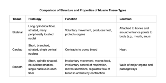
Muscle TissuesSkeletal Muscle: attached to bones; motion, thermal homeostasis
- myocyte bundles with striations of actin and myosin
- multinucleated
- AKA “voluntary” muscle & activated by nervous systemCardiac Muscle: heart walls; contract without stimulation
- cardiomyocytes with single nuclei connected by intercalated discs, striatedSmooth Muscle: involuntary movements of internal organs
- spindle-shaped cells, single nuclei, no striations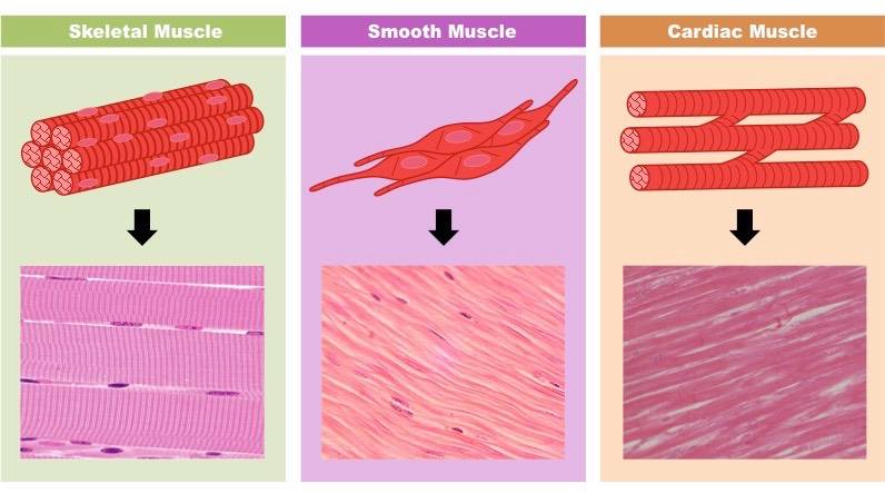
Nervous TissueNeuron: propagates information via electrochemical signals (action potential)
- sheathed in myelin (myelin sheath)
- Synapse: gap between neuronsNeuroglia: support neurons
- Astrocytes: homeostasis of CNS
- Oligodendrocytes: produce myelin for CNS
- Schwann Cells: produce myelin for PNS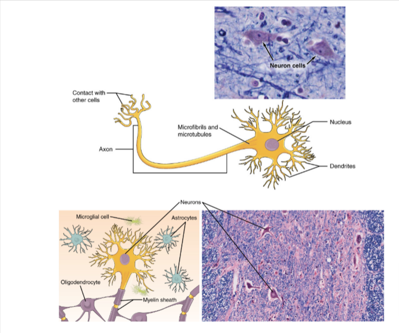
MembranesCutaneous Membrane: skin, keratinized epidermis attached to this connective tissue (dermis)
- exposed to air, is very dryMucous Membrane: AKA mucosae, line all body cavities that open to outside (e.g., digestive & respiratory tracts)
- bathed in secretions → “wet”
- epithelial sheets or lamina propria
- adapted for absorption & secretionSerous Membranes: AKA serosae, closed ventral body cavities
- visceral & parietal layers separated by serous fluid
- simple squamous epithelium over thin areolar connective tissue
Tissue RepairRegeneration: replacement of destroyed tissue with correct tissue type
Fibrosis: replacement of destroyed tissue with scar tissue
1. Inflammation: chemicals cause white blood cells, fluid, clotting proteins to leak into space; clot seals off injury
2. Organization: restored blood supply by new capillaries, new collagen production, macrophages clear debris
3. Permanent Repair: new tissue matures