Histo slides w/ short descrip | Quizlet
1/35
There's no tags or description
Looks like no tags are added yet.
Name | Mastery | Learn | Test | Matching | Spaced |
|---|
No study sessions yet.
36 Terms
Catarrhal Bronchopneumonia
· thick bronchide walls
· catarral exudate in alveoli lumen (RBCs, leukocytes + mucous)
· congested vessels
· edema separates good + bad
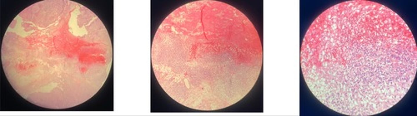
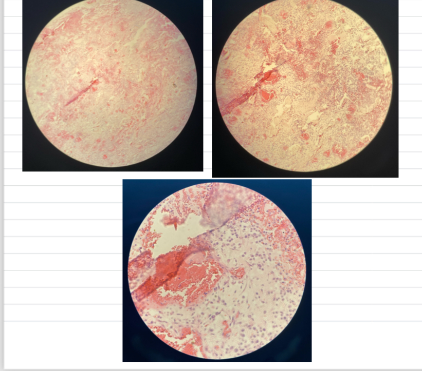
Fibrinous Bronchopneumonia => mosaic appearance
· Acute = capillaries congested + alveoli filled w/light pink fluid
· Red = alveoli filled w/ exudate = fibrin + RBCs
· Grey = lots of neutrophils in lumen
· Resolution = fibrin in strands due to neutrophils enz release
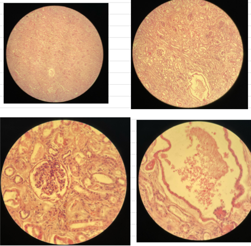
Bacterial embolic purulent nephritis
- bacterial emboli formed in capillary vessels and bv’s
- degen of tubular epi cells
- at edge of bacteria emboli => microabscess (neutrophils w/ debris)
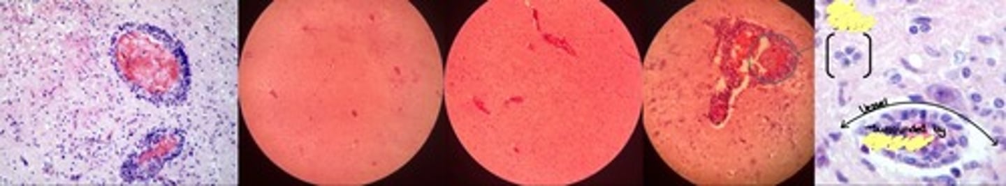
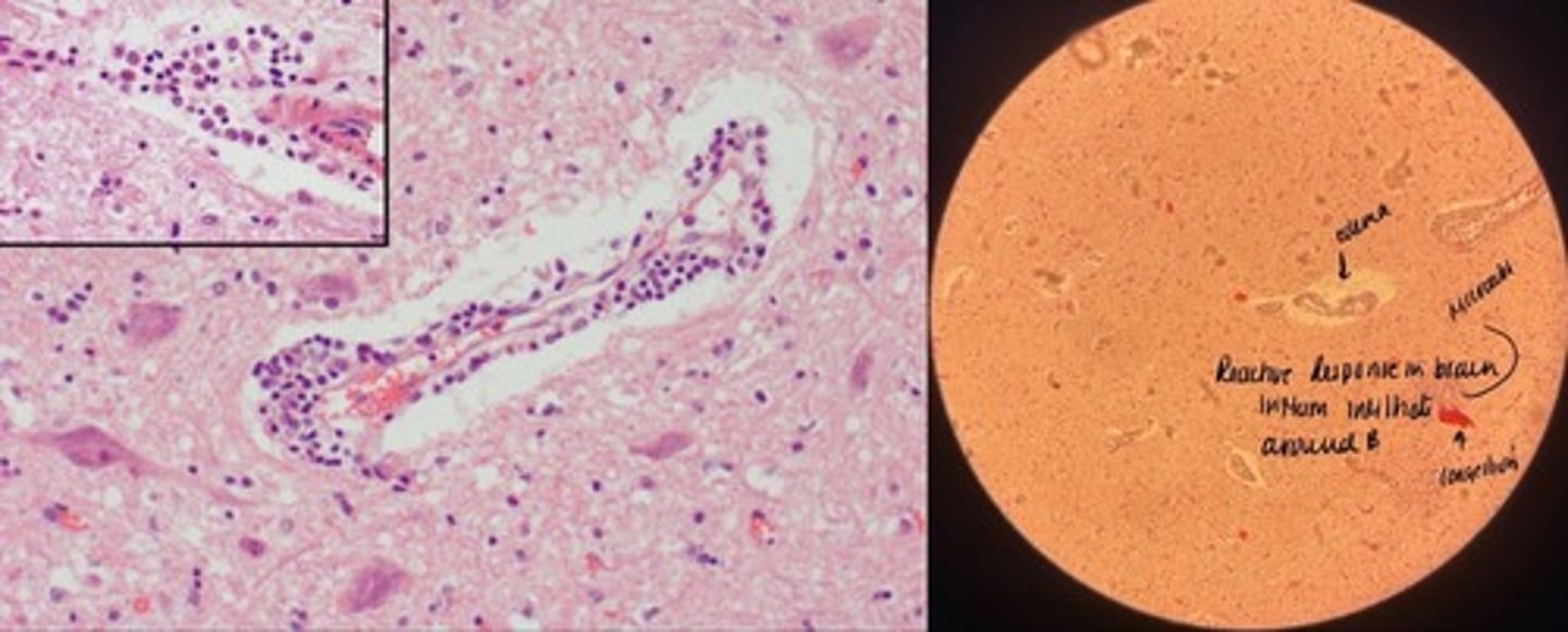
Non-purulent encephalitis
- perivascular cuffing w/ lymphocytes of congested vessel
- increased glial cells => glialosis rosettes
- focal neuronal loss
- increased plasma + macrophages
Purulent encephalitis
- congestion of bv + neuronal degen
- perivascular cuffing + edema
- microabscess in necrotic brain tissue
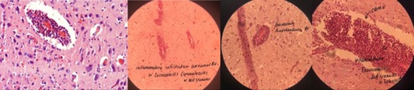
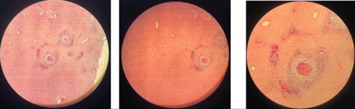
Eosinophilic encephalitis
- MANY eosinophils in perivascular space
- cerebral edema => empty space around bvs + neurons
- neuronal degen + necrosis
Organisation of fibrin during post-traumatic pericarditis
- heart is covered in fibrin w/ vilious like projections
- young + old granulation tissue
- fat, inflam cells, myocardium
Chronic interstitial nephritis
- inflam (lymphocytes, plasma cells, macrophages) infiltrates in interstitial tissue
- hyperplasia of fibrous tissue
- retention cysts in lower parts of tubules
- atrophy of renal tubules
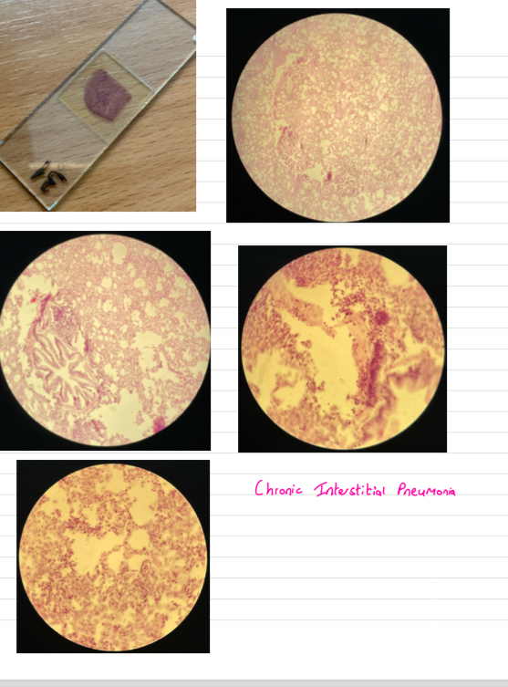
Chronic interstitial pneumonia
- thickened alveolar septa …narrowed lumen
- lymphoid follicles around bvs
- prolif of s.m cells
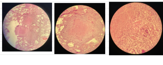
Pulmonary tuberculosis
- central necrosis surrounded by epitheloid cells w/ giant cells btw
- infiltrates of lymphocytes, plasma, fibrosis
Actinomycosis => Gram+ filamentous anaerobicrods
- accumulation of bacteria
- pyogranulomas separated w/ fibrous CT
- basophilic stained masses in centre .. surrounded with clubbed corona (eosinophilic material) then neutrophils then macrophages
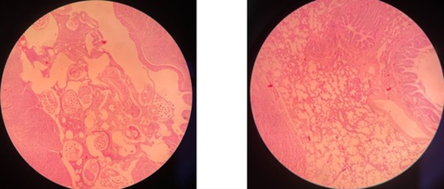
Parasitic granuloma (of liver)
- dead parasites in centre surrounded by multinucleated giant cells
- regressive lesions of adjacent hepatocytes
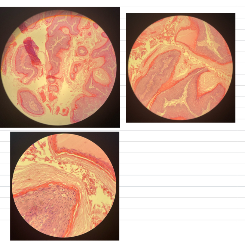
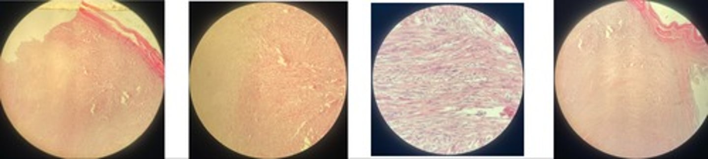
Papilloma => benign epithelial neoplasm
- CT core w/ neoplastic squamous epi
- papilloma is thick bc… acanthotic, hyperplasia, hyperkeratotic
Cornified layer is vacuolated
Squamous cell carcinoma => malignant epithelial neoplasm
- irregular masses + cords proliferating downwards
- ulcerations on surface
- keratin pearls
- coag. necrosis
- mitotic figures + neoplastic cells
Adenoma => benign epithelial neoplasm
- tumour contains 1 layer of epi … neoplastic cells and cyst is empty
- hepatocytes @ edge + degen
- parenchyma is demarcated from liver tissue
- increased fibrous CT
Canine mammary carcinoma => simple tubular carcinoma
- neoplastic epi arranged tubular … 2 cell thick
- mitotic figures + nuclear pleomorphism
- neoplastic cells invade BM
Pulmonary metastasis of adenocarcinoma
- in alveoli and lymphatic system
- solid, papillary and tubular pattern
- metastasis nodules composed of neoplastic epi cells
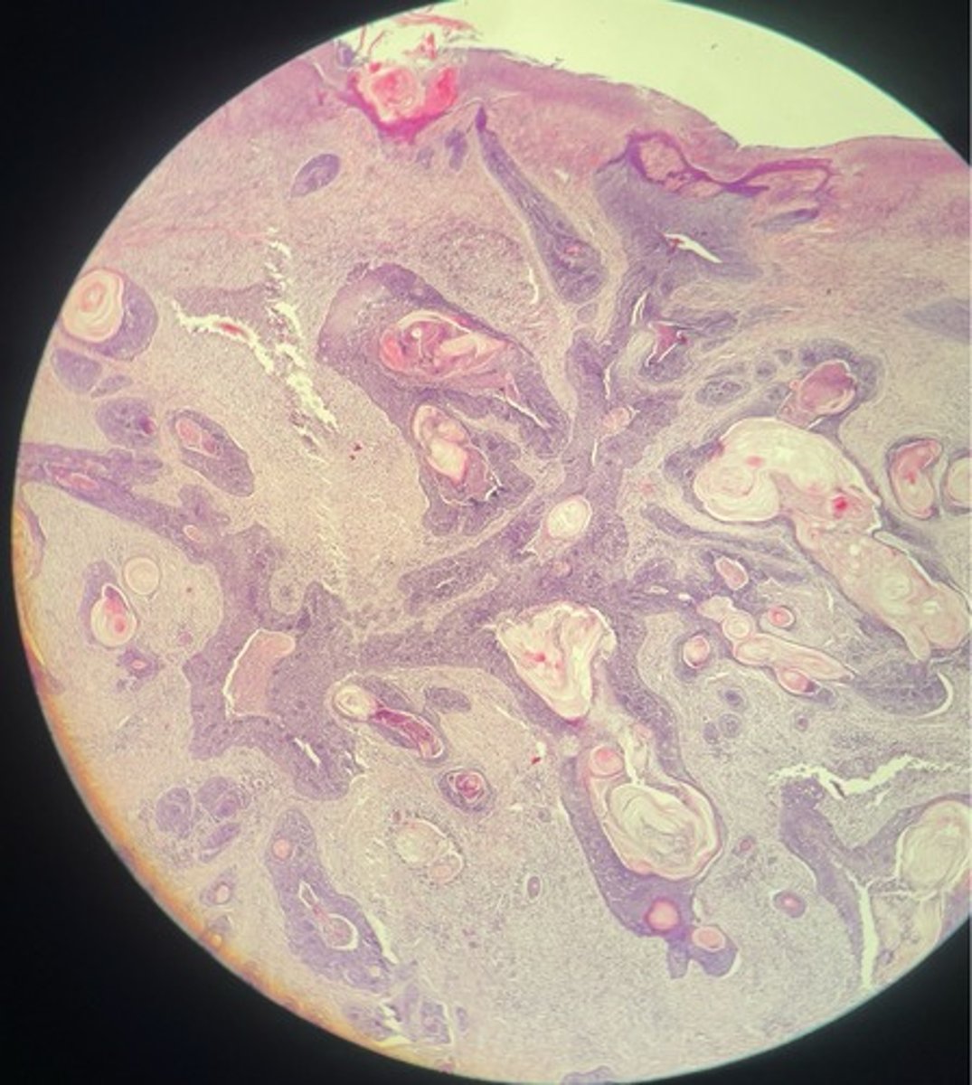
Perianal gland tumour
- multilobulated masses
- basal reserve cells @ periphery lobules
- CT in stroma
Melanoma
- mitotic figures
- cellular cytoplasm … melanin pigment => Fontana staining
- nuclei pleomorphism + hyperchromasia
Fibroma
- whorls + interlacing bundles of fibroblasts, fibrocysts, collagen fibres
- may be firm or soft
- soft => fibromyxoma… collagen mixed w/ mutinous ground substance
Fibrosarcoma
- highly vascular but poorly formed
- interwoven ⭐️ immature fibroblasts
- collagen fibres
- nuclei elongated + mitotic figures

Feline injection site sarcoma FISS
- neoplastic spindle cells separated by fibromyxoma matrix (soft!)
- nuclei elongated + mitotic figures
- area of necrosis
- multinucleated giant cell
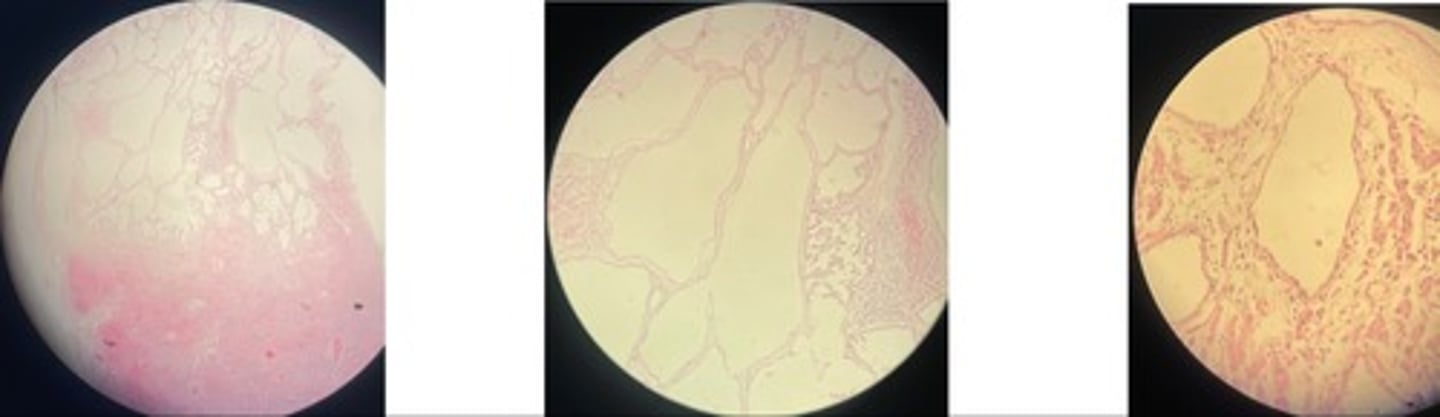
Lipoma
- mature adipocytes (appearance like normal fat)
- NO mitotic figures + rare CT w/ bvs
- nucleus eccentric (@ side)
Liposarcoma
- small + large adipocytes
- haemorrhages + necrosis
- highly cellular w/ mitotic figures
Mast cell tumour MCT
- cells in sheets + cords b/w collagen
- deep blue granules in cytoplasm
- round neoplastic cells are mixed w/ eosinophils
Lymphoma
- many lymphoblasts infiltrating muscle fibres => atrophy + degen
- mitotic figures
Hepatic coccidiosis … intracell parasites
- dilated bile ducts
- hyperplasia of biliary epi => papillary projections
- oocysts in lumen + inflam cells
- atrophy + degen
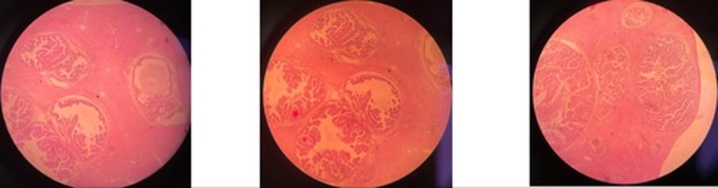
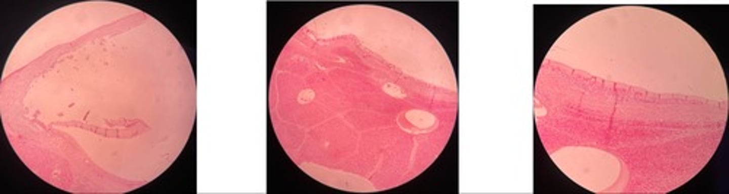
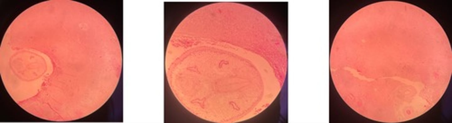
Hepatic echinococcus
- cystic structure lined with chitin mem
- may have protoscolices (parasite larvae)
- liver parenchyma => atrophy, fatty degen, necrosis, haemorrhage
- infiltrate cells: macrophages, epitheliod cells and giant cells
Hepatic fascioliasis
- coag. necrosis + haemorrhages
- hemosiderin pigments + inflam cells
- fibrosis @ portal area
- prolif of biliary ducts
- scar formation in migration pathway
Parasitic pneumonia
- distended alveoli, bronchi + bronchioles
- degenerated + mononuclear cells
- s.m + epi hyperplasia … thickened alveolar septa
- exudate + emphysema … congestion
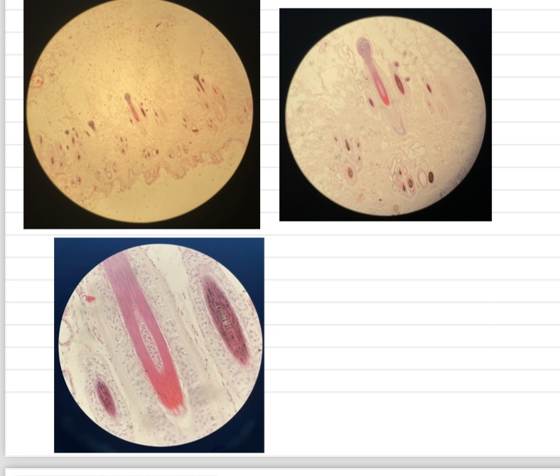
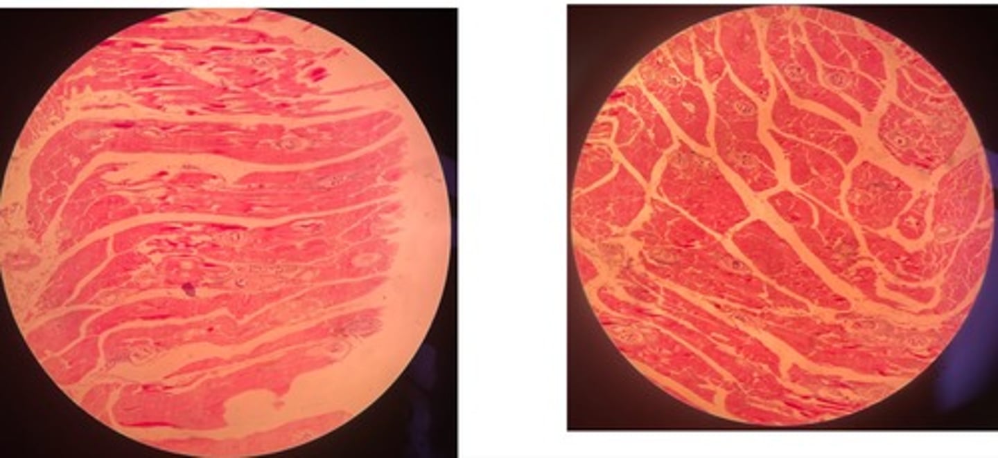
Trichinellosis
- Early: parasite within myocytes +infiltrate cells
- Late: normal morph w/ thick capsule and possible accumulation of Ca salt
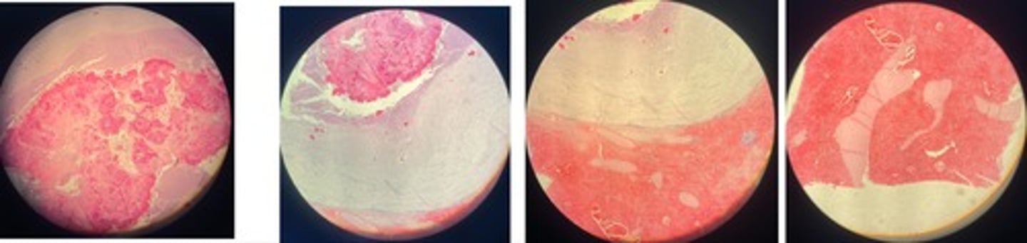
Contagious Bovine Pleuropneumonia (CBPP)
- fibrous pneumonia = acute, red, grey (increase neutrophils), resolution
- area of necrosis w/ bv @ centre + inflam cells surrounding
- marginal organisation…. granulation grows to surviving lobes
Equine Infectious Anemia (EIA)
- necrosis of central lobes
- congestion of c.v + sinuses
- hemosiderin + inflam cells in triad
- hyperplasia of Browicz-Kupfur cells
Infectious Canine Hepatitis (ICH)
- hepatic necrosis
- congestion + inflam infiltrate
- intranuclear viral inclusion bodies (basophilic)
Canine Distemper Virus (CDV)
- interstitial pneumonia => narrow lumen
- bacterial/catarrhal-purulent bronchopneumonia => inflam cells in bronchioles + alveoli
- intracellular viral inclusion bodies
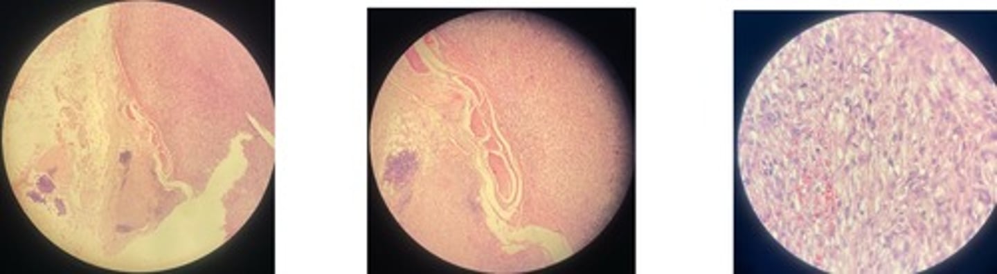
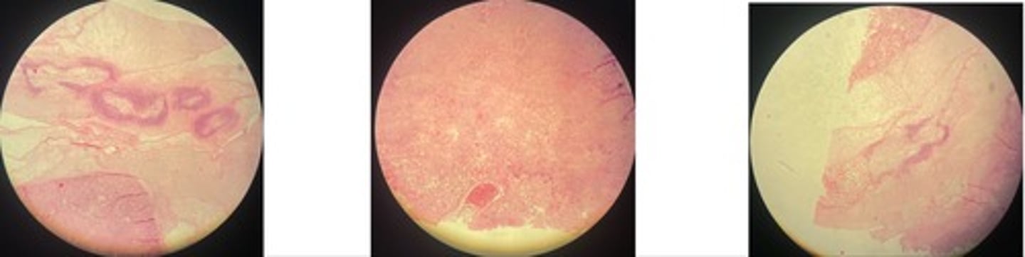
Feline Panleukopenia.... catarrhal inflam of intestinal mm
- looks like worm on slide
- fibrin accumulation + crypt necrosis
- hemoarrhages + inflam cells
- basophilic intranuclear inclusion bodies in crypt epi