BSC2085L E.14 The Spinal Cord, Spinal Nerves, Reflexes, and the Autonomic Nervous System
0.0(0)
Studied by 0 peopleCard Sorting
1/37
Last updated 6:33 PM on 4/7/23
Name | Mastery | Learn | Test | Matching | Spaced | Call with Kai |
|---|
No analytics yet
Send a link to your students to track their progress
38 Terms
1
New cards
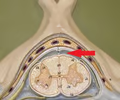
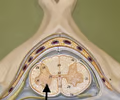
epidural space
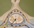
• The space deep to the vertebral canal and superficial to the dura mater.
• Filled with fat.
• Filled with fat.
2
New cards
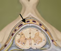
meninges
• Three layers of protective membrane that surround the spinal cord.
• Composed of the dura mater, arachnoid mater, pia mater.
• Composed of the dura mater, arachnoid mater, pia mater.
3
New cards
dura mater
• The outer layer of the meninges.
• Deep to the epidural space and superficial to the arachnoid mater.
• Deep to the epidural space and superficial to the arachnoid mater.
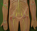
4
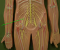
New cards
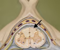
arachnoid mater
• The intermediate layer of the meninges.
• Deep to the dura mater and superficial to the subarachnoid space.
• Deep to the dura mater and superficial to the subarachnoid space.
5
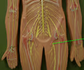
New cards
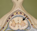
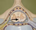
subarachnoid space
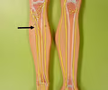
• The space deep to the arachnoid mater and superficial to the pia mater.
• Contains cerebrospinal fluid.
• Contains cerebrospinal fluid.
6
New cards
pia mater
• The inner layer of the meninges.
• Deep to the subarachnoid space and superficial to the spinal cord.
• Emerges denticulate ligaments.
• Deep to the subarachnoid space and superficial to the spinal cord.
• Emerges denticulate ligaments.
7
New cards
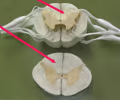
denticulate ligament
Lateral folds of the pia mater that holds the spinal cord.
8
New cards
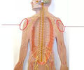
cervical enlargement
• Located at C3-T1 of the spinal cord.
• A bulge that houses the neurosomas whose fibers supply the upper limbs.
• A bulge that houses the neurosomas whose fibers supply the upper limbs.
9
New cards
lumbar enlargement
• Located at T9-T12 of the spinal cord.
• A bulge that houses the neurosomas whose fibers supply the lower limbs.
• A bulge that houses the neurosomas whose fibers supply the lower limbs.
10
New cards
conus medullaris
• Located at L1-L2 of the spinal cord.
• The cone-shaped tip termination of the spinal cord.
• The cone-shaped tip termination of the spinal cord.
11
New cards
cauda equina
• Located beyond the conus medullaris.
• The continuation of nerves from the spinal cord extending downward from the conus medullaris.
• The continuation of nerves from the spinal cord extending downward from the conus medullaris.
12
New cards
filum terminale
• Located beyond the conus medullaris.
• Fibrous extensions of the pia mater that continue to S2, located on the midline.
• Fibrous extensions of the pia mater that continue to S2, located on the midline.
13
New cards
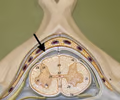
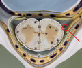
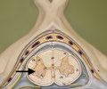
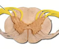
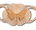
ventral horns
• Gray matter in the spinal cord.
• Located on the ventral (anterior) side of the spinal cord.
• Contains somatic motor neurosomas.
• Also called anterior horns.
• Located on the ventral (anterior) side of the spinal cord.
• Contains somatic motor neurosomas.
• Also called anterior horns.
14
New cards
dorsal horns
• Gray matter in the spinal cord.
• Located on the dorsal (posterior) side of the spinal cord and contacts the surface.
• Contains the axon terminals of peripheral sensory neurons and their synapses with interneurons.
• Also called posterior horns.
• Located on the dorsal (posterior) side of the spinal cord and contacts the surface.
• Contains the axon terminals of peripheral sensory neurons and their synapses with interneurons.
• Also called posterior horns.
15
New cards
lateral horns
• Gray matter in the spinal cord.
• Located laterally on the spinal cord only in the thoracic region.
• Contains visceral motor neurosomas.
• Located laterally on the spinal cord only in the thoracic region.
• Contains visceral motor neurosomas.
16
New cards
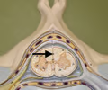
gray commissure
• Gray matter in the spinal cord.
• The bridge connecting the gray matter on both sides of the spinal cord.
• Surrounds the central canal.
• The bridge connecting the gray matter on both sides of the spinal cord.
• Surrounds the central canal.
17
New cards
central canal
• Located in the gray commissure of the spinal cord.
• Channels cerebrospinal fluid.
• Channels cerebrospinal fluid.
18
New cards
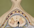
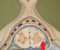
ventral median fissure
• Located between the ventral columns on the ventral side of the spinal cord.
• The larger of the two grooves in the spinal cord.
• Also known as the anterior median fissure.
• The larger of the two grooves in the spinal cord.
• Also known as the anterior median fissure.
19
New cards
ventral column
• White matter in the spinal cord.
• Located on the ventral (anterior) side of the spinal cord.
• Located on the ventral (anterior) side of the spinal cord.
20
New cards
lateral column
• White matter in the spinal cord.
• Located laterally on either side of the spinal cord.
• Located laterally on either side of the spinal cord.
21
New cards
dorsal column
• White matter in the spinal cord.
• Located on the dorsal (posterior) side of the spinal cord.
• Located on the dorsal (posterior) side of the spinal cord.
22
New cards
dorsal median sulcus
• Located between the dorsal columns on the dorsal side of the spinal cord.
• The smaller of the two grooves in the spinal cord.
• Also known as the posterior median sulcus.
• The smaller of the two grooves in the spinal cord.
• Also known as the posterior median sulcus.
23
New cards
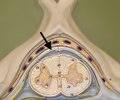
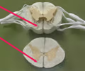
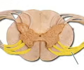
dorsal root
• A division of the spinal nerves external to the spinal cord.
• Connects to the dorsal horns of the spinal cord.
• Contains sensory fibers.
• Also known as the posterior root.
• Connects to the dorsal horns of the spinal cord.
• Contains sensory fibers.
• Also known as the posterior root.
24
New cards
ventral root
• A division of the spinal nerves external to the spinal cord.
• Contains motor fibers.
• Also known as the anterior root.
• Contains motor fibers.
• Also known as the anterior root.
25
New cards
dorsal root ganglion
• A bulge of the dorsal root external to the spinal cord.
• Contains sensory neurosomas bringing information from the periphery to the spinal cord.
• Contains sensory neurosomas bringing information from the periphery to the spinal cord.
26
New cards
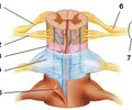
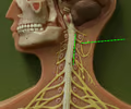
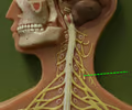
cervical plexus
• A branched network of spinal nerves C1-C5.
• Innervates muscles of the shoulder and neck.
• Gives rise to the phrenic nerve.
• Innervates muscles of the shoulder and neck.
• Gives rise to the phrenic nerve.
27
New cards
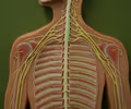
brachial plexus
• A branched network of spinal nerves C5-T1.
• Innervates muscles and skin of the shoulder and upper limbs.
• Gives rise to the axillary, radial, median, and ulnar nerves.
• Innervates muscles and skin of the shoulder and upper limbs.
• Gives rise to the axillary, radial, median, and ulnar nerves.
28
New cards
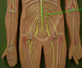
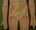
lumbar plexus
• A branched network of spinal nerves L1-L4.
• Innervates the lower abdominopelvic region and anterior thigh.
• Gives rise to the femoral nerve.
• Innervates the lower abdominopelvic region and anterior thigh.
• Gives rise to the femoral nerve.
29
New cards
sacral plexus
• A branched network of spinal nerves L4-S4.
• Innervates the rear pelvis. posterior side of thigh, and the lower limbs.
• Gives rise to the sciatic, common fibular, and tibial nerve.
• Innervates the rear pelvis. posterior side of thigh, and the lower limbs.
• Gives rise to the sciatic, common fibular, and tibial nerve.
30
New cards
phrenic nerve
• A nerve arising from the cervical plexus.
• Travels downward and innervates the diaphragm.
• Travels downward and innervates the diaphragm.
31
New cards
axillary nerve
• A nerve arising from the brachial plexus.
• Wraps around the head of the humerus.
• Wraps around the head of the humerus.
32
New cards
radial nerve
• A nerve arising from the brachial plexus.
• The lateral of the three major nerves innervating the upper limbs.
• The lateral of the three major nerves innervating the upper limbs.

33
New cards
median nerve
• A nerve arising from the brachial plexus.
• The intermediate of the three major nerves innervating the upper limbs.
• The intermediate of the three major nerves innervating the upper limbs.
34
New cards
ulnar nerve
• A nerve arising from the brachial plexus.
• The medial of the three major nerves innervating the upper limbs.
• The medial of the three major nerves innervating the upper limbs.
35
New cards
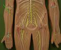
femoral nerve
• A nerve arising from the lumbar plexus.
• Travels anterior to the coxal bone and innervates the anterior thigh.
• Travels anterior to the coxal bone and innervates the anterior thigh.
36
New cards
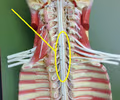
sciatic nerve
• A nerve arising from the sacral plexus.
• Travels down the posterior leg and divides into the common fibular and tibial nerves.
• Travels down the posterior leg and divides into the common fibular and tibial nerves.
37
New cards
common fibular nerve
• A nerve arising from the sacral plexus.
• The lateral of the two nerves innervating the lower leg.
• Best seen on the posterior side.
• The lateral of the two nerves innervating the lower leg.
• Best seen on the posterior side.
38
New cards
tibial nerve
• A nerve arising from the sacral plexus.
• The medial of the two nerves innervating the lower leg, travelling along the tibia.
• Best seen on the posterior side.
• The medial of the two nerves innervating the lower leg, travelling along the tibia.
• Best seen on the posterior side.
