Pathology
1/70
There's no tags or description
Looks like no tags are added yet.
Name | Mastery | Learn | Test | Matching | Spaced | Call with Kai |
|---|
No analytics yet
Send a link to your students to track their progress
71 Terms
What is liver agenesis?
Absence of liver formation
What is partial situs inversus?
When abdominal contents are reversed
What is complete situs inversus?
When chest and abdominal contents are reversed
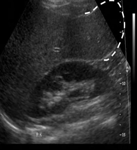
What is a Reidel lobe?
When right lobe of live extends below lower pole of right kidney
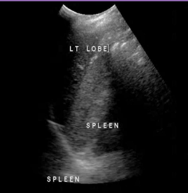
What is an elongated left lobe?
When left lobe of liver extends into LUQ
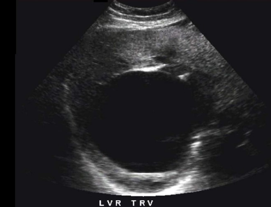
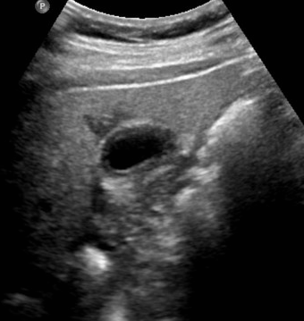
What is a hepatic cyst?
Fluid filled space with an endothelial lining
What is a hemorrhagic hepatic cyst?
When blood leaks into an existing cyst due to hemorrhage or infection and creates a complex appearance
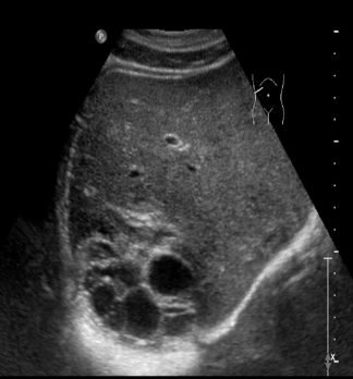
What is polycystic liver disease (PCLD)?
Prescence of multiple cysts that do NOT communicate with biliary tree
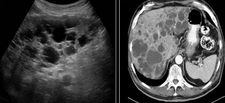
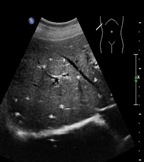
Identify this image.
Polycystic liver disease (PCLD)
What is a biliary hamartoma or von Meyenburg Complexes?
Dilated intrahepatic ducts that appear as small echogenic nodules with ringdown and twinkle artifact
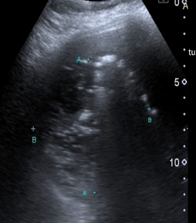
Identify this image.
Biliary hamartoma or von Meyenburg Complexes
What is fatty infiltration of the liver or hepatic steatosis?
Triglyceride accumulation in liver cells most commonly caused by obesity
What is alcoholic fatty liver disease?
Fatty liver caused by chronic alcohol intake that can lead to alcoholic hepatitis
What is the most common chronic liver disease in Western countries?
Nonalcoholic fatty liver disease
What is nonalcoholic fatty liver disease?
Ftty liver caused by diabetes, obesity, and TB that can lead to nonalcoholic steatohepatitis (NASH)
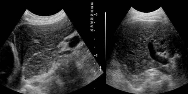
What is the sonographic appearance of fatty liver disease or hepatic steatosis?
Increase in liver echogenicity
Hepatomegaly
Rounding of inferior liver border
Fatty sparing appears as focal areas of normal echogenicity near GB
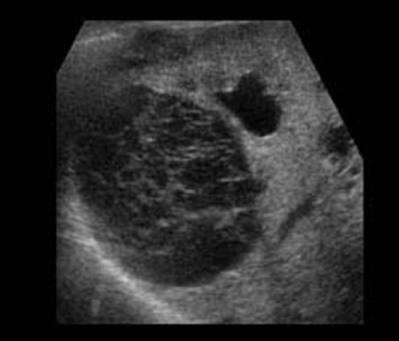
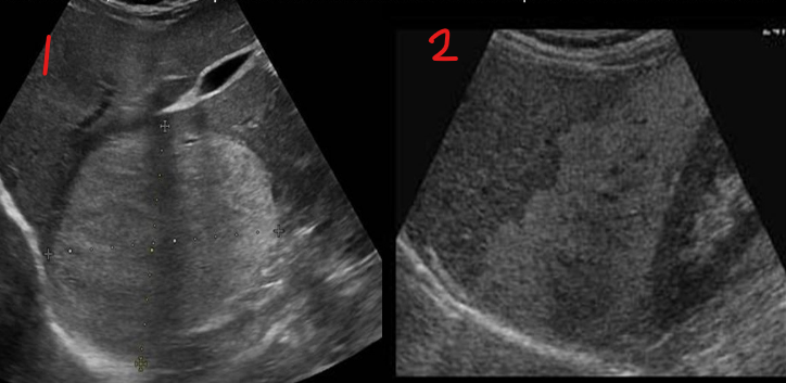
Identify this image.
Mild fatty liver
Identify this image.
Severe fatty liver
Identify this image.
Fatty sparing
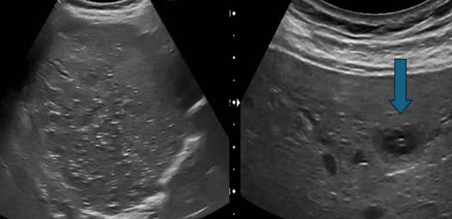
Identify this image.
Hemangioma seen with “mass effect” on other structures and vessels
Fatty infiltration seen with no displacement of other tissues
What is Reye Disease?
Disease associated with aspirin use during viral infection that causes excessive fat accumulation in liver with an acute increase in cranial pressure
What is amyloid disease?
Deposition of amyloid protein in vessel walls leading to organ failure
What is glycogen storage or von Gierke disease?
Excessive or reduced storage of glycogen within hepatocytes
What is the sonographic appearance of excessive glycogen storage or von Gierke disease?
Increased liver echogenicity
Hepatomegaly
Adenoma formation
What is the sonographic appearance of reduced glycogen storage or von Gierke disease?
Decreased liver echogenicity
Prominent appearance of portal walls
“Starry sky” appearance
What is hemochromatosis?
Abnormal iron deposition in organs that leads to fibrosis and cirrhosis with NO change in liver reflectivity
What is Wilson disease?
Excessive deposition of copper in liver and brain that leads to hepatic dysfunction, hepatitis, and cirrhosis
What are the symptoms associated with Wilson disease?
Jaundice
Hematemesis
Portal HTN
Ascites
Kayer-Fleischer rings or brown colored rings around iris of eyes
What is the sonographic appearance of Wilson disease?
Increased liver echogenicity
Hepatomegaly
Fibrotic periportal thickening
Nodular cirrhotic changes
What is the difference between fatty liver disease and cirrhosis?
Fatty liver disease is reversible while cirrhosis is irreversible
What is cirrhosis?
Diffuse liver disease caused most commonly caused by alcoholism and hepatitis C
Cirrhosis is associated with…
Hepatocellular carcinoma (HCC)
What are the symptoms of cirrhosis?
Increased abdominal girth from ascites
Jaundice
Weight loss
What is the sonographic appearance of early cirrhosis?
Increased liver echogenicity
Hepatomegaly
Heterogenous parenchyma
What is the sonographic appearance of late stage cirrhosis?
Decreased liver size commonly sparing caudate lobe
Coarse texture
Nodule formation (micro < 1 cm, macro 1 - 5 cm)
Ascites
Splenomegaly
Portal HTN or thrombosis
Narrowed hepatic veins
Identify this image.
Cirrhosis
What is the most common cause of intrahepatic cholestasis?
Hepatitis
What is hepatitis?
Inflammation of liver most commonly spread by IV drug users
What is the most common chronic bloodborne infection in the US?
Hepatitis C
What are the different types of hepatitis?
Hepatitis A: Fecal-oral
Hepatitis B: Blood-borne
Hepatitis C: Blood-borne
Hepatitis D, E, G are less common
What is alcoholic hepatitis?
Acute liver inflammation caused by prolonged heavy alcohol abuse
What is the sonographic appearance of acute alcoholic hepatitis?
Diffuse increase in liver echogenicity
Hepatomegaly
CHA PSV > 100 cm/sec
Dilated CHA
What is the sonographic appearance of chronic alcoholic hepatitis?
Atrophied liver with micronodular cirrhosis
What is nonalcoholic steatohepatitis (NASH)?
Consequence of nonalcoholic fatty liver disease
What is fulminant hepatitis?
Inflammation that leads to acute liver failure caused by acetaminophen overdose
What is the sonographic appearance of acute hepatitis?
Decreased liver echogenicity
Hepatomegaly
“Starry sky” appearance
Ascites
Cholecystitis

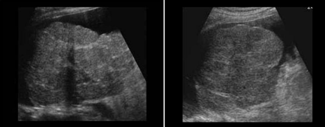
Identify this image.
Acute hepatitis
Identify this image.
Acute hepatitis
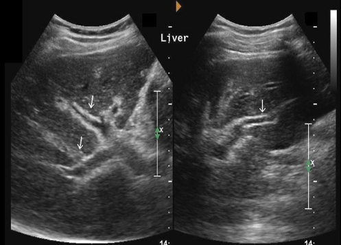
What is chronic hepatitis?
Persistence of hepatitis infection for more than 6 months
What is the sonographic appearance of chronic hepatitis?
Diffuse increase in liver echogenicity
Atrophied liver
Decreased echogenicity of portal vein walls
Granuloma formation
Identify this image.
Chronic hepatitis
What is a echinococcal or Hydatid cyst?
Cysts formed from tapeworms found in feces of infected animals
What is the sonographic appearance of a echinococcal or Hydatid cyst?
Early stages seen as cyst with debris or Hytadid sand
Late stage honeycomb sign seen as fluid collections with septa
Late stage water lily sign seen as fluid collection with split wall
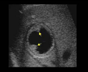
Identify this image.
Early stage echinococcal or Hydatid cyst
Identify this image.
Late stage echinococcal or Hydatid cyst
What is the most common parasitic infection in humans?
Schistosomiasis
What is schistosomiasis?
Periportal fibrosis caused by parasitic infection from polluted waters or shellfish
What is the sonographic appearance of schistosomiasis?
Periportal fibrosis seen as thick, echogenic portal vein walls
Bull’s eye sign with portal veins
Identify this image.
Schistosomiasis
What is pneumocystis Jirovecci or Carinii?
Yeast-like fungus that commonly affects AIDS patients
What is the sonographic appearance of pneumocystis Jirovecci or Carinii?
Multiple non-shadowing echogenic foci within liver tissue
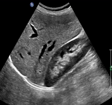
What are granulomatous infections?
Fungal respiratory disease caused by histoplasmosis and TB
Identify this image.
Granulomas
What is the most common form of liver abscess?
Pyogenic abscess
What is the most common cause of a pyogenic abscess?
Biliary disease
What is a pyogenic abscess?
Bacterial infection located in right lobe of liver
What is the sonographic appearance of a pyogenic abscess?
Complex cystic mass
Cluster sign seen as cluster of small abscesses
Reverberation or ringdown artifact
What is a fungal abscess or candidiasis?
Fungal infection seen in immunocompromised patients that causes a LOW WBC
What is the sonographic appearance of a fungal abscess or candidiasis?
Wheel within wheel
Hypoechoic ring around hyperechoic center
What is an amebic abscess?
Parasitic infection commonly located in right dome of liver
What is the sonographic appearance of an amebic abscess?
Round mass with indistinct walls
Low level internal echoes
Elevated diaphragm
Echogenic foci