ap II lab- types of WBCs (exam 1)
1/7
There's no tags or description
Looks like no tags are added yet.
Name | Mastery | Learn | Test | Matching | Spaced | Call with Kai |
|---|
No analytics yet
Send a link to your students to track their progress
8 Terms
platelets always stain
dark (dark red, or purple)
how can you tell the difference between RBC’s and WBC’s
look at the nucleus- RBC’s dent have a nucleus
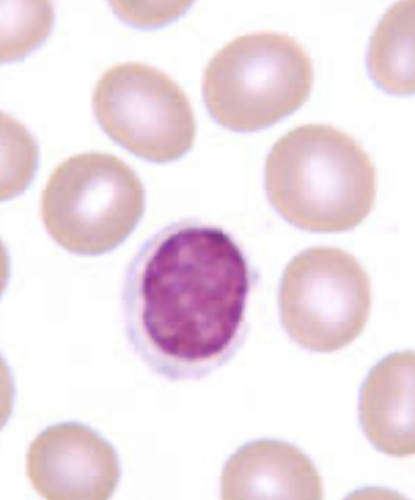
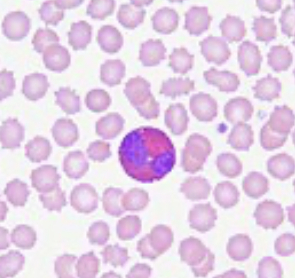
nucleus fills up almost the whole cell and leaves a sliver of cytoplasm
dont confuse with basophil (these are smooth)
lymphocyte
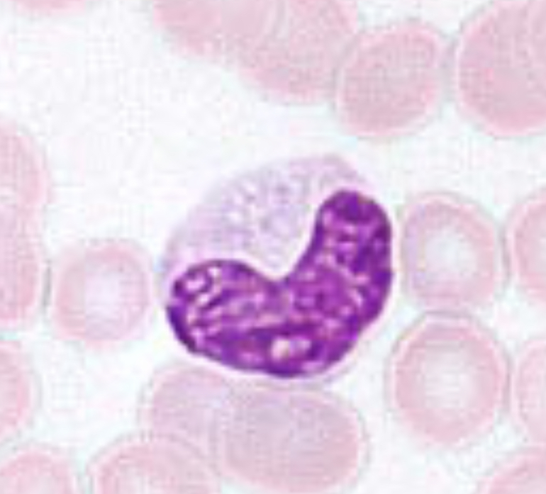
nucleus is horseshoe or kidney bean shaped
nucleus can spin so it might be “c” or “n” shaped
dont confuse with neutrophil… look for the UNIFORM thickness of this nucleus
monocyte
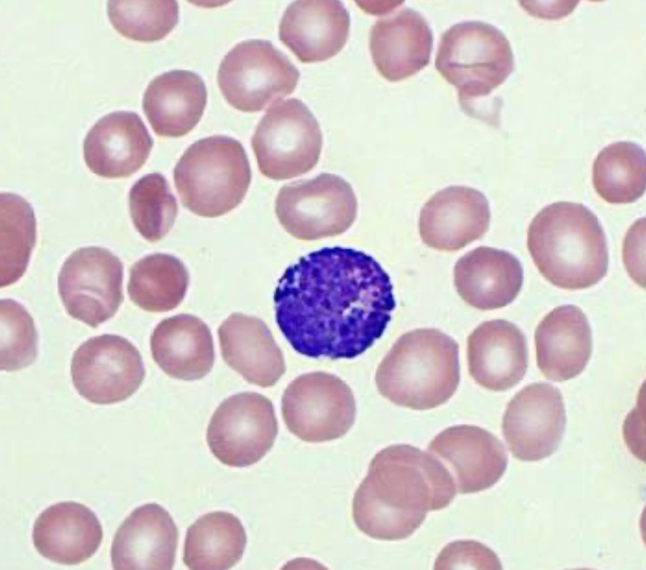
the nucleus looks like a chain of sausages “thick-thin-thick-thin”
no 2 of these look exactly alike
somethings the thin area of the sausage might not stain so the sausage looks detached
dont confuse with monocyte… look at the thickness of nucleus sausage
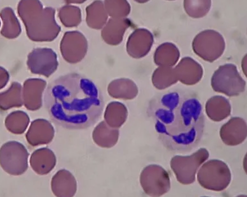
neutrophil
full of granules- can’t see the nucleus because the granules cover it
lumpy texture
bumpy on the sides of the cell because of granules
dont confuse with lymphocyte
basophil
bilobed nucleus (looks like lungs)
2 dark sections in its nucleus
has orange/ red granules in the cytoplasm
eosinophils
order of abundance for WBCs
neutrophils, lymphocytes, monocytes, eosinophils, basophils
never let monkeys eat bananas