Exam 2 BIOl 223 Structure of skeletal muscle cells
1/132
There's no tags or description
Looks like no tags are added yet.
Name | Mastery | Learn | Test | Matching | Spaced |
|---|
No study sessions yet.
133 Terms
What is muscle tissue specialized for?
Contraction
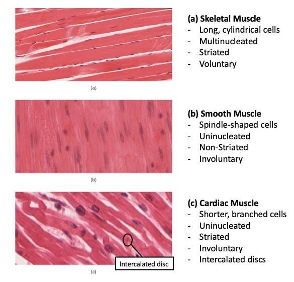
What are the 3 types of muscle tissue? Are they striated, nonstriated, voluntary, or involuntary?
Skeletal — attached to bone
Striated, voluntary
Cardiac — Found in the heart
Striated, involuntary
Smooth — Lines hollow organs
Nonstriated, involuntary
What type of muscle tissue lines hollow organs?
Smooth muscle tissue

What are the functions of the skeletal muscle?
Produce skeletal movement
Maintain posture & body position
Support soft tissues
Guard entrances & exits
Maintain body temp
Nutrient reserves
T/F: The skeletal muscle produces skeletal movement.
T
T/F: The skeletal muscle maintains posture and body position.
T
T/F: The skeletal muscle supports hard tissues.
F, they support soft tissues
T/F: The cardiac muscle guides entrances & exits.
F, that is a job for the skeletal muscle.
T/F: The skeletal muscle can help maintain body temp.
T
T/F: The skeletal muscle helps with nutrient reserves.
T
Regarding the gross anatomy of skeletal muscle, what is the origin? What is the insertion?
Origin – attached to bone that remains relatively stationary during movement
Insertion — attached to the bone that moves
What are the different parts of antagonistic muscles?
Flexors (close) and extensors (open)
What is the exception to the rule that most muscles require an opposing muscle to reverse an action?
Circular sphincter muscles. While most muscles require an opposing muscle to reverse an action, a sphincter performs both actions—opening and closing—on its own.

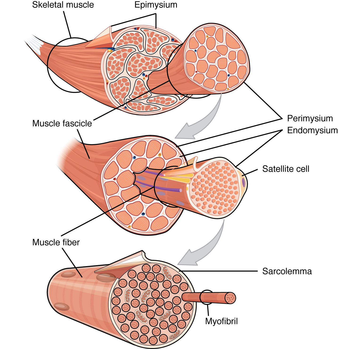
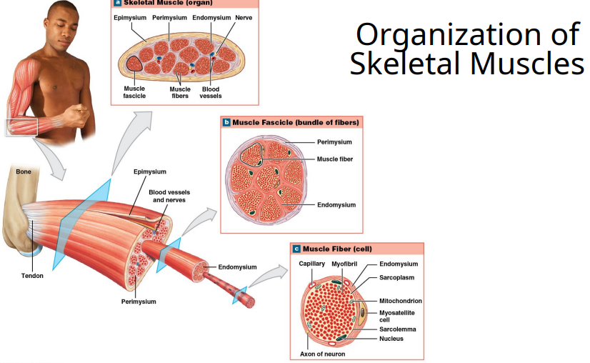
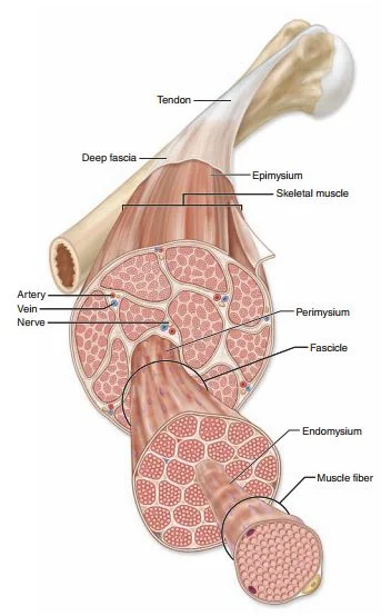
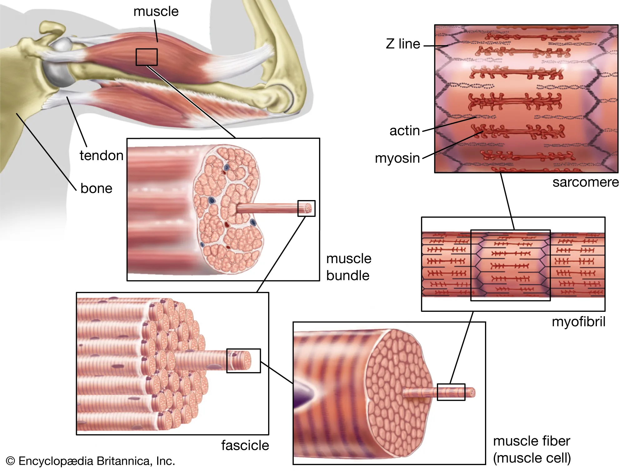
What are the different types of connective tissues in skeletal muscles and what they do?
• Endomysium - covers individual muscle fibers (muscle cells)
• Perimysium - sheathes bundles of muscle fibers (muscle fasicle)
• Epimysium - surrounds a muscle (endomysium and perimysum contain blood vessels and nerves)
• Deep fascia – wrap groups of cooperating muscles together
What is endomysium?
• Endomysium - covers individual muscle fibers (muscle cells)
What is perimysium?
• Perimysium - sheathes bundles of muscle fibers (muscle fasicle)

What is epimysium?
• Epimysium - surrounds a muscle (endomysium and perimysium contain blood vessels and nerves)
What is deep fascia?
• Deep fascia – wrap groups of cooperating muscles together

endomysium and perimysium contain
blood vessels and nerves
T/F: Muscle cell is a synonym for muscle fiber.
T
T/F: Muscle cell is not the same thing as muscle fiber.
F. Muscle cell is a synonym for muscle fiber.
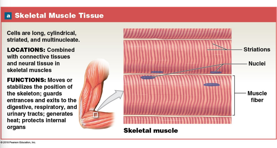
A skeletal muscle cell is multinucleate, meaning it is
A very long cell. Each muscle cell is as long as the muscle.
When is the skeletal muscle cell formed?
Formed during embryogenesis by end-to-end fusion of uni-nucleate myoblasts
Why is adult muscle repair limited?
New skeletal muscle cells come from stem cells called satellite cells.
Once skeletal muscle fibers are formed, they are multinucleated and very large, meaning they’re not able to undergo mitosis (they can’t divide and make new cells). So, if a muscle fiber is damaged, it can’t just “make more of itself.”
Even though mature muscle fibers can’t divide, there are a few special cells called satellite cells that sit just outside the muscle fiber, under its protective layer (the basal lamina).
When injury happens:
These satellite cells are activated.
They divide (mitosis) and differentiate into new muscle cells or fuse with damaged fibers to help repair them.
Your teacher’s phrase —
“New skeletal muscle cells come from stem cells called satellite cells” —
is key because:
👉 There aren’t very many satellite cells in adult muscle.
👉 As you age, their number and activity decrease.
👉 They can only handle small injuries — not large-scale muscle loss or chronic damage.
Myo & Sarco indicate what type of muscle cell?
skeletal muscle cell
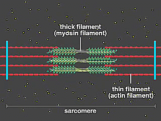
The structure of skeletal muscle fiber contains large quantities of protein filaments called ____, which are composed of what 2 components?
myofilaments
Actin are THIN myofilaments
Myosin are THICK myofilaments

What are myofibrils (skeletal muscle fiber)?
bundles of myofilaments (which are composed of actin and myosin)
Is actin a thick or thin myofilament?
Actin are THIN myofilaments
Myosin are THICK myofilaments
Is myosin a thick or thin myofilament?
Actin are THIN myofilaments
Myosin are THICK myofilaments

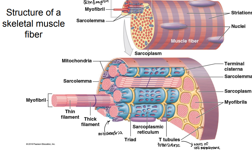
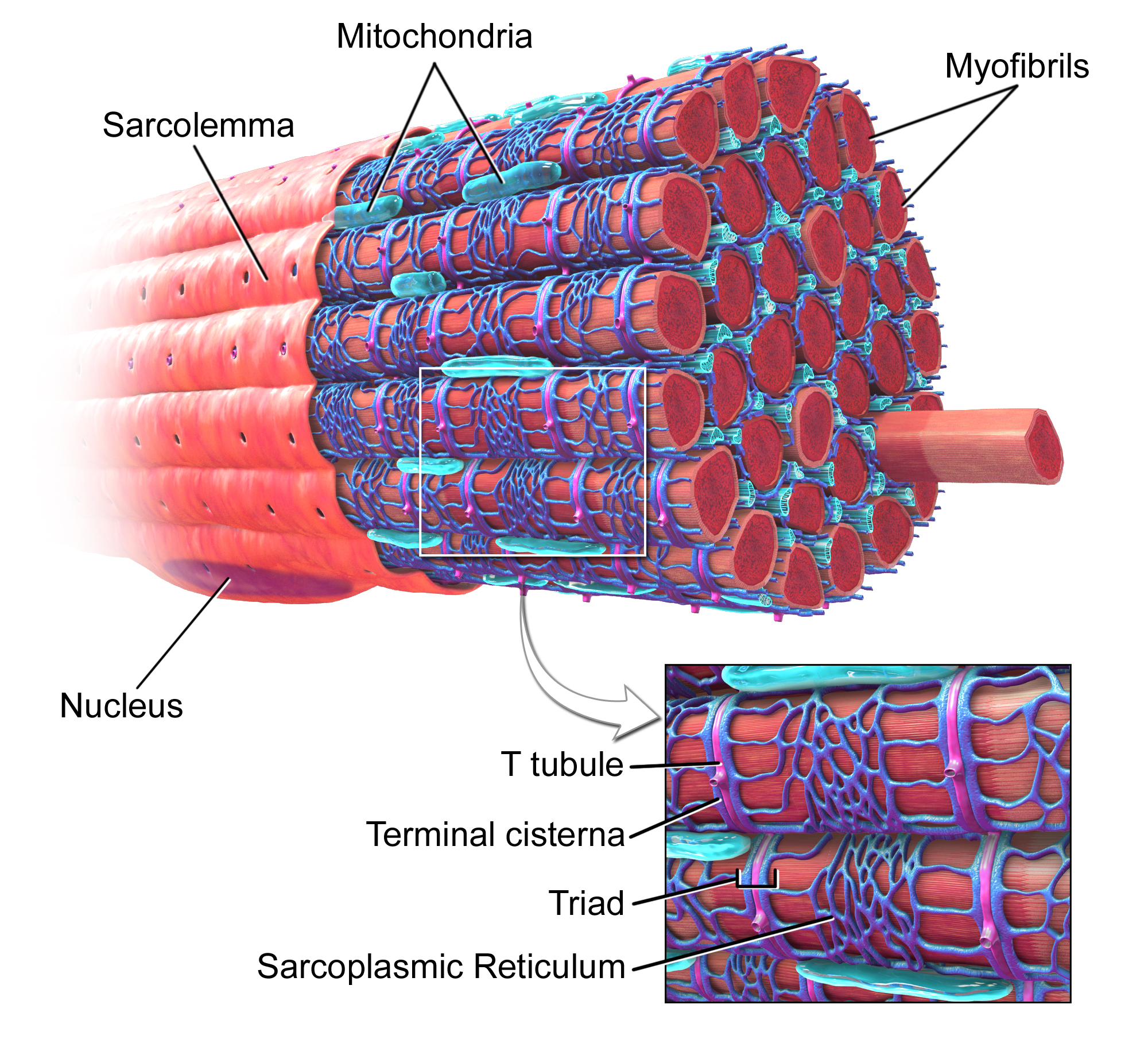
What is the sarcoplasm?
skeletal muscle cell cytoplasm

What is the sarcolemma?
skeletal muscle cell membrane

What is the sarcoplasmic reticulum?
skeletal muscle cell modified ER

The sarcolemma (muscle cell membrane) is an excitable membrane, which means it conducts action potentials. Narrow tubes of sarcolemma extend into the cell at right angles to the cell surface. What are these tubes called? What are their functions?
Transverse Tubules (T-tubules)
Transverse tubules conduct action potential deep into the cell, and comes in close contact with the sarcoplasmic reticulum.
It can also be thought of an extension of a cell membrane.
Here’s how it works:
Sarcolemma receives the electrical signal (action potential) from a neuron. (ach)
The signal travels along the sarcolemma and then down the T-tubules (inward tunnels).
The T-tubules carry the signal deep into the muscle fiber.
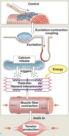
This signal reaches the sarcoplasmic reticulum, which releases its storage of Ca²⁺.
Calcium triggers the muscle contraction due to depolarization.

Depolarization is the process where a cell's membrane potential becomes less negative (more positive) due to the influx of positive ions, which does what to the muscle?
muscle contraction or nerve signal generation
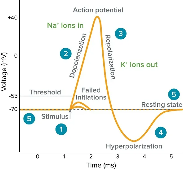
Repolarization is the opposite process of depolarization, where positive ions flow out of the cell to return the membrane potential to its negative resting state, allowing the cell to
relax or reset for another signal

What is the sarcoplasmic reticulum and its function?
Similar to smooth ER
Forms a tubular network around each myofibril
Terminal cisternae form triads with T tubules
Stores a high concentration of Ca+ ions needed for muscle contraction
Fill in the blanks!!
______ receives the electrical signal (action potential) from a neuron.
The signal travels along the _____ and then down the _______ (inward tunnels).
The _______ carry the signal deep into the _______ _______ .
This signal reaches the _______ _______ , which releases its storage of ___.
_______ triggers the_______ _______ due to depolarization.
Sarcolemma receives the electrical signal (action potential) from a neuron.
The signal travels along the sarcolemma and then down the T-tubules (inward tunnels).
The T-tubules carry the signal deep into the muscle fiber.
This signal reaches the sarcoplasmic reticulum, which releases its storage of Ca²⁺.
Calcium triggers the muscle contraction due to depolarization.
What does this picture represent.
Thick myosin (myofilament) and thin actin (myofilament)
Where are myofibril, which are bundles of myofilaments, located?
They are anchored to inner surface of sarcolemma at either end of cell.
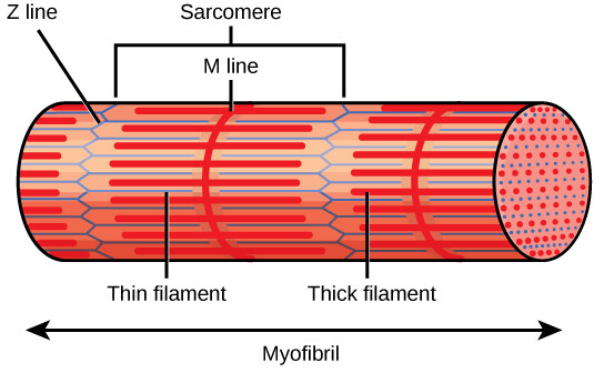
What are sarcomeres?
repeating units of myofilaments in myofibril.
It can actively shorten.
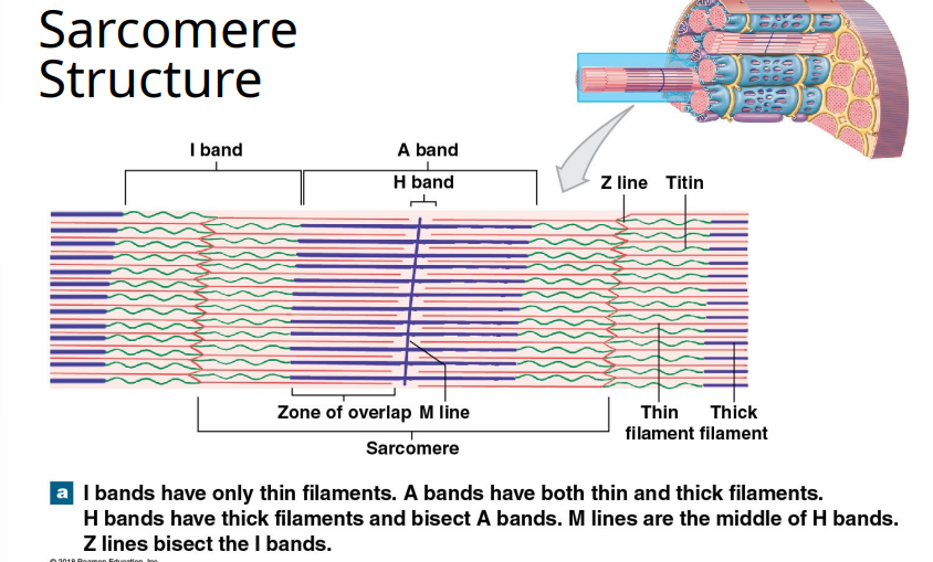
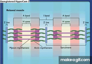
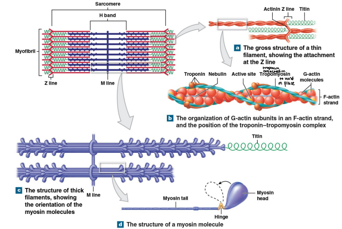
Differences in distribution of thick & thin myofilaments gives a banded appearance in striated sarcomeres. Describe the bands of the sarcomere structure.
I bands – LIght band
contains only thin filaments
A bands – DArk band
contains thick filaments, and some overlap with thin filaments
H band contains only thick filaments
Z disk (line) – border between sarcomeres

What are the levels of functional organization in skeletal muscle fiber?
Skeletal muscle → Muscle fasicle → muscle fiber → myofibril → sarcomere

The skeletal muscle is surrounded by ____ and contains ____ __
epimysium; muscle fasicles

The muscle fasicle is surrounded by ____ and contains ____ __
perimysium; muscle fibers
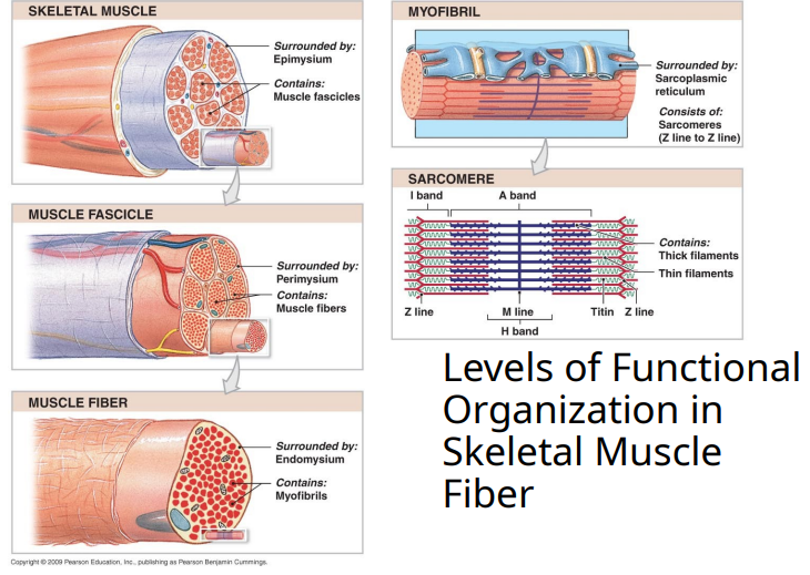
The muscle fibers is surrounded by ____ and contains ____
endomysium; myofibrils

The myofibrils is surrounded by ____ ____ and consists of ____
sarcoplasmic reticulum; sarcomeres (Z line to Z line)

What do sarcomeres contain?
Thick and thin filaments
Order of organization in “mysiums” go in what order
Epimysium (skeletal muscle)
Perimysium (muscle fasicicle)
Endomysium (muscle fiber)

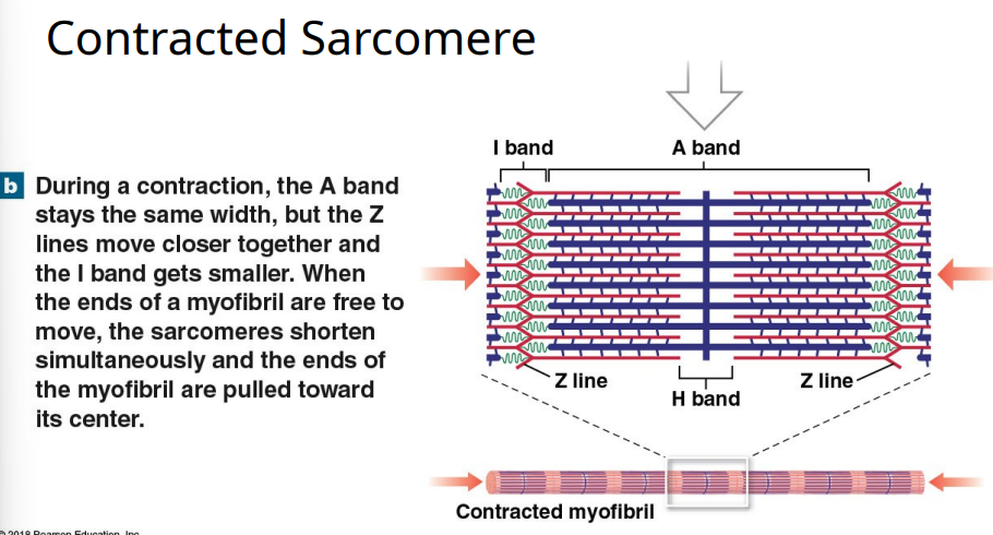
Muscles shorten during contraction (as seen above) because
Muscles shorten during contraction because actin (thin) filaments slide over myosin (thick) filaments, pulling the Z lines closer together and shortening each sarcomere — the sliding filament theory of contraction.
The myosin and actin filaments slide between each other to shorten each sarcomere
Myofibril in muscle cell consists of thousands of ____ end to end
sarcomeres
Interactions between thin filaments (actin) and thick filaments (myosin) are responsible for
muscle contraction
Thin filaments (Actin) slide over thick filaments (myosin), shortening the
sarcomere
Shortening occurs in every sarcomere in the myofibril, thus
shortens the myofibril
Describe the Sliding Filament Model of Muscle Contraction.
Thin actin filaments – attached to the Z disk: Each sarcomere (the functional unit of a muscle fiber) is bordered by Z disks (or Z lines).
The thin filaments made of actin are anchored to these Z disks and extend toward the middle of the sarcomere.As thin filaments move toward the center of the sarcomere: When the muscle contracts:
The myosin heads on the thick filaments grab onto actin (thin filaments) and pull them inward toward the M line (the center of the sarcomere).
This pulling action uses ATP and repeats many times (cross-bridge cycling).
Thin filaments slide over thick filaments: The actin filaments slide past the myosin filaments — they don’t shorten, they just move.
Because they overlap more and more, the sarcomere itself shortens, and the muscle contracts.Z lines are pulled closer together: As actin slides inward:
The Z lines at each end of the sarcomere are drawn closer together.
That means the entire sarcomere shortens, and since sarcomeres are arranged end to end, the whole muscle fiber shortens.
I bands and H band narrow: I band and H band both get smaller during contraction.
A band stays the same width: Since myosin filaments don’t shorten, the A band length stays constant throughout contraction.
Sarcomere is at maximum shortening when it’s the width of the A band, no I band or H band are visible.
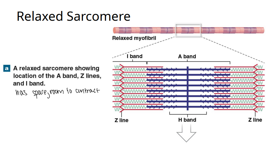
A relaxed sarcomere has spare room to ____.
contract
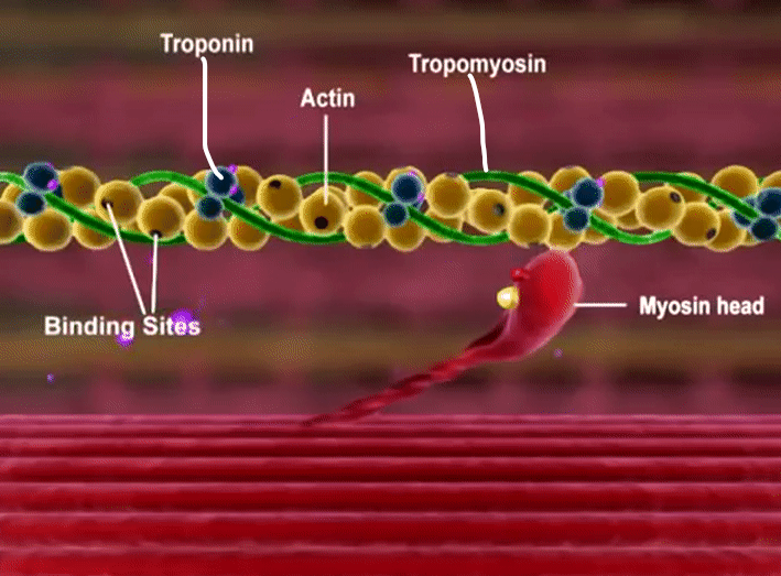
What causes thin & thick myofilaments to slide across each other?
Myosin filaments have many short projections extending out from the filament
These projections can bind to sites on actin filaments, forming cross-bridges
Cross-bridges, once formed, change shape pulling the actin past the myosin
Cross bridges use energy of ATP to change shape and pull the actin - converting chemical energy to mechanical energy
just look at this beautiful diagram
yay

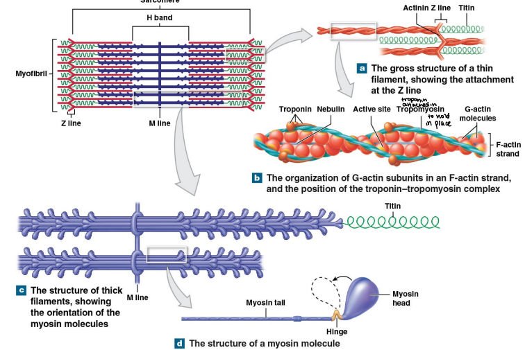
What is the molecular anatomy of thick myosin?
Composed of many (~100) identical myosin molecules bundled side by side, in staggered bipolar array
Myosin molecules have elongate tail, globular head – golf club shape
Arrayed with half facing each end, center is just tails
Heads form cross-bridges during
Interactions between myosin head and actin prevented by tropomyosin during rest
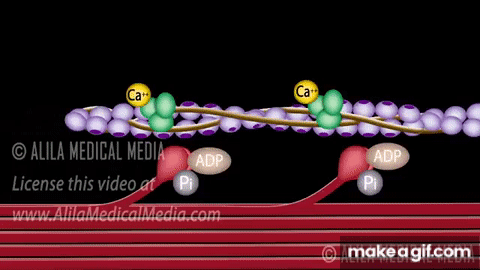
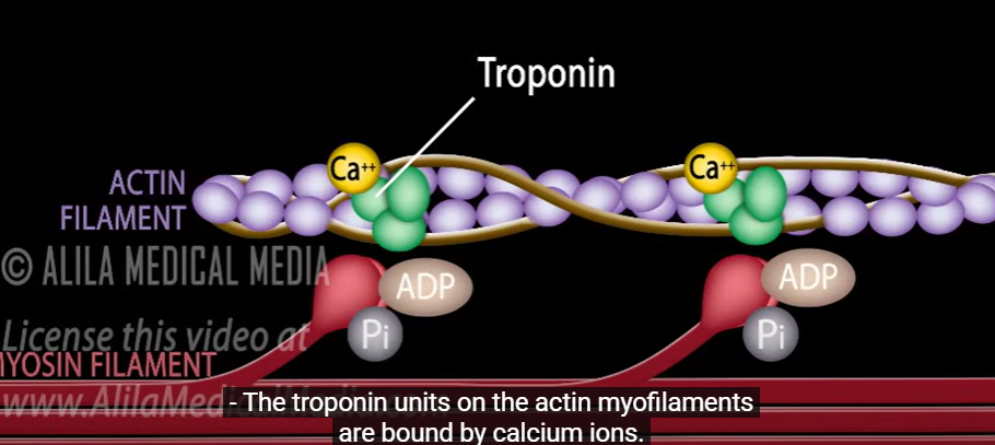
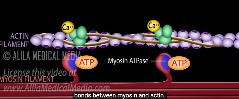
Describe the sliding filament theory.
Explains the relationship between thick & thin filaments as contraction proceeds.
Cyclic process beginning with calcium release from SR
Calcium binds to troponin
Troponin moves, moving tropomyosin and exposing the actin active site
Myosin head forms crossbridge to actin, bends toward the center of sarcomere, pulling the actin
ATP allows release of cross bridge
Draw a sarcomere.
good job
Myosin filaments have many short projections extending out from the filament. These projections can bind to sites on actin filaments, which then form
cross bridges.
What do cross bridges do once formed?
Change shape pulling the actin past the myosin
What is the energy source that cross bridges use to change shape and pull the actin - convert chemical energy to mechanical energy?
ATP duhh
Myosin is a golf club lookin ahhh because
The anatomy of it consists of an elongate tail and globular head
Muscle contraction is initiated when
muscle fibers are stimulated by a nerve impulse calcium ions are released
When do the myosin heads form cross-bridges? Why?
during muscle contraction; because interactions between myosin head and actin are prevented by tropomyosin during rest, and the tropomyosin covers the actin bonding site
Tropomyosin strands are attached to actin by
troponin
The sliding filament theory and its cyclic process begins with what release?
calcium ion release from SR (sarcoplasmic reticulum)
(First step) In the sliding filament theory, what does Ca bind to?
Troponin
(Second step) In the sliding filament theory, what happens after calcium ions bind to troponin?
The troponin moves, which moves tropomyosin and exposes the actin active site
(Third step) In the sliding filament theory, what happens after the calcium binds to troponin and troponin moves to move the tropomyosin to expose the actin active site?
The myosin head forms the cross bridge to actin, bends towards the center of the sarcomere, and pulls the actin.
(Fourth step) In the sliding filament theory, what allows the release of the cross bridge between myosin and actin?
ATP
What is the role of ATP in the molecular mechanism of contraction?
ATP supplies the energy for the movement of the myosin head, converting chemical energy to mechanical energy of movement.
What happens to the myosin head in an energized position?
It binds to an actin active site and releases ADP & P. It then pivots to pull on actin and moves to an unenergized state.
ATP binds to un-energized myosin head. What happens to the myosin?
Detaches myosin from actin. The ATP is then split and the head is energized again
What is the role of calcium ions in the molecular mechanism of contraction?
The concentration of Ca2+ around the sarcomere controls sarcomere contraction.
Ca2+ is low around the sarcomere at rest. The action potential in the sarcolemma and T-tubules then results in contractions, which causes the voltage-gated Ca2+ channels of the SR to open and release Ca2+ into the sarcoplasm around sarcomere.
T/F: Ca2+ is low around the sarcomere at rest.
T
t/F: Ca2+ is high around the sarcomere at rest.
F, it’s low
What happens when Ca2+ binds to troponin?
Causes troponin to change shape and pull tropomyosin off of actin active sites
When Ca2+ binds to troponin, it causes troponin to change shape and pull tropomyosin off of actin active sites. Then what happens?
The myosin heads bind to the now available actin active sites over and over until the Ca2+ level falls
When Ca2+ binds to troponin, it causes troponin to change shape and pull tropomyosin off of actin active sites. Then, the myosin heads bind to the now available actin active sites over and over until what?
the Ca2+ level falls. When the Ca2+ level falls, tropomyosin covers actin active sites, ending contraction
What ends muscle contraction?
When the Ca2+ level falls, tropomyosin covers actin active sites, ending contraction
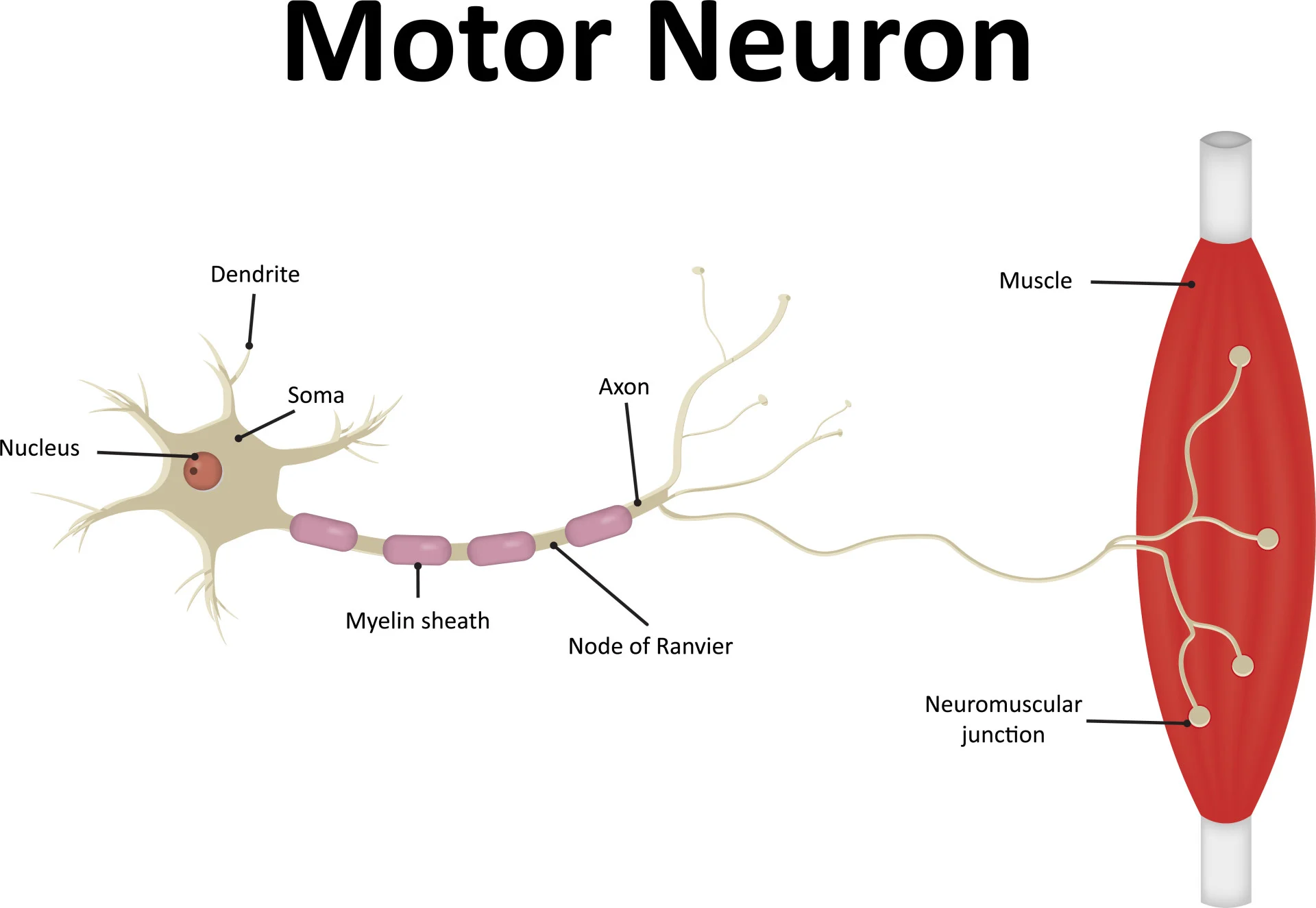
What is a motor neuron?
nerve cell that controls muscle contraction
What is a neuromuscular junction?
a synapse between a motor neuron and a muscle cell
A nerve cell that controls muscle contraction is called
motor neuron

A synapse between a motor neuron and a muscle cell is called
A neuromuscular junction

In response to central nervous system commands, what does the motor neuron initiate, and where does it travel?
the action potential gets initiated in the motor neuron in responsen to CNS commands, which travels through the motor neuron and arrives at the synaptic terminal

Control of skeletal muscle activity occurs where?
at the neuromuscular junction
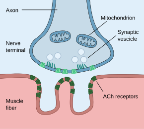
The action potential in a motor neuron causes what neurotransmitter to be released from the motor neuron terminal?
Acetylcholine (ACh)

The action potential in a motor neuron causes Acetylcholine (ACh) to be released from the motor neuron terminal. Where does the ACh go? What adventures must it traverse?
ACh diffuses across the synaptic gap
ACh binds to the receptors on chemically-gated sodium channels in muscle membrane

When sodium ions flow into the muscle cell, does it repolarize or depolarize the muscle cell membrane?
It depolarizes the muscle cell membrane and starts an action potential in the muscle cell.
This influx of positive charge causes the inside of the muscle cell membrane to become less negative (or more positive), a process called depolarization.
If the depolarization is strong enough to reach a certain threshold, it triggers an action potential, an electrical signal that travels along the muscle cell membrane and leads to muscle contraction.

An action potential is triggered in the muscle cell. Where does the electrical signal go after? What does it lead to?
The electrical signal travels along the muscle cell membrane and leads to muscle contraction.
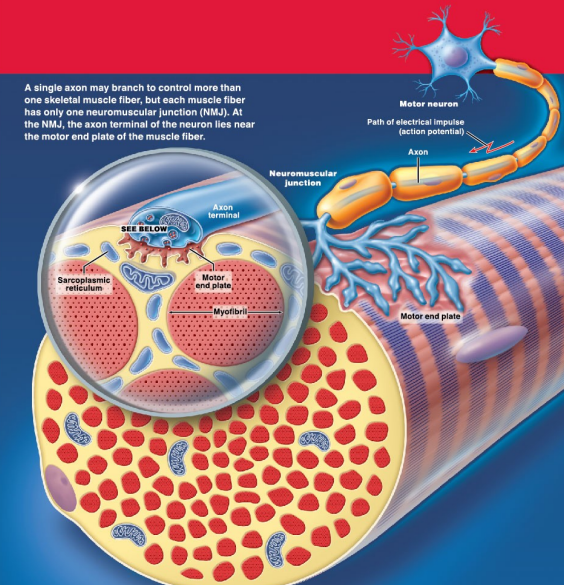
T/F: Each muscle fiber has only one neuromuscular junction.
T!
What has to be present for chemically regulated gates to stay open?
ACh
Chemically regulated gates stay open as long as
ACh is present
Where is acetylcholine esterase (AChE) located? What isits function in the control fo skeletal muscle activity?
Located in synaptic gap and rapidly breaks down Acetylcholine
What rapidly breaks down Acetylcholine?
acetylcholine esterase (AChE), located in the synaptic gap
When a muscle cell gets excited, what becomes initiated to spread across the entire muscle cell membrane (sarcollemma) including the T-Tubules?
Action potential
The excitation of a muscle cell leads to action potential, which leads to
The signal getting spread to the entire muscle cell membrane (sarcolemma), including the T-Tubules