ANAT 100 Module 12
1/71
There's no tags or description
Looks like no tags are added yet.
Name | Mastery | Learn | Test | Matching | Spaced | Call with Kai |
|---|
No analytics yet
Send a link to your students to track their progress
72 Terms
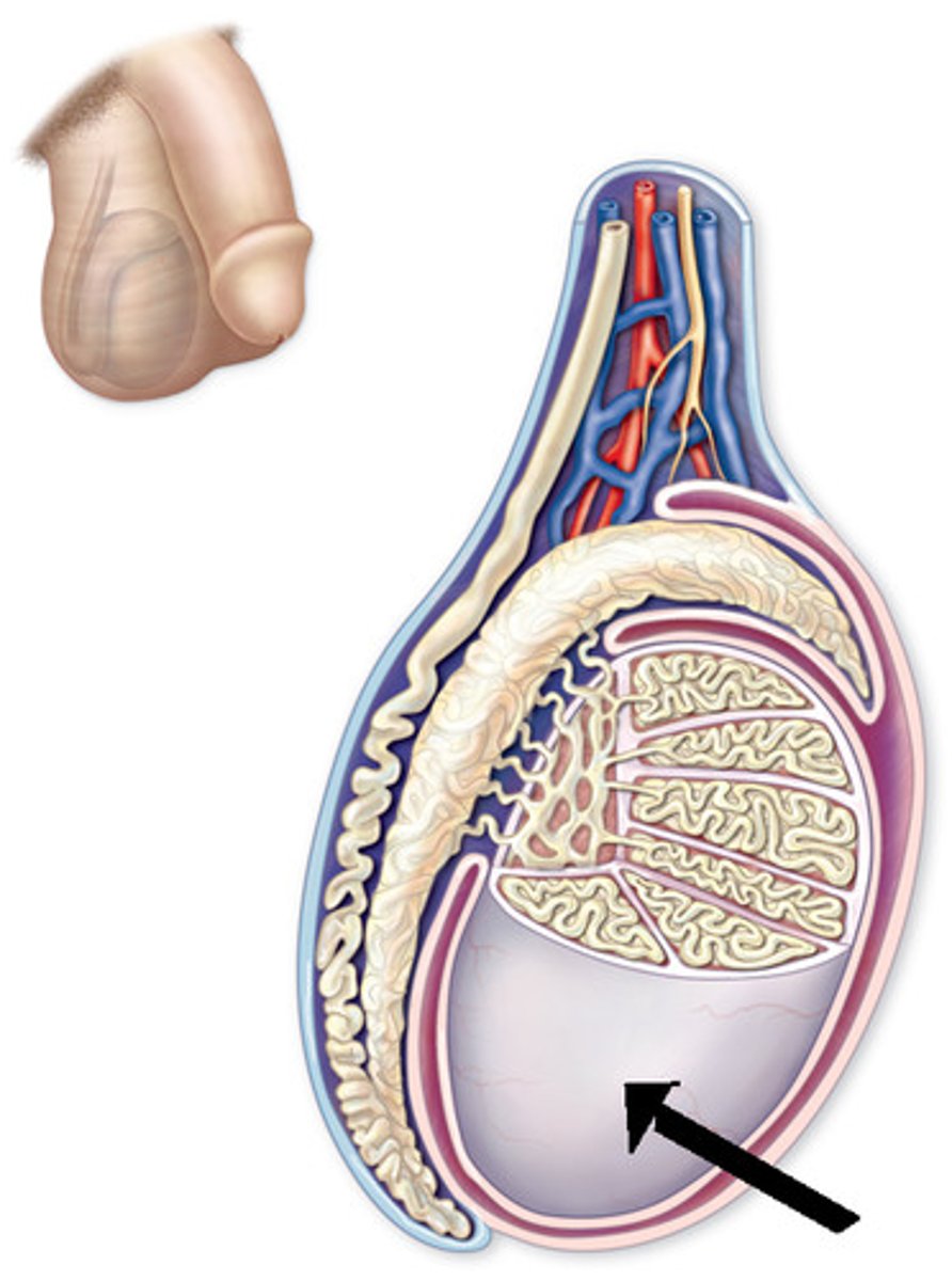
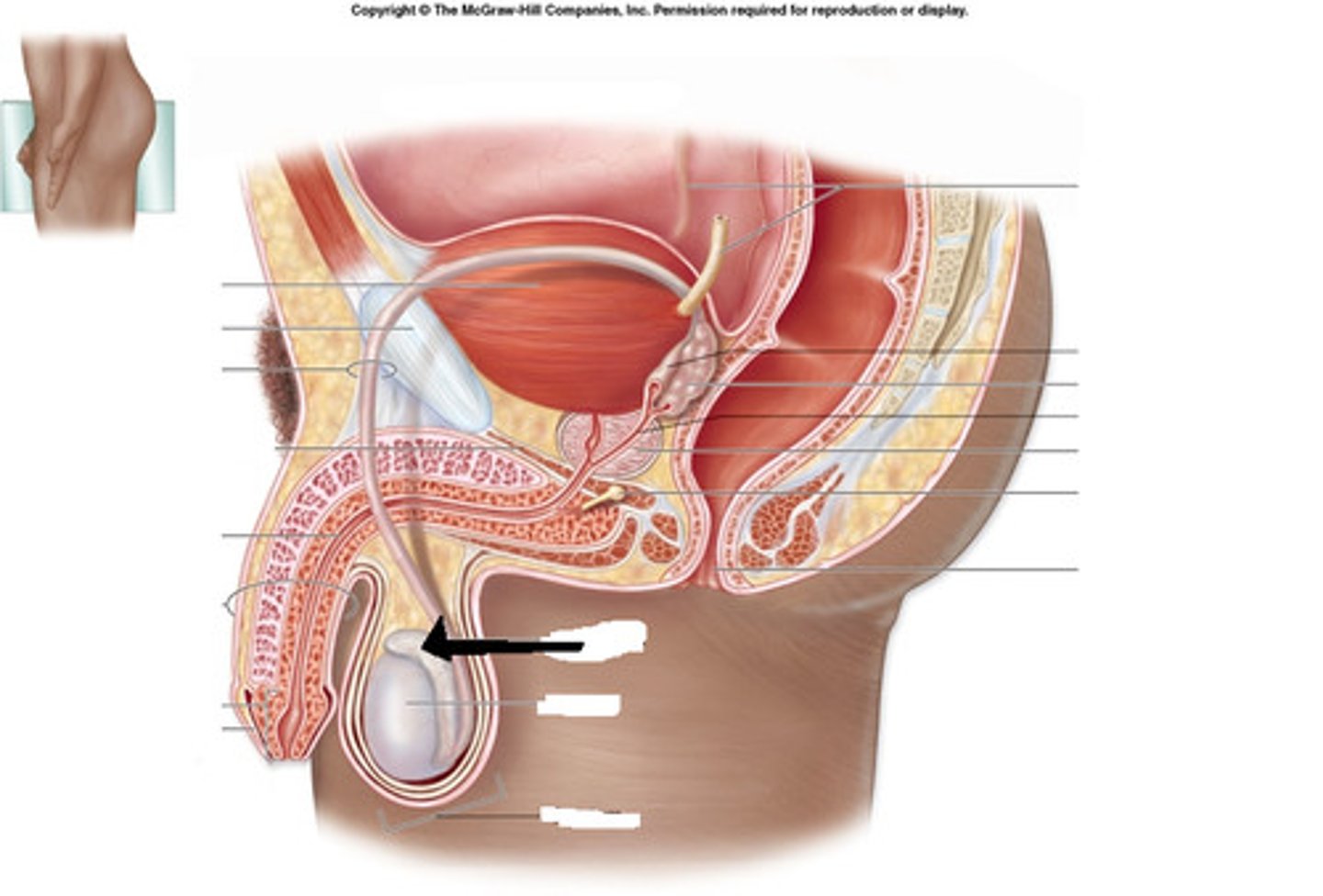
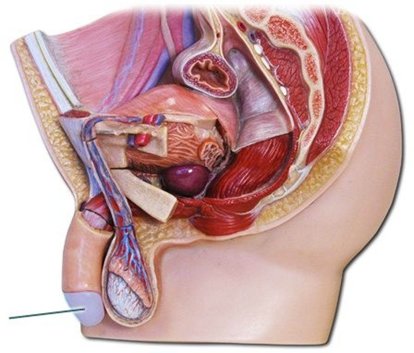
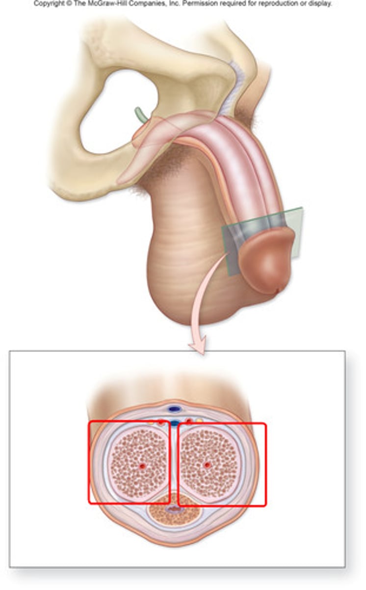
Testes
sperm creation and development
Tunica Vaginalis
outer protective covering of testes
Tunica Albuginea of Testes
inner fibrous capsule, extensions divide testes into lobules
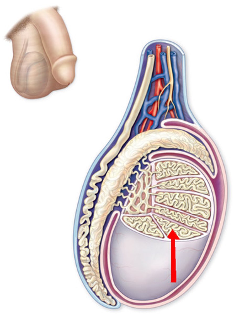
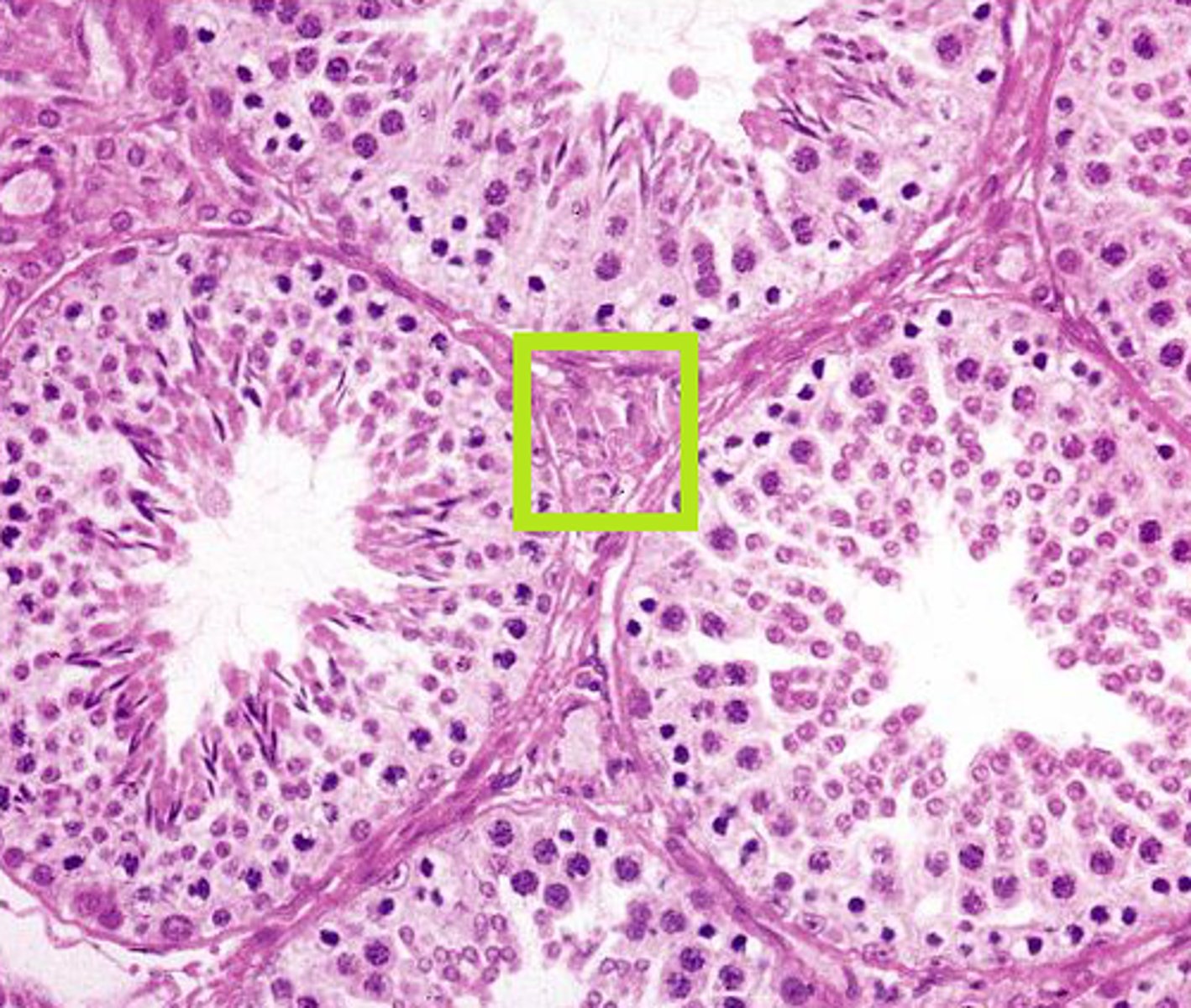
Seminiferous Tubules
tightly coiled tubules produce sperm
~600-1000/testes
Leydig cells
produce and secrete testosterone
found in loose connective tissue between seminiferous tubules
Head of sperm
has X or Y chromosome
acrosomal cap has enzymes for egg penetration
(A + B)

Midpiece of sperm
mitochondrial collar helps produce energy
©
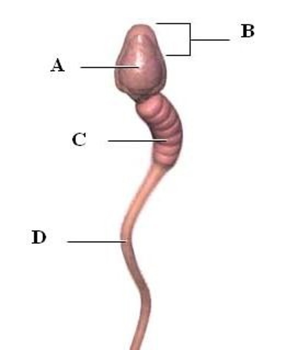
Tail of Sperm
flagellum is movement source
(D)

Scrotum
sac of skin and fascia around testes
controls temp of testes
derived from abdominal wall
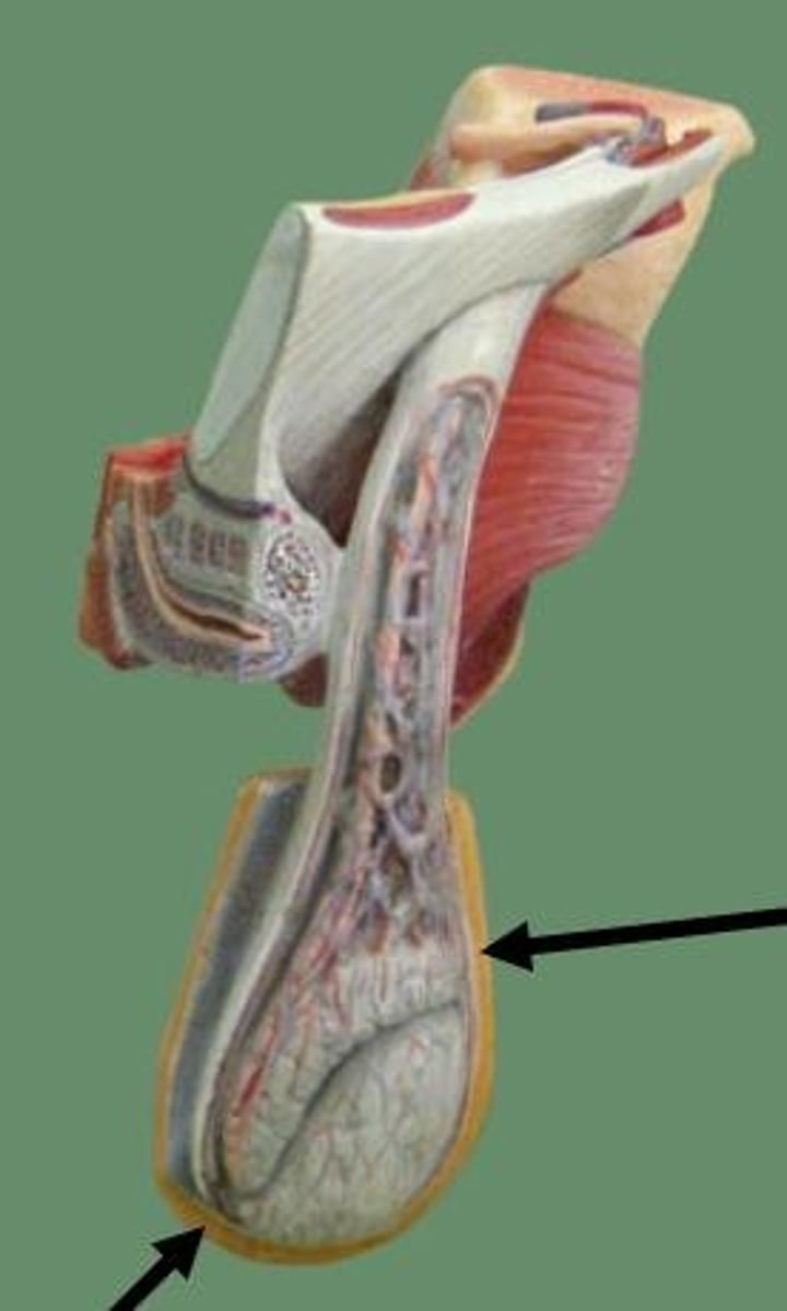
Dartos Muscle
regulates temp by changing exposed SA
superficial (in skin), gives wrinkly appearance
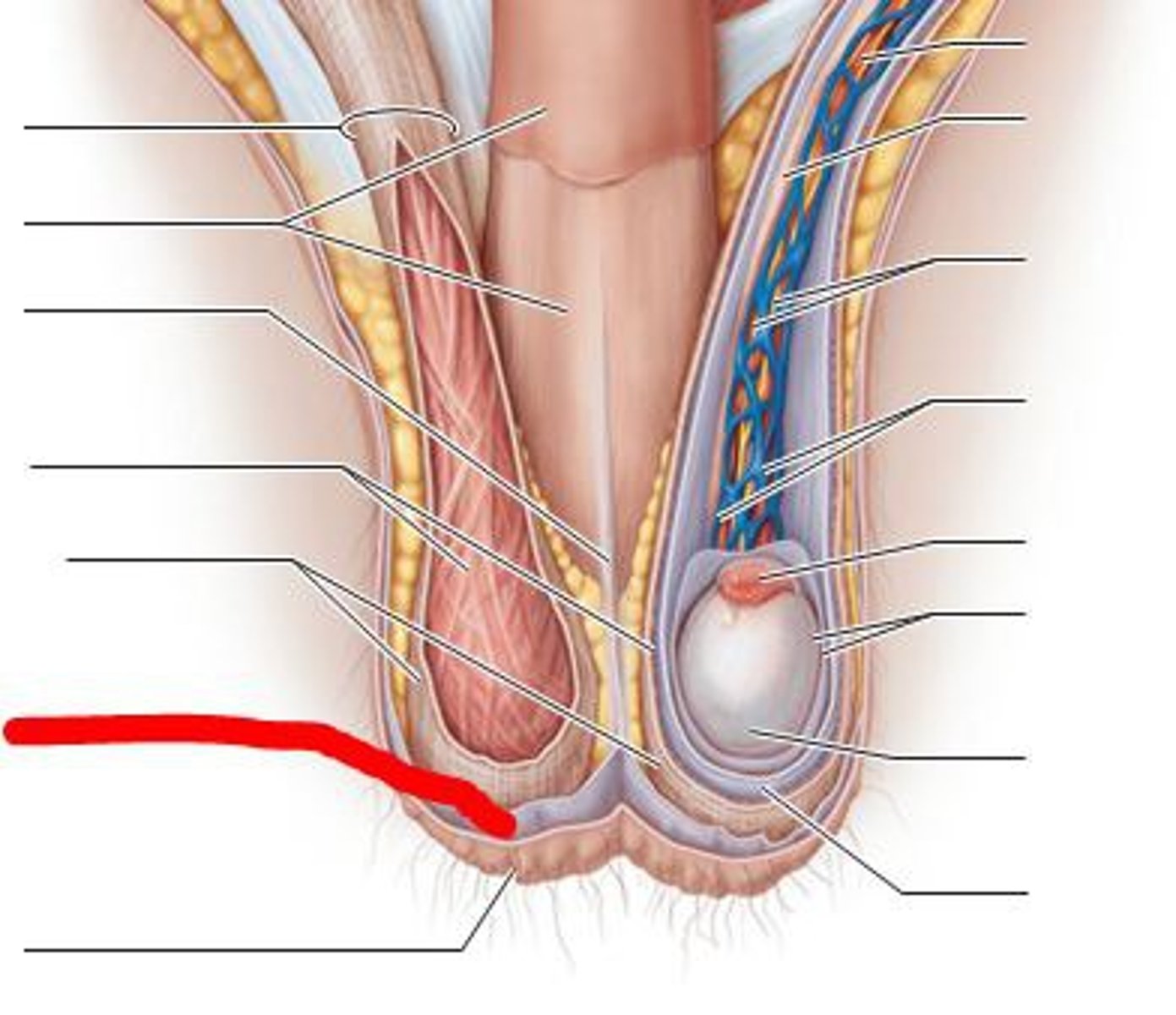
Cremastor Muscle
contracts in cold to bring testes superiorly (closer to body heat)
deep in scrotal wall
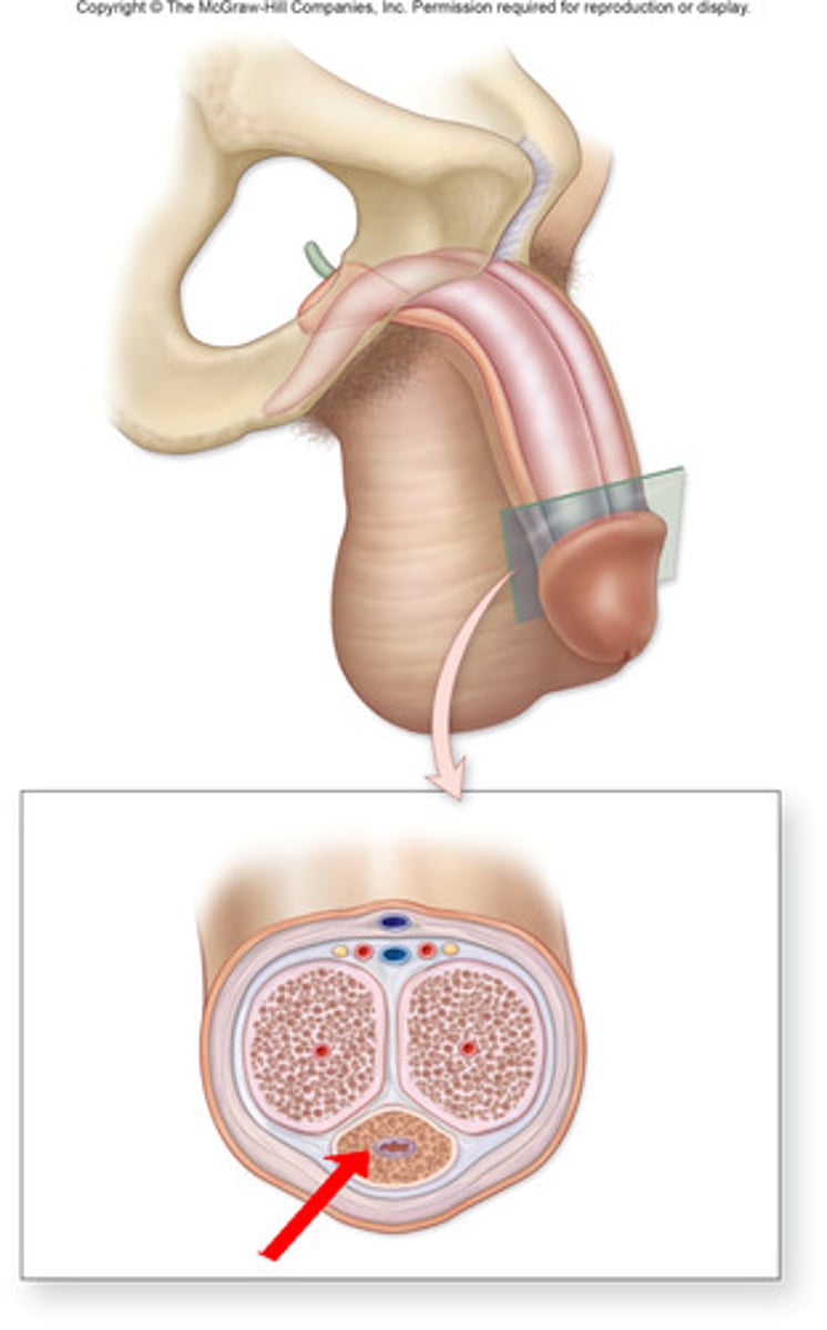
Epididymis
A long, coiled duct on the outside of the testis in which sperm mature.
Epididymis - Epithelium
ducts lined with pseudostratified columnar epithelium
Epididymis Head
receives and stores sperm from seminiferous tubules
Epididymis Neck
contains coiled duct of epididymis
Epididymis Tail
coiling diminishes, ascends into vas deferens, stores sperm before ejaculation
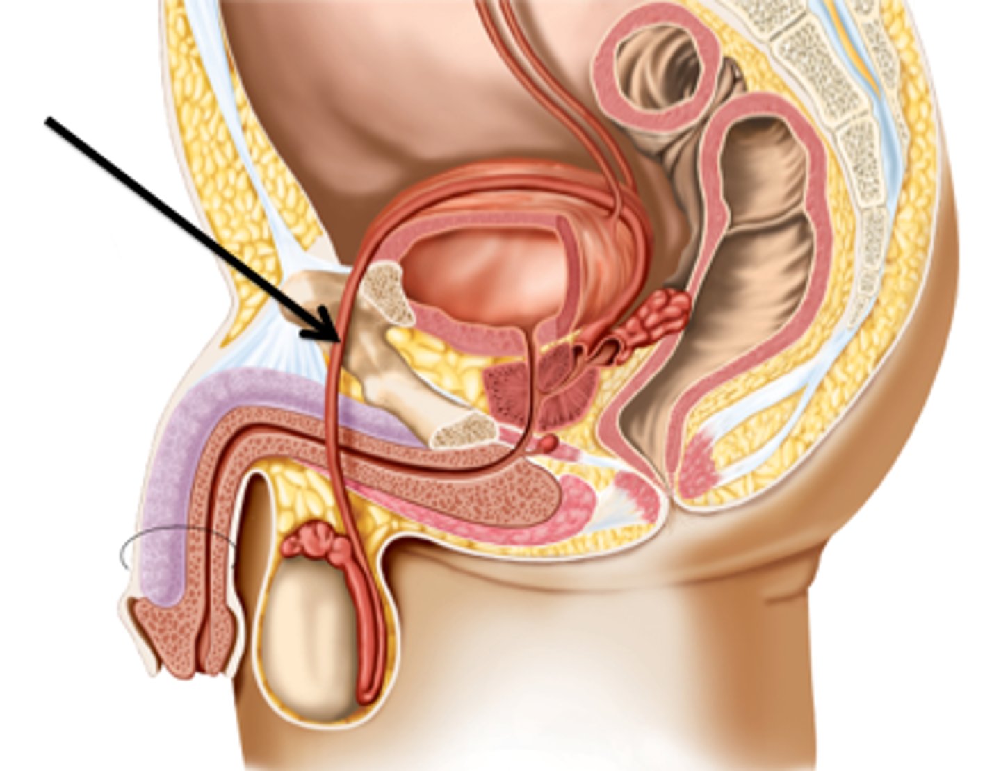
Vas Deferens
stores and transport sperm from epididymis to urethra
Ampulla of Vas Deferens
expanded distal portion
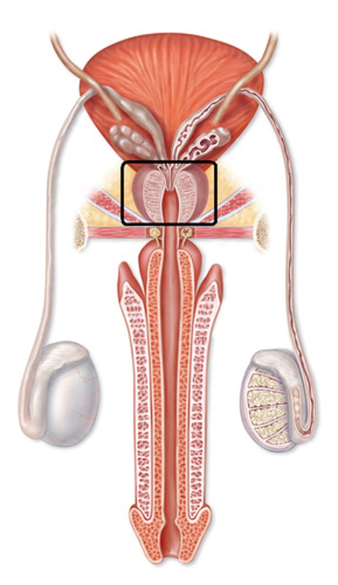
Ejaculatory Duct of vas deferens
ampulla + seminal vesicle converge
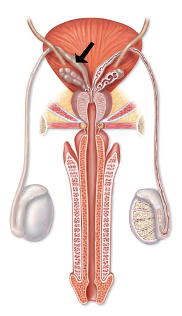
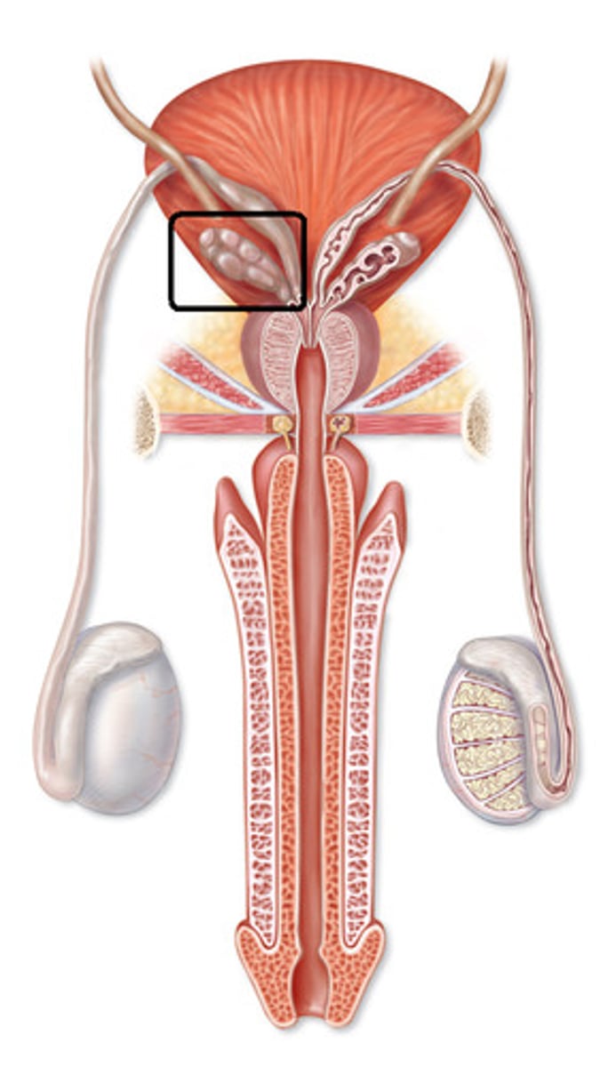
Seminal Vesicles
secretes nutritive fluid for sperm
Vas Deferens Histology
pseudostratified columnar epithelium with stereocilia
small lumen, thick muscularis with smooth muscle to propel semen
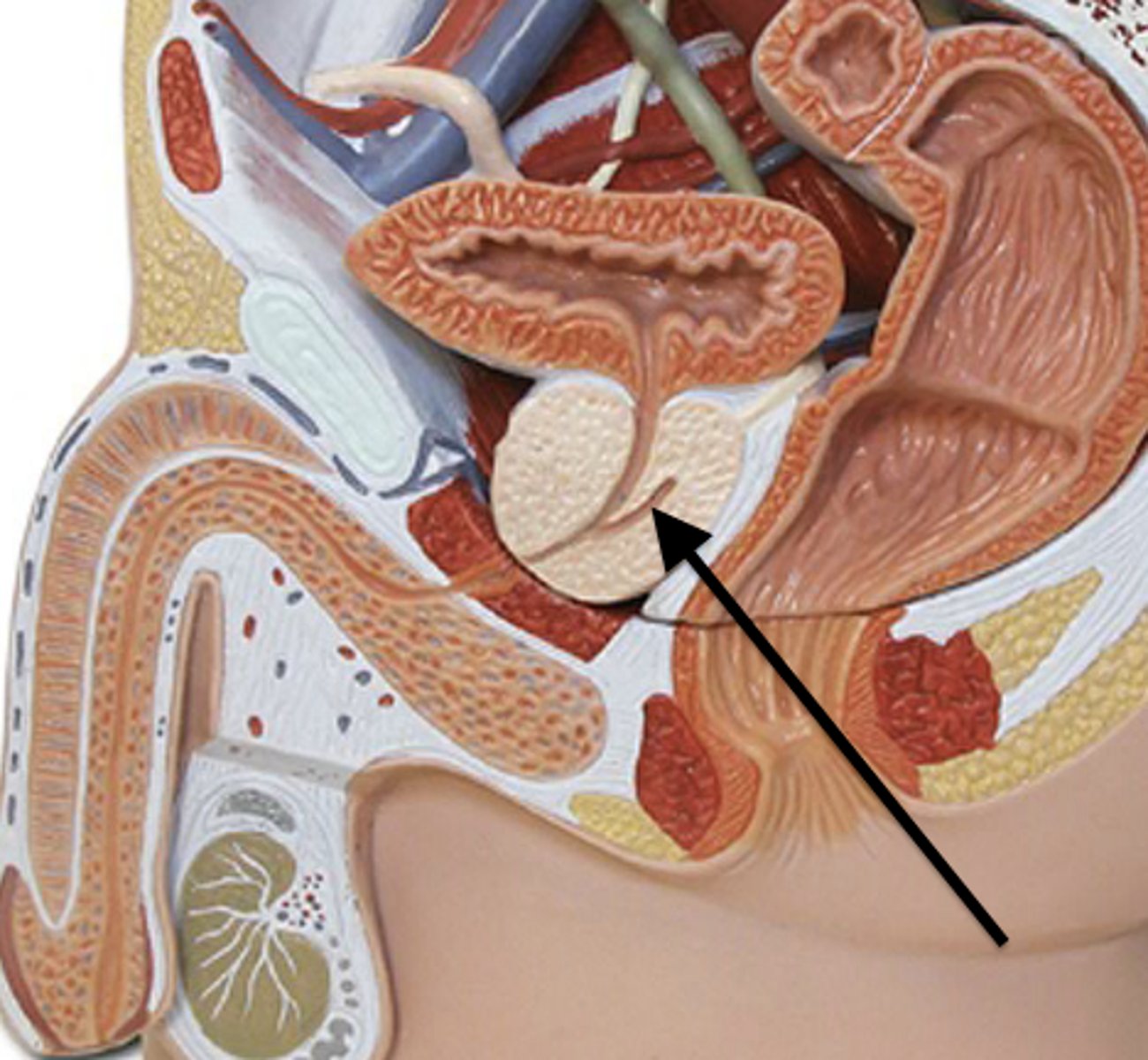
Prostate Gland
tubular glands in mass smooth muscle and connective tissue
produces and secetes sugars and enzymes
proteases dissolve cervical mucus
Bulbourethral Glands
secrete thick, clear mucus into penile urethra
released before ejaculation to neutralize acidic urine in urethra
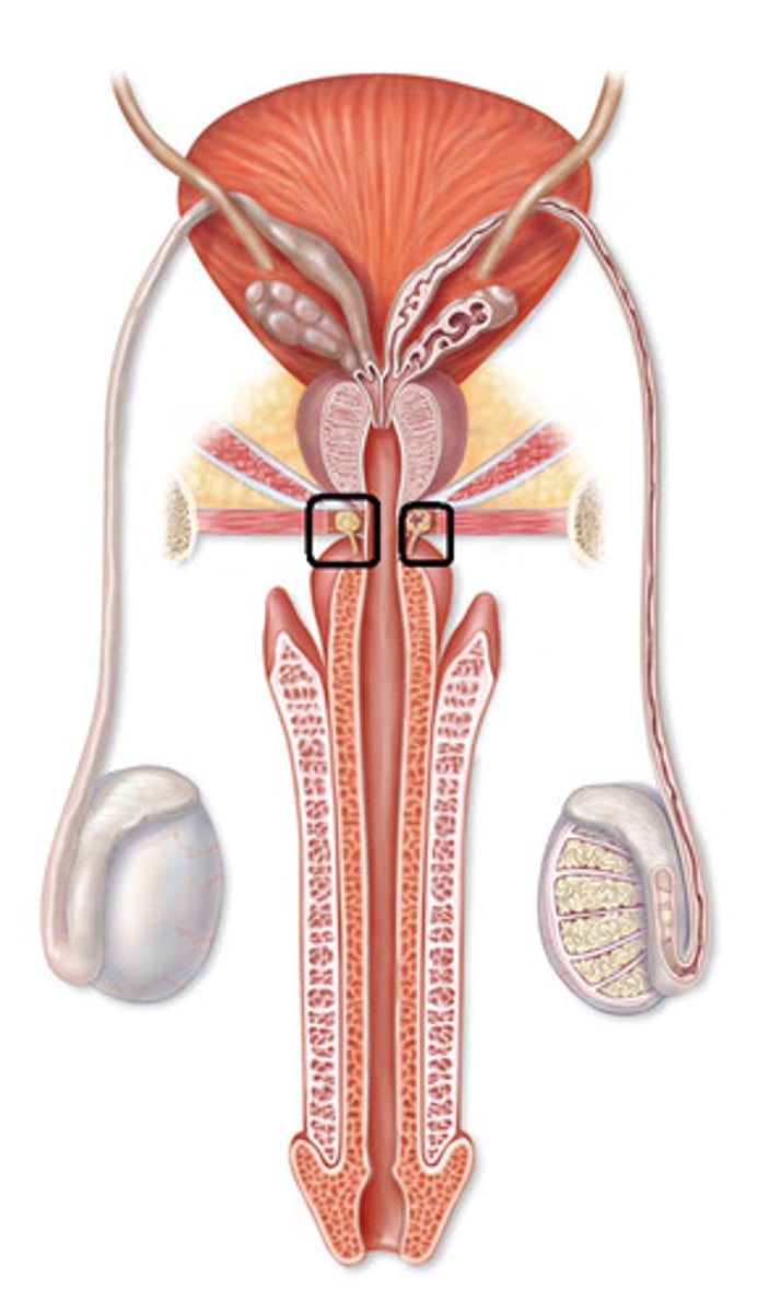
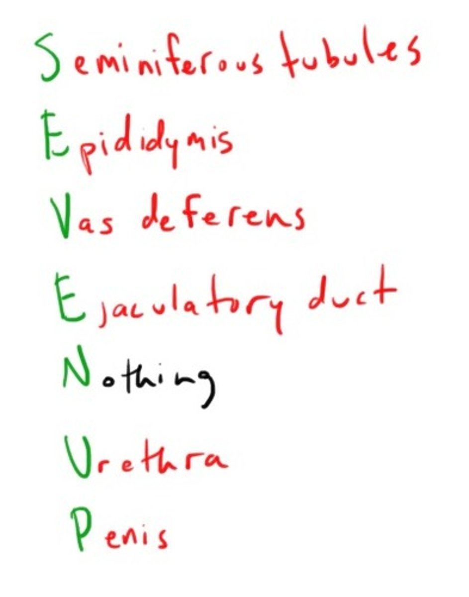
Pathway of Sperm
seminiferous tubules of testes --> epididymis --> vas deferens --> ejaculatory duct --> urethra
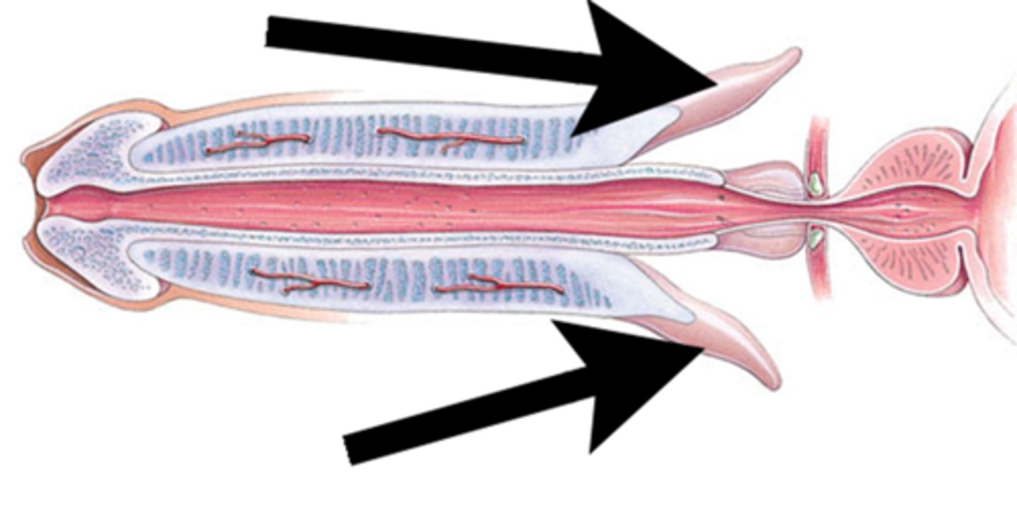
Root of Penis
fixed portion attaches penis to ischium
body of penis
3 cylinders erectile tissue, contains urethra
glans of penis
expanded distal end, contains external urethral opening
Corpora Cavernosa
erectile cylinders on dorsal surface
Corpus Spongiosum
cylinder contains urethra, expands to form glans
Crura
proximal portion corpa cavernosa, attached to bony pelvis by muscles
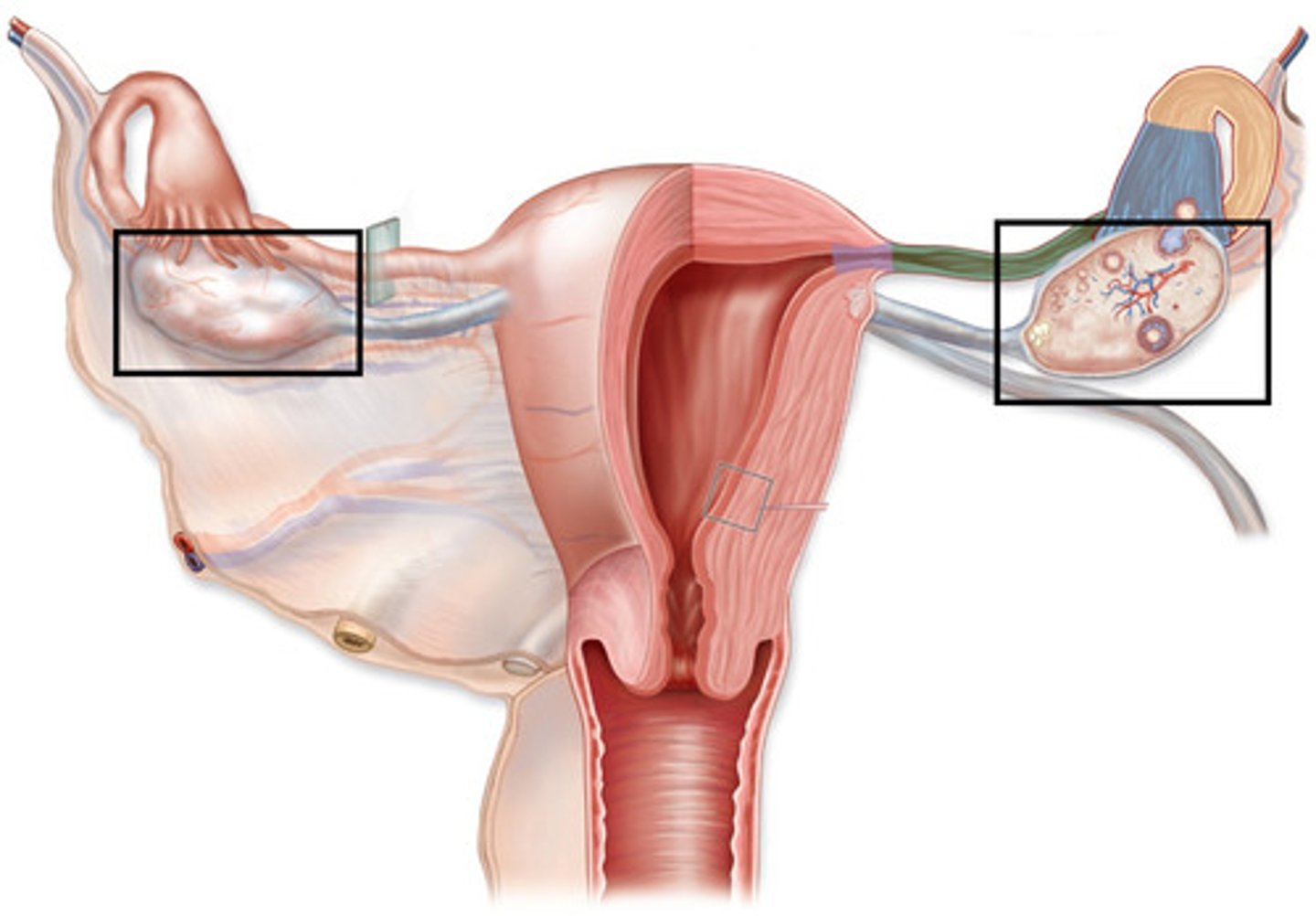
Ovaries
where eggs produced and stored before ovulation
Ligaments of Ovaries
protect and anchor ovaries, covered by dense connective tissue
Ovarian ligament
anchors ovaries to uterus
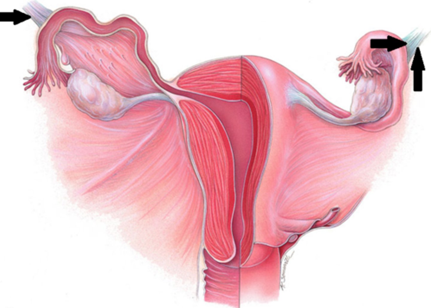
Suspensory Ligament
anchors ovaries to pelvic wall
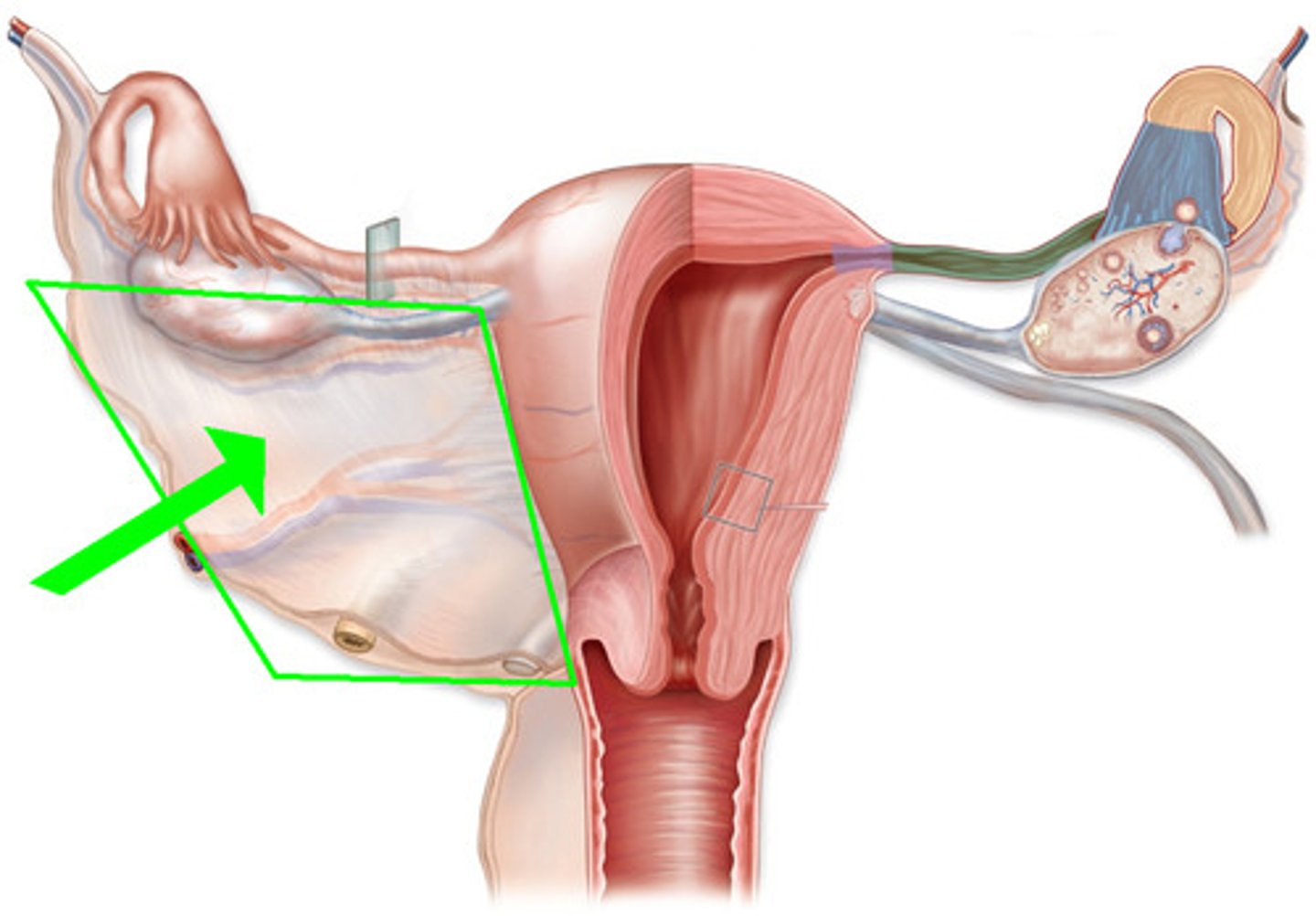
broad ligament
drapes over uterus
Tunica Albuginea of Ovaries
connective tissue capsule surrounding ovaries
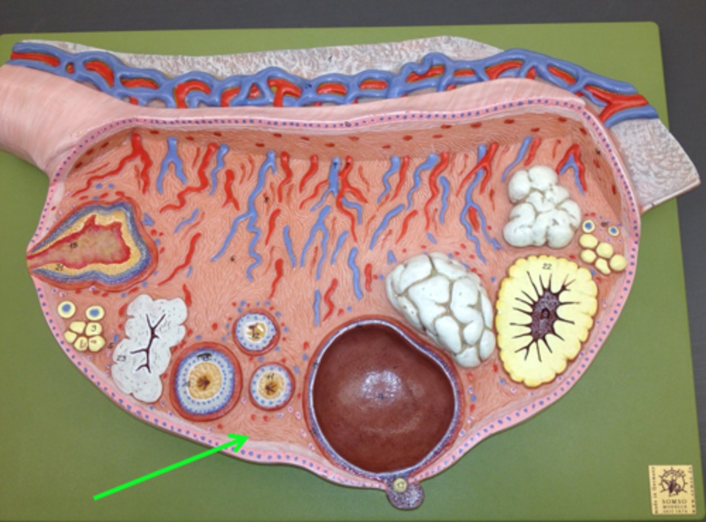
Medulla of Ovaries
contains blood vessels, nerves, lymphatics supplying ovary
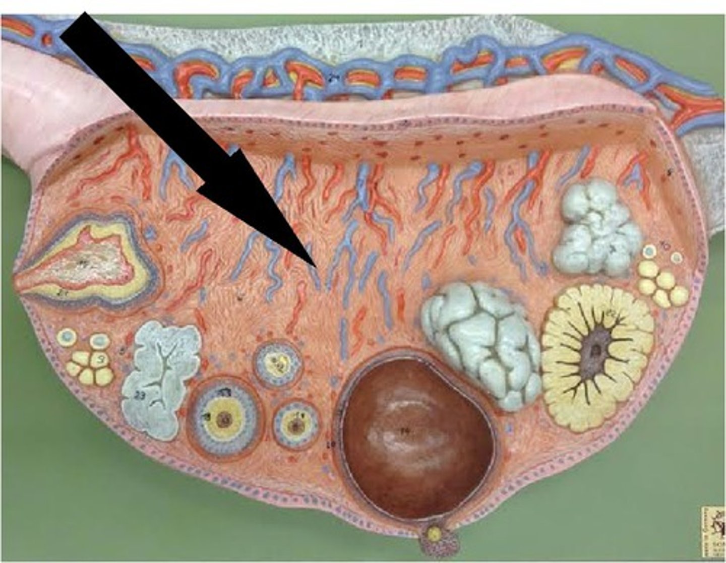
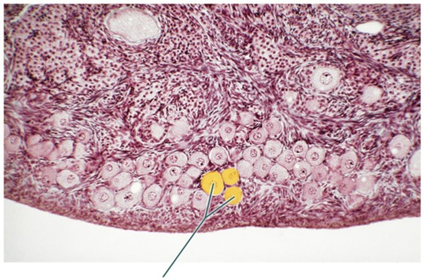
Cortex of ovaries
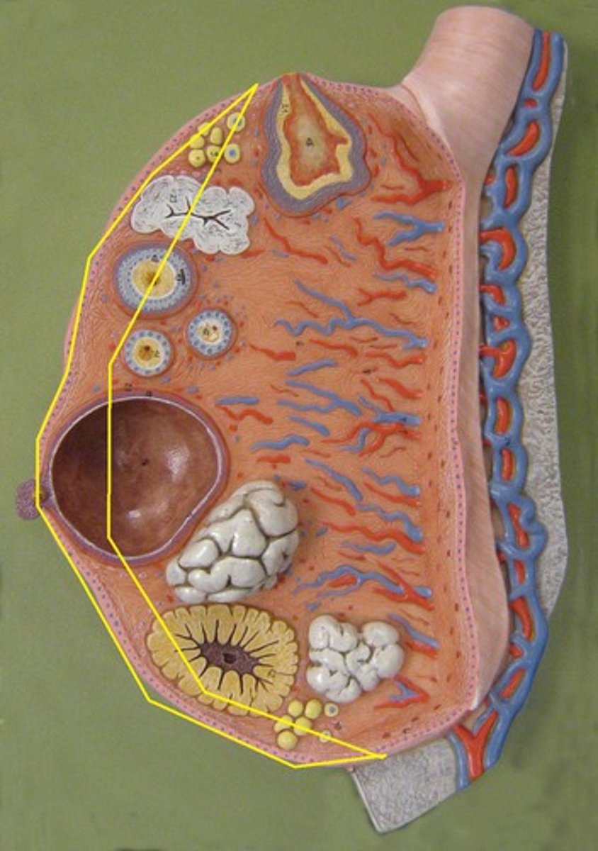
Primordial Follicle
most immature, made of single layer squamous cells surrounding oocyte
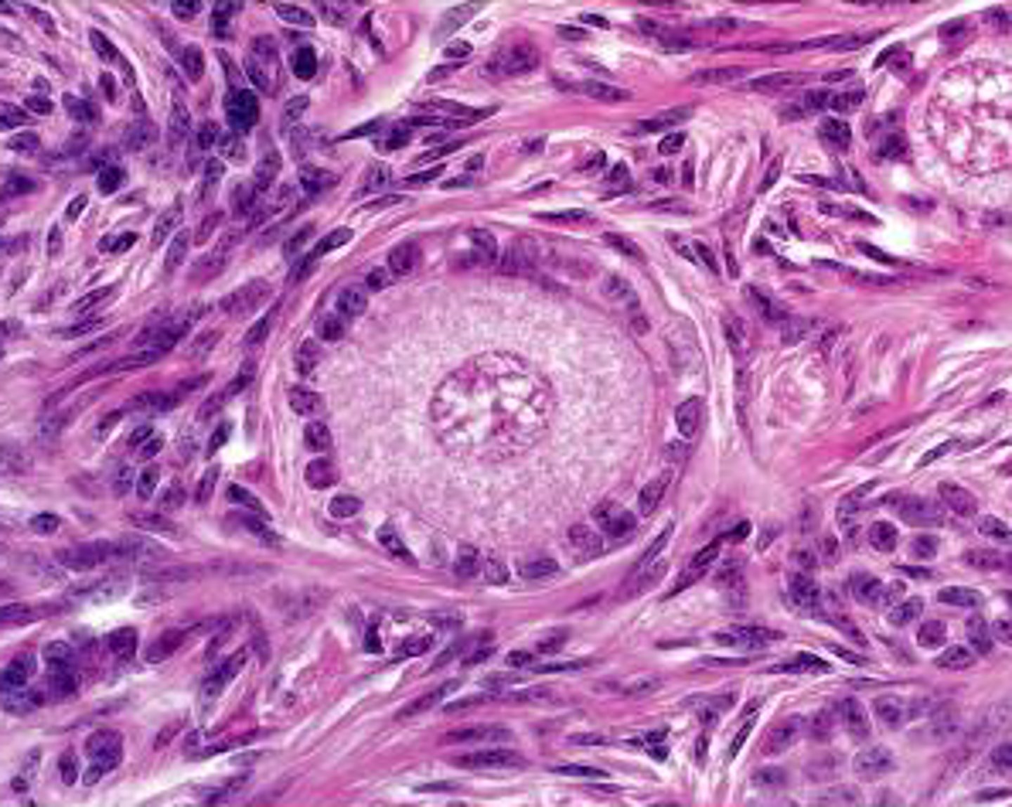
Primary Follicle
two or more layers surrounding egg
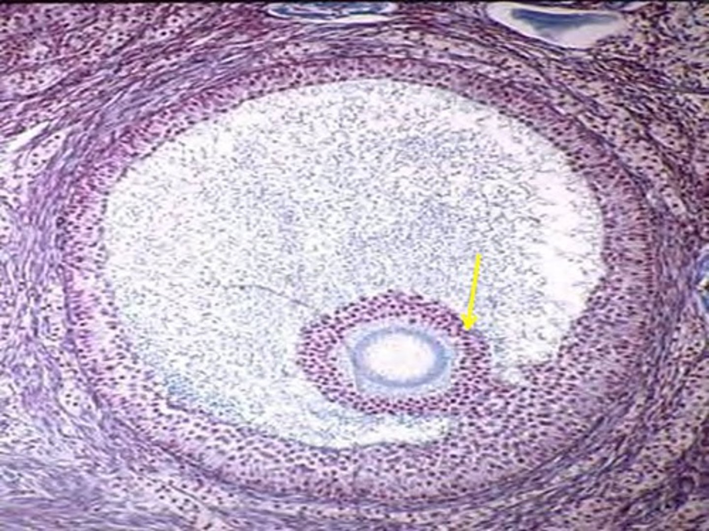
Secondary Follicle
surrounded by many layers, contains antrum (fluid filled space)
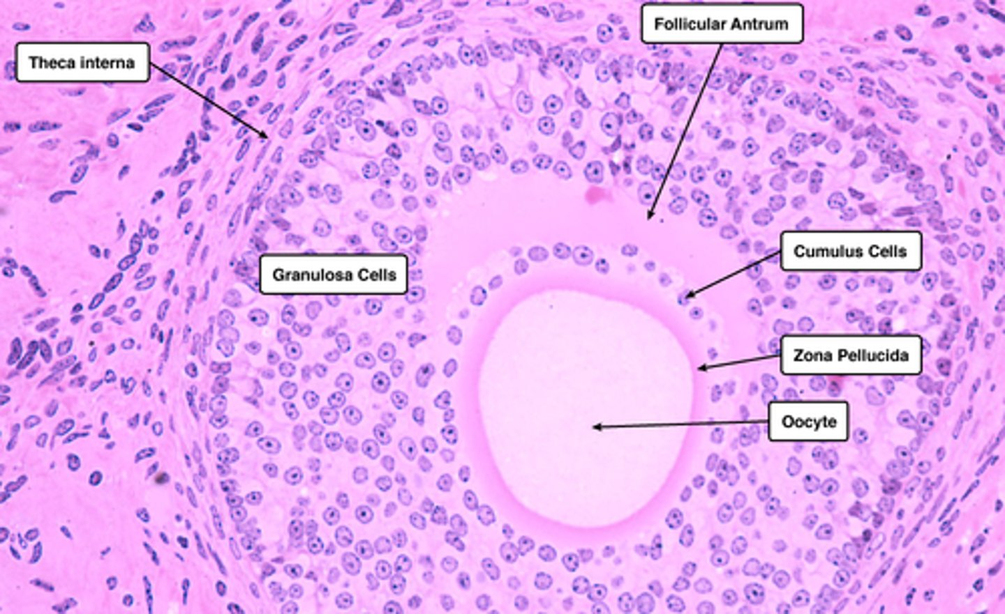
Graafian Follicle
mature follicle ovulated when ruptured, surrounded multiple layers cells (antrum)
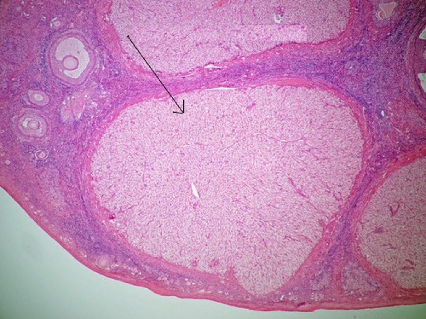
Corpus Luteum
develops from remnants mature follicle, produces progesterone and some estrogen to support fertilization
without fertilization, degenerates
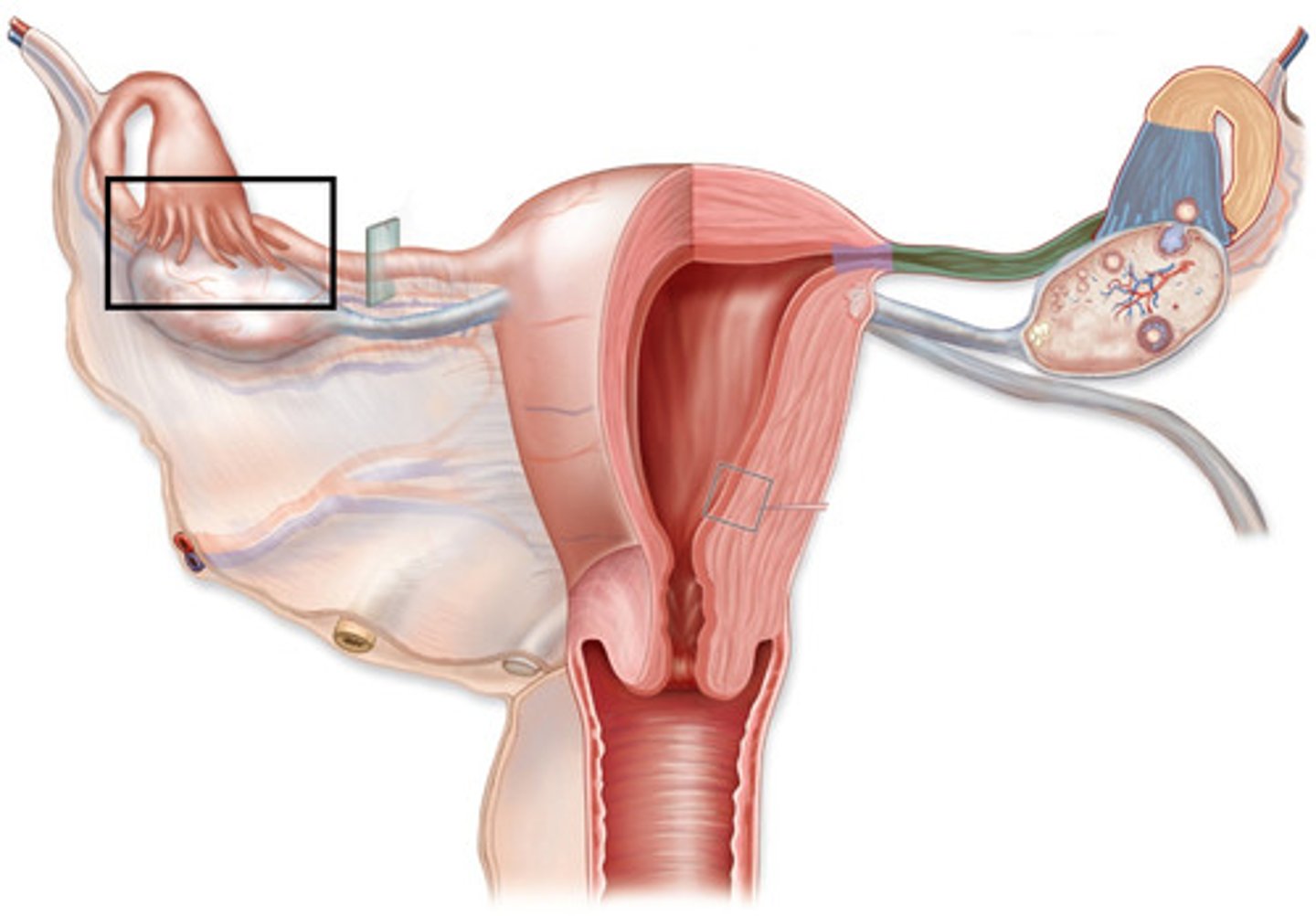
Infundibulum of Ovaries
funnel shaped, captures egg after release from ovary
has fimbriae
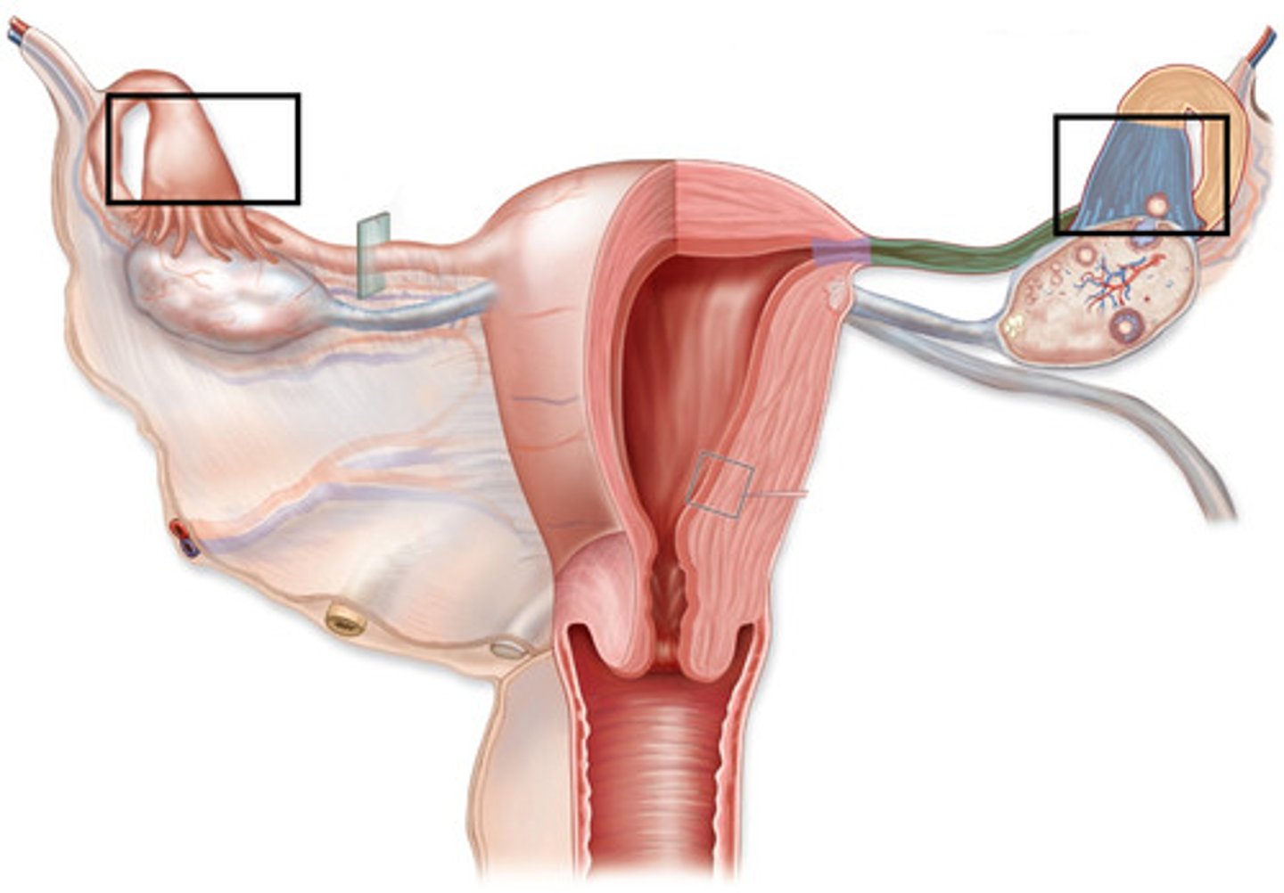
Fimbriae
Ampulla of Ovaries
where fertilization normally occurs
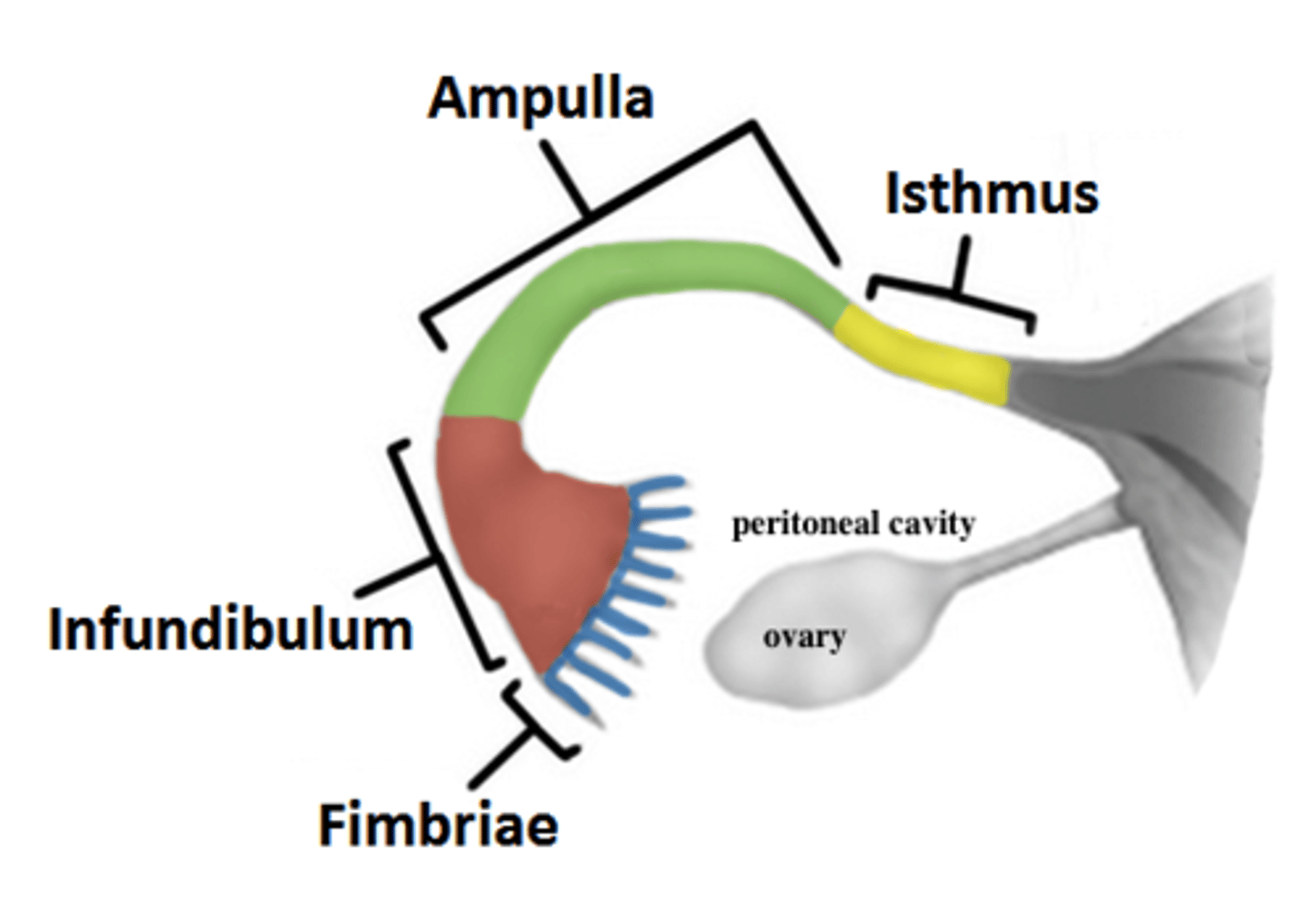
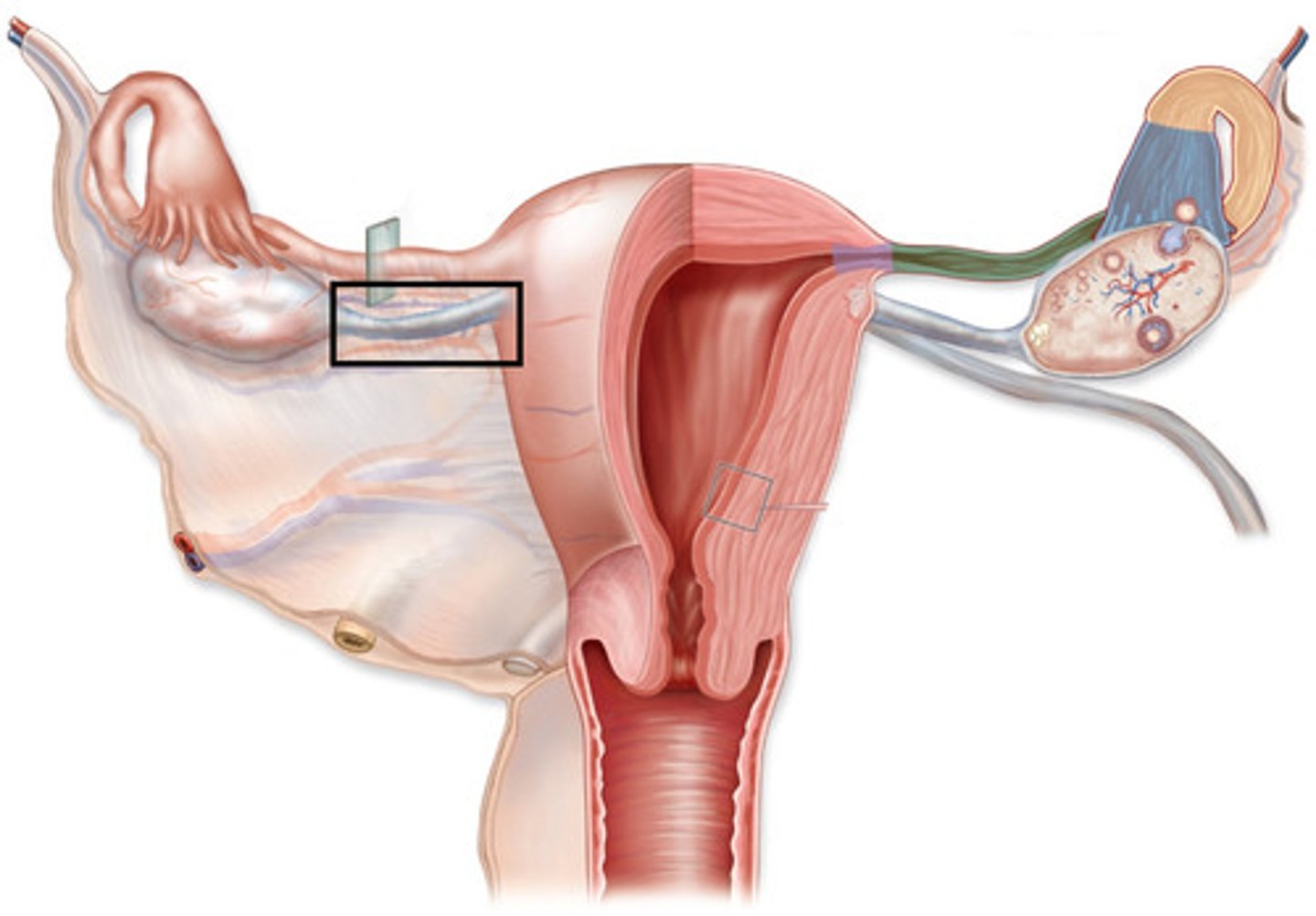
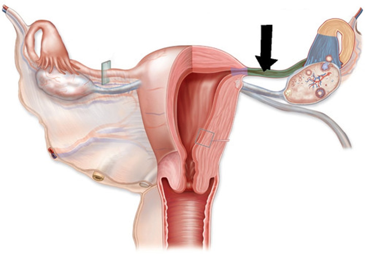
Isthmus
passes through uterine wall, opens into lumen of uterus
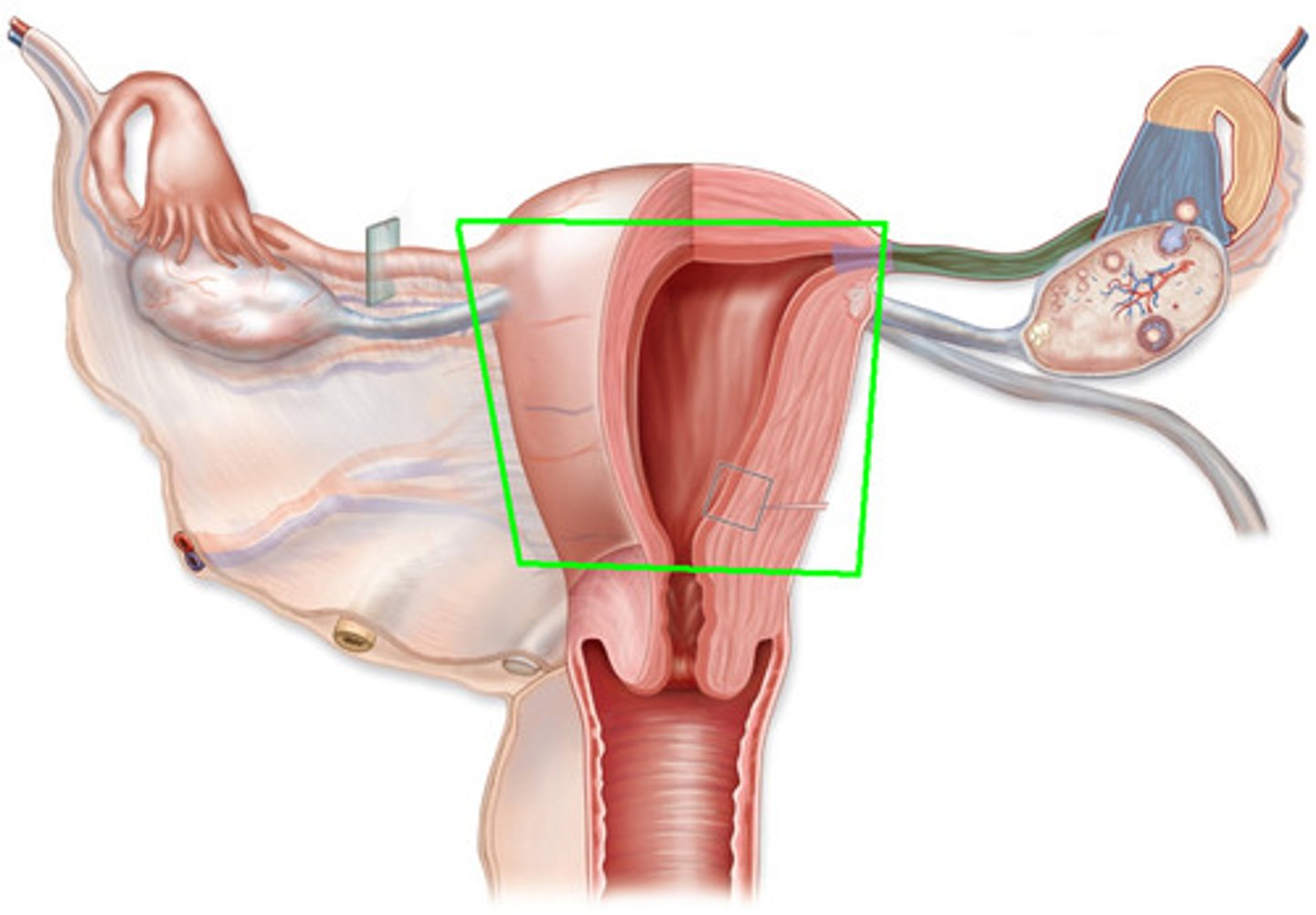
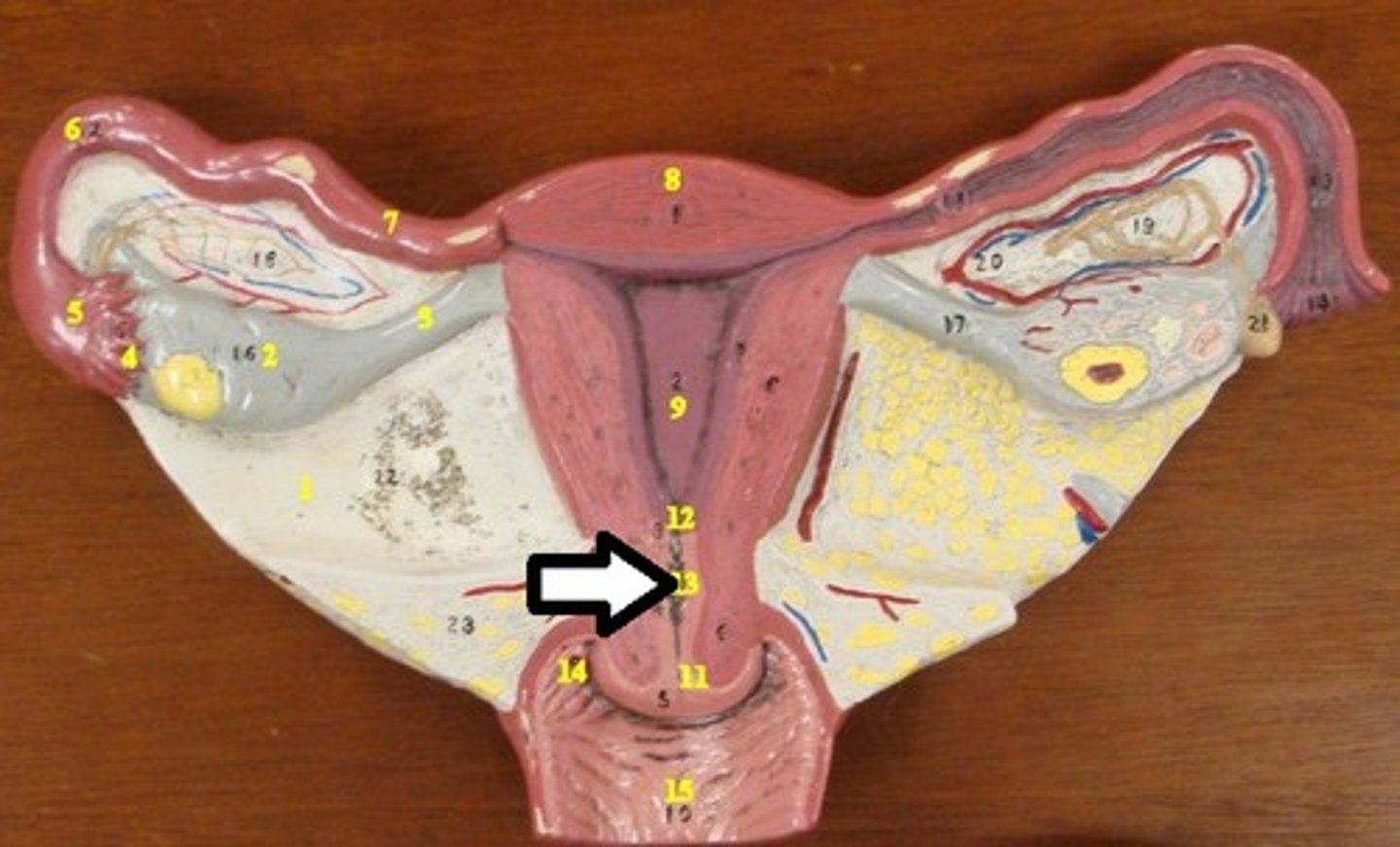
uterus
carries and supports fetus, contracts to eject fetus
Fundus of Uterus
most superior, extends between uterine tubes

Body of Uterus
main part, smooth muscle
Cervix
inferior, projects into vagina
contains cervical canal
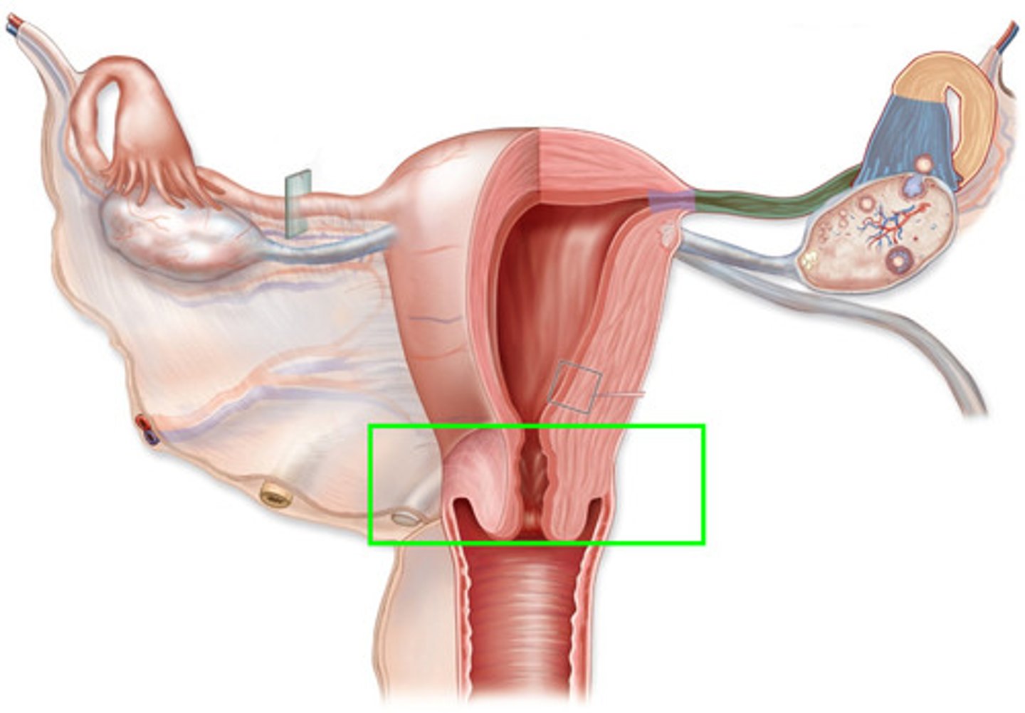
Cervical Canal
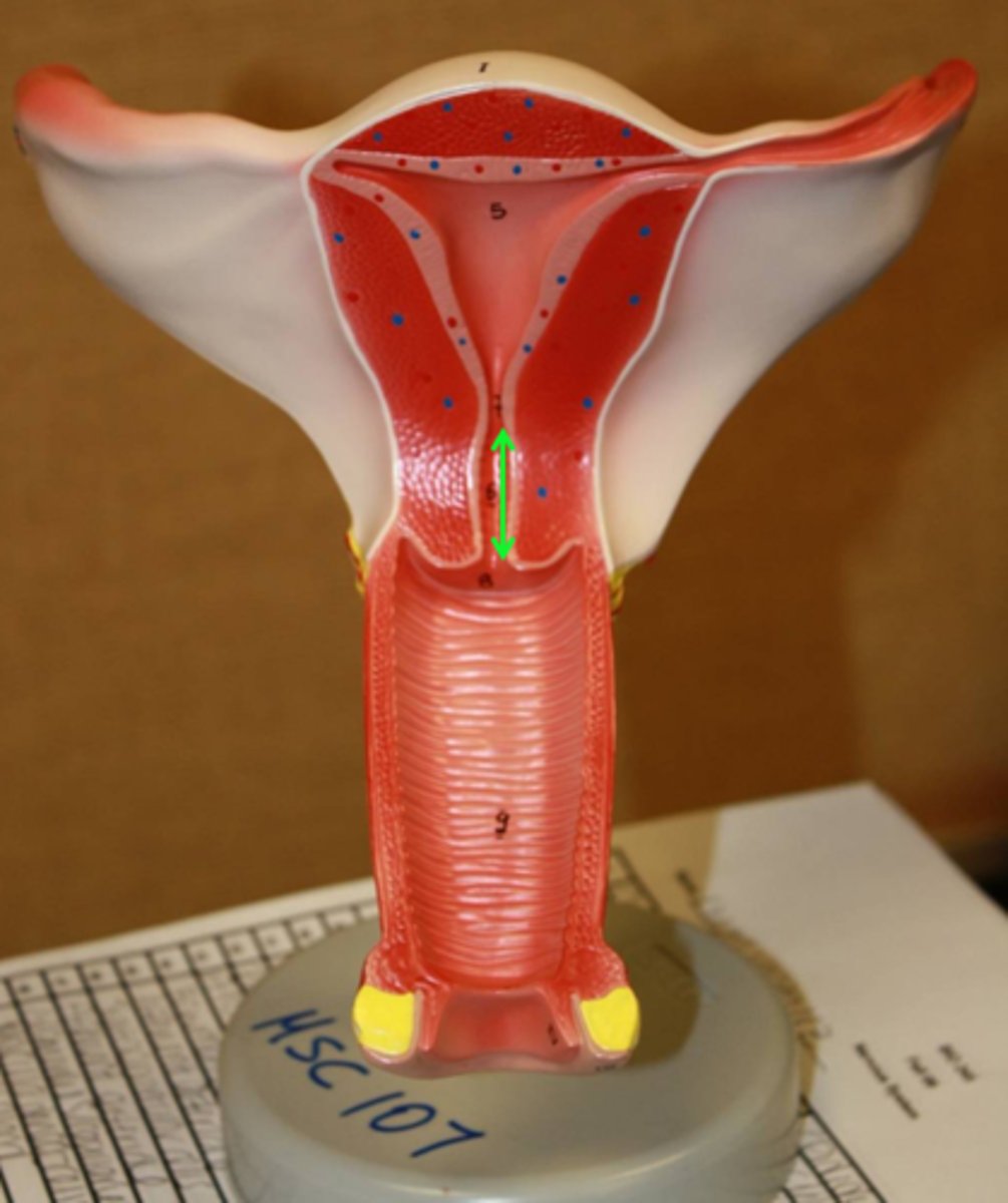
Internal os of cervical canal
connects uterus and cervix
External Os of cervical canal
connects cervix and vagina
Endometrium
glandular inner lining uterus, changes throughout menstrual cycle
Myometrium
middle layer uterus smooth muscle
Perimetrium
thin outer layer serosa of uterus
Vagina
connects uterus to outside world, birth canal
Vagina - Mucosa
stratified squamous, produces glycogen to be metabolized bybacteria into lactic acid
protects vagina by creating acidic environment
Vagina - Muscularis
elastic and loose connective tissue, inner circular outer longitudinal smooth muscle
Vagina - Serosa/Adventitia
adventitia
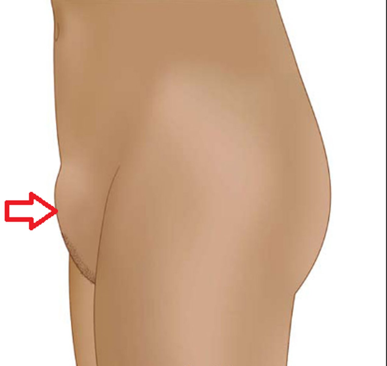
Mons pubis
adipose tissue over pubic bone
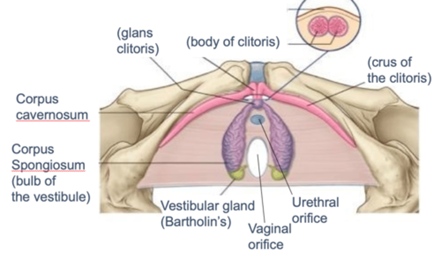
Clitorus
sensitive area erectile tissue
Labia Majora
elongated, fatty folds of skin with pubic hair (outside)
Labia minora
thin, fat free folds of skin without pubic hair (inside)
Vestibule of Vagina
space bounded by labia minora, has external urethral and vaginal opening
Glans of Clitorus
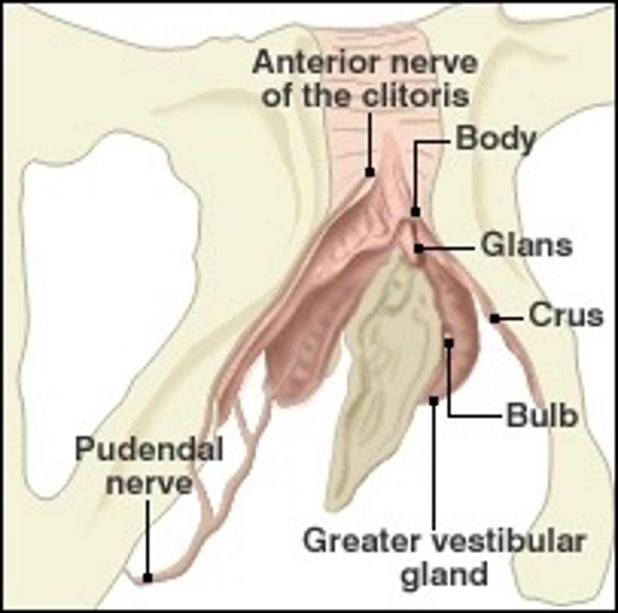
Crus of Clitorus
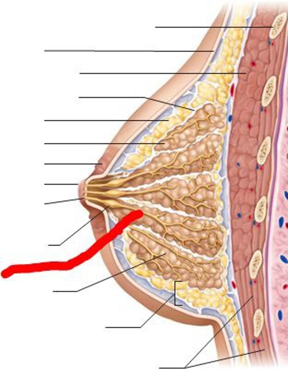
Mammary Glands
modified sweat glands produce and secrete milk
connective tissue separates lobes, atach breast to underlying muscle and overlying skin
Suspensory Ligaments
Mammary Lobes
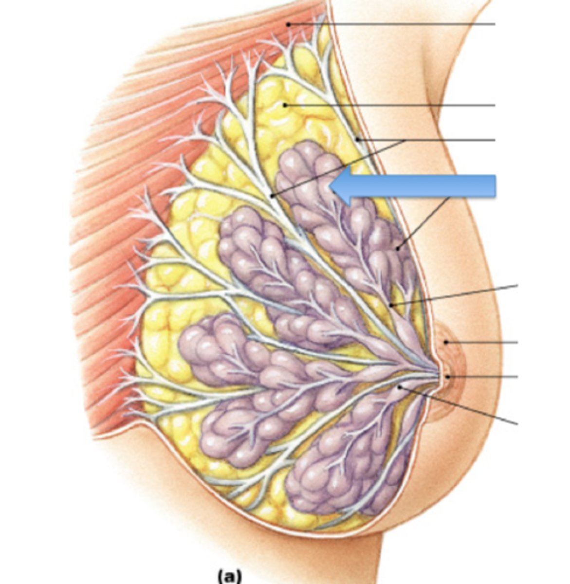
Lactiferous Ducts