nuclear cardiology delaney
1/81
There's no tags or description
Looks like no tags are added yet.
Name | Mastery | Learn | Test | Matching | Spaced | Call with Kai |
|---|
No analytics yet
Send a link to your students to track their progress
82 Terms
Angina
chest pain usually due to insufficient oxygen supply to the heart.
Aortic Valve
A semilunar valve between the left ventricle and aorta that prevents flow from the aorta back into the heart.
Apnea
No breathing.
Arrhythmia
variation of the heartbeat.
Asystole
No ventricular activity is present.
Atherosclerosis
Build up of cholesterol and fatty deposits inside arterial walls and can lead to ischemia.
Atrial Fibrillation
Multiple ectopic areas in the atria fire causing random impulses to reach the ventricles resulting in an irregular rhythm.
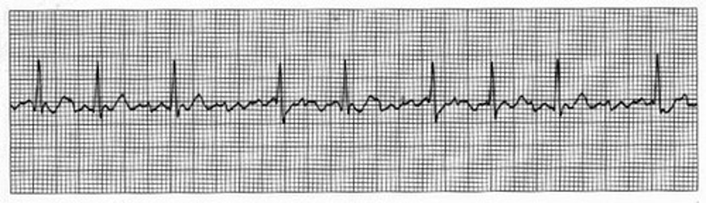
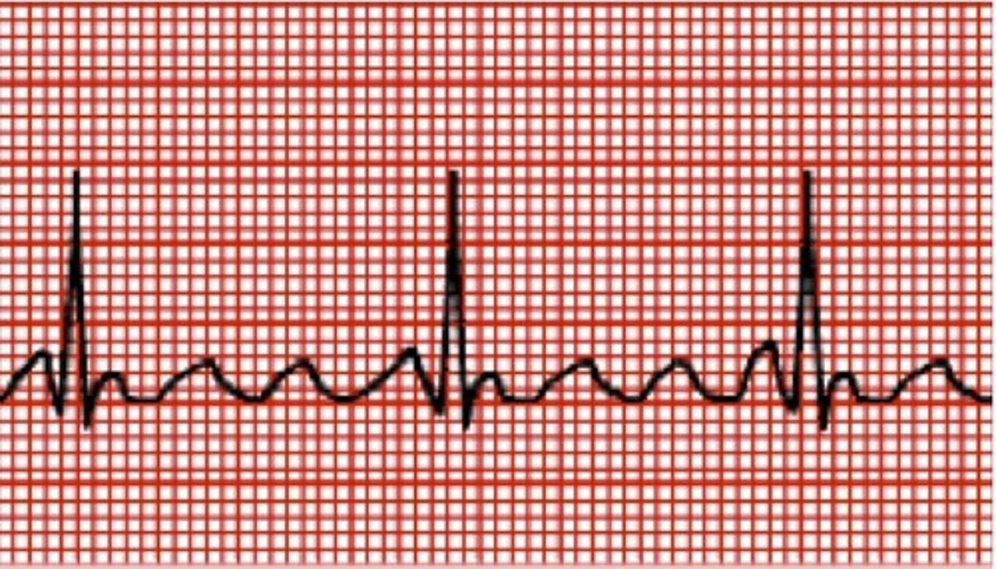
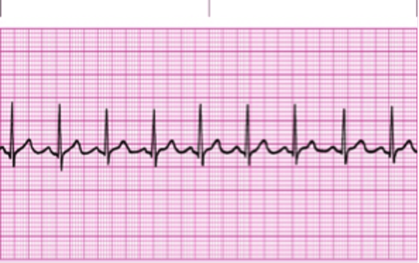
Atrial Flutter
Varying impulses reach the ventricles but in a more regular pattern, P-waves have a characteristic 'saw-toothed' configuration.
Atrial Pacemaker
The pacemaker lead is located in the right atrium, usually in the coronary sinus. It acts as a P-wave and causes the atria to contract.
AV Node
Receives the impulse from the SA Node, pauses for a fraction of a second to allow the ventricles time to fill with blood then passes the impulse down the His Bundle.
Blood Pressure
Force exerted on the walls of the blood vessels as the blood flows through them.
Bradycardia
Lower than normal heart rate, less than 60 beats per minute.
Bundle of His
Spreads impulse from apex to base of the heart to control the direction of the contraction.
Cardiac Cycle
One beat of the heart, which consists of systole, the period of ventricular contraction, and diastole, the period of ventricular relaxation.
(Cardiac) Ejection Fraction
The fraction of the total volume of blood of the left ventricle ejected per contraction.
Cardiac Output
The quantity of blood ejected by the heart over a one-minute interval. Usually 5 L/min at rest and 15 L/min during exercise in normal adults.
Congestive Heart Failure
inability of the heart to pump sufficient blood to meet the demands of the body.
Coronary Artery Bypass Graft (CABG)
Process in which blood flow is rerouted through a new artery (grafted from another vessel in the body) to areas of the heart muscle where diseased coronary arteries have reduced or blocked blood flow.
Coronary Artery Disease (CAD)
A narrowing or blockage of the arteries and vessels that deliver oxygen and nutrients to the areas of the heart
what can eventually lead to ischemia or infarct?
coronary artery disease
Diastolic Pressure
The heart in a state of relaxation, the bottom number of the blood pressure reading.
Dyskinesis
impairment in the movement of the heart wall.
Dyspnea
Difficulty breathing.
Ejection Fraction
is a measurement to how well the heart is functioning, it is the fraction of the total volume of blood that the heart can hold that is pushed out of the heart with each beat.
Electrocardiogram
Records the electrical activity of the heart and is a valuable record of the heart's function.
End-diastolic Volume
The quantity of blood in the ventricle at the end of diastole.
End-systolic Volume
The amount of blood in the ventricle at the end of a contraction.
Endocardium
inner most layer of tissue in the heart, membrane of epithelium and underlying connective tissue, including blood vessels and specialized fibers; functions as a protective inner lining of the chambers and valves.
what are the three layers of the heart
endocardium, myocardium, and epicardium
Heart Block
A disruption in the impulses from the atrium to the ventricles with varying degrees, 1st, 2nd, and 3rd degree.
Hypertension
elevated blood pressure.
Hypokinesis
Slow/decreased movement of the heart wall.
Hypotension
Low Blood Pressure. Is both a contraindication and a side effect for stress drug studies using a vasodilator.
Infarction
Area of tissue death due to local lack of oxygen.
Ischemia
decreased blood supply from the coronary arteries to the heart muscle, characterized by inverted T-waves.
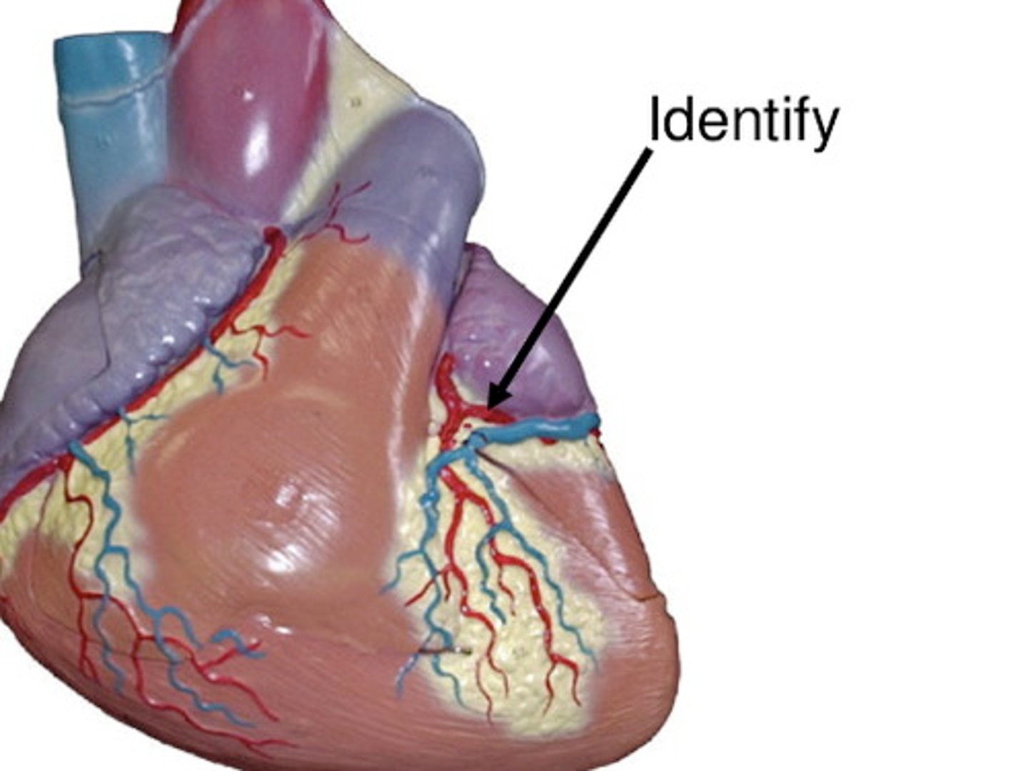
Left Anterior Descending Artery
Supplies blood to the septal, apex, and anterolateral wall of the left ventricle.
Left Circumflex Artery
Supplies blood to the posterior, lateral walls of the left ventricle.
Left Ventricle
The chamber on the left side of the heart that receives blood from the left atrium and pumps it into the aorta, which pumps the blood to the rest of the body.
Mitral Valve
Valve between left atrium and ventricle, also called the bicuspid valve.
Myocardium
Muscle layer of the heart (imaged in MPI).
Myocardial Infarction
death of heart muscle, caused by a prolonged, severe reduction of blood flow, usually as a result of occluded arteries.
Normal Sinus Rhythm
impulse originates in the sinus node, generating a P wave in front of each QRS on an ECG.
Pacemaker
A small device that's placed under your skin near the heart to help control your heartbeat.
Pulmonary Valve
a valve located between the right ventricle and the pulmonary artery. Blood is pumped through the pulmonary valve to the lungs, where it becomes oxygenated, and returns to the heart via the pulmonary vein.
Purkinje Fibers
A specialized cardiac muscle fiber that is part of the conduction system of the heart that initiates ventricular contraction.
Repolarization
Recovery of the ventricles after contraction; return of the heart to the resting state.
Right Coronary Artery
Supplies blood to the posterior and inferior walls of the left ventricle.
Right Ventricle
The chamber on the right side of the heart that receives blood from the right atrium and pumps it into the pulmonary trunk, which brings blood to the lungs.
SA Node
Stimulates both atria to contract, the normal pacemaker of the heart.
Semilunar Valves
The valves within the arteries leaving the heart, aortic valve and pulmonary valve.
Septum
This is the muscular wall that divides the right atrium from the left atrium and the right ventricle from the left ventricle. Ventricular conduction goes through the septum.
Stenosis
A narrowing of a canal or vessel.
Stroke Volume
The amount of blood ejected in a single heartbeat.
Systolic Pressure
The heart in a state of contraction, the top number of the blood pressure reading.
Thrombus
the formation of a blood clot inside a blood vessel, obstructing the flow of blood through the circulatory system. Myocardial infarctions are often caused by the obstruction of a coronary artery by a thrombus.
Tricuspid Valve
Valve between right atrium and ventricle.
myocardial infarctions are often caused by
a thrombus
short axis
images from apex to base *
horizontal long axis
images from inferior to anterior *
vertical long axis
images from septal to lateral
ventricular conduction goes through the
septum
pericardium
outer membranous sac encasing the heart
myocardium
muscular tissue, what is imaged in nuc med
endocardium
lines inner tissues and chambers
how does blood flow in the heart
superior vena cava, right atrium, tricuspid valve, right ventricle, pulmonary valve, pulmonary artery, lungs, pulmonary vein, left atrium, bicuspid valve, left ventricle, aortic valve, aorta
pacemaker of the heart
SA node
transmits pulse to the ventricles
AV node
spreads pulse from apex to base to control direction of contraction
bundle of HIS
right coronary artery (RCA)
A
supplies blood to the right atrium, right ventricle, bottom portion of the left ventricle and back of the septum
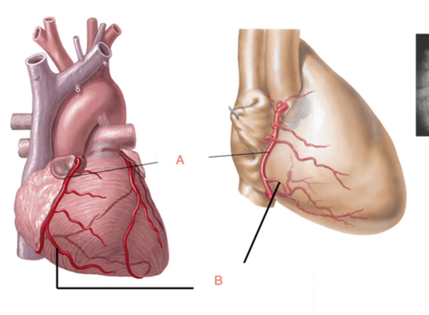
left coronary artery (LCA)
travels through the coronary sulcus under the left auricle and divides into two branches
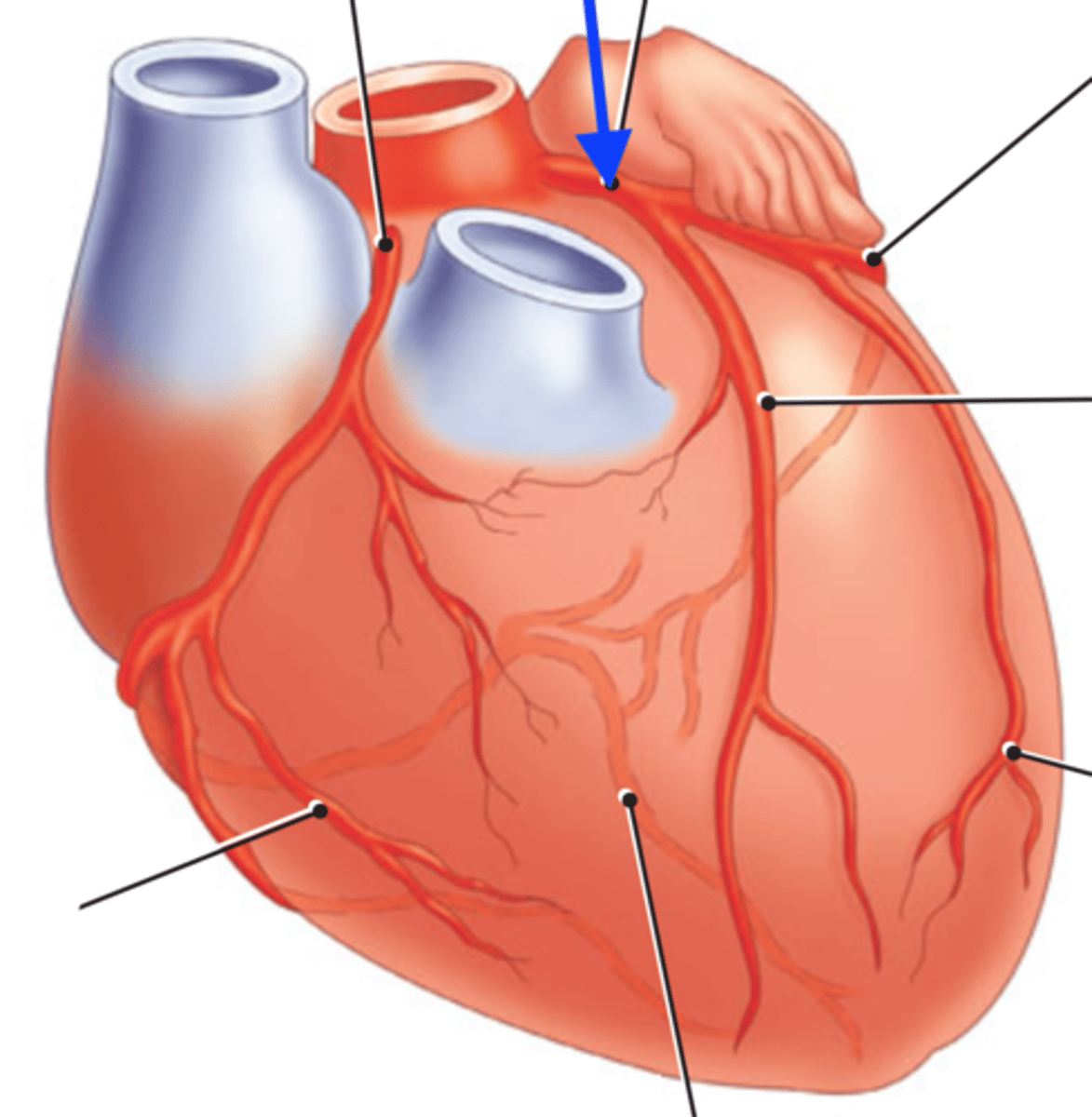
left anterior descending artery (LAD)
supplies blood to the front and bottom of the left ventricle and the front of the septum
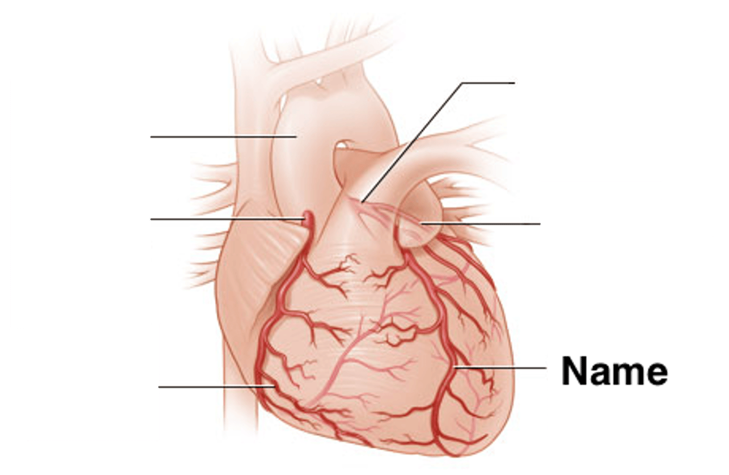
left circumflex artery (LCX)
follows the atrioventricular groove (coronary sulcus) and supplies the atria and the posterior left ventricle
Tc99m sestamibi MOL
passive diffusion
Tc99m sestamibi clearance
hepatobiliary system (liver, gallbladder,gut)
Tc99m sestamibi biological half life in myocardium
3 hours
how much mibi localizes in myocardium
only 1-2%
patient exposure of mibi
16mRad/mCi
dose for mibi if <100kg
8mCi rest
32 mCi stress
dose for mibi if >/= 100kg
10mCi
40mCi
why should patient drink water before mibi scan
to separate GI system from the heart in images
what are the DSPECT images acquired with mibi scan
supine gated rest
supine gated stress
upright nongated stress
Still learning (7)
You've started learning these terms. Keep it up!