Urinalysis #2: Sediment
1/96
There's no tags or description
Looks like no tags are added yet.
Name | Mastery | Learn | Test | Matching | Spaced | Call with Kai |
|---|
No analytics yet
Send a link to your students to track their progress
97 Terms
Best samples to use for sediment
morning samples
Samples after hours of water deprivation
Why use these samples for sediment
sample is more concentrated
will contain more sediment
recommended standard volume of urine
3-10ml
Centrifuge the urine at what speed and for how long
1500 rpm for 5 minutes
Why do we centrifuge urine at a low speed
Higher speeds disrupt the cells and cast
Remove the majority of urine from the tube and leave what in the bottom
sediment
what should you do if you have a lot of sediment
get a USG from the centrifuged urine before discarding
On the other end of the slide, add another drop of urine and one drop of what stain
New methylene blue stain
The stain aids in identifying what 3 things in the urine
RBC
WBC
Bacteria
view at lowpower (10x objective: Yellow) with the light intensity:
down low
which objective are sediment counts done
high power objective (40x objective: blue)
At least how many microscopic fields must be evaluated
10
what things per highpower field (40x hpf) are reported
the average number of various cells
Following wet-mount preparation, what should be done with urine
re-centrifuge
Use a second clean microscope slide to spread the material in a monolayer via what 3 possible options
the blood smear
line prep
compression prep techniques
Allow the dry mount slide to:
air-dry
Heat fixation is not necessary and will alter cell morphology
3 types of epithelial cells can be found in urine sediment:
Squamous
Transitional
Renal tubular
Each epithelial cell originates from a different area of ____ and helps to diagnosis the damage
urinary tract
Squamous Epithelial Cells
large
angular borders
a small nuclei
where do Squamous Epithelial Cells originate from lining of what 3 places
distal urethra
vagina
prepuce
Is Squamous Epithelial Cells indicative of disease
generally not indicative of disease
Transitional Epithelial Cells
centrally located nucleus
spherical, polyhedral, or pear shaped cell
Transitional Epithelial Cells lining what 4 structures
renal pelvis
ureters
bladder
upper portion of the male urethra
The only abnormal finding would be Transitional Epithelial Cells exhibiting these 2 abnormal morphology with:
vacuoles
irregular nuclei
vacuoles and irregular nuclei may indicate what 2 things:
malignance or viral infection
Renal tubular epithelial cells
Vary in size and shape depending on where they come from
Nuclei are eccentrically placed and they are not totally round.
More than 2 Renal tubular epithelial cells per hpf indicates:
tubular injury
The most clinically significant epithelial cell
Renal tubular epithelial cells
Renal tubular epithelial cells may indicate:
renal tubular necrosis.
Heavy metal exposure
Drug induced toxicity
Hemoglobin and myoglobin toxicity
Viral infections
Pyelonephritis
Allergic reactions
Malignant infiltrations
Allogenic transplant rejections
Glomerular disorders
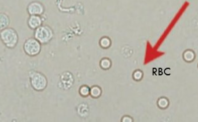
unstained Red Blood Cells are:
colorless or yellowish
Red Blood Cells
round and slightly refractile
can see biconcave disk & central pallor
Uniform in size
In concentrated urine, rbcs will do 3 things:
shrink
loose fluid
crenated
In dilute urine, rbcs will do 2 things:
absorb water
swell
Normal values of RBCs in urine
2-4 RBC/hpf
RBCs in urine indicate what pathological thing
bleeding somewhere in the urinary or genital tract.
RBCs in urine indicate what nonpathological thing
trauma from collection methods
White Blood Cells in urine
round
granular
larger than rbcs
The presence of how many WBC/hpf indicate a pathologic condition
more than 5-8WBC/hpf
The presence of more than 5-8WBC/hpf indicate ____ depending on the method of collection
inflammation of the urinary or genital tract
what is pyuria
excessive amount of WBC in the urinary tract
urine samples with increased numbers of Bacteria & WBCs should be:
cultured for bacteria
what cells on left
WBCs
casts are elongated structures composed of what 2 things
protein from plasma
mucoprotein secreted from the tubules
where do casts form
lumen of the distal and collecting tubules of the kidney
Increased number of casts helps to:
localize the renal disease
casts usually have what types of outlines
parallel sides with a definite outline
5 general types of Casts:
Hyaline
Granular
Cellular
Waxy
Fatty
Hyaline Cast
Clear, colorless
somewhat transparent
highly refractile and difficult to see
what should you do to view hyaline casts better
reduce light to lowest setting
Hyaline Cast indicates:
mild renal irritation
Hyaline Cast may increases with what conditions
fever
poor renal perfusion
strenuous exercise
general anesthesia
Most common form of casts seen in animals
Granular Cast
types of Granular Cast
Coarse or fine granular
Granular Cast is seen with:
acute nephritis
Granular Cast indicates what kidney condition, compared to hyaline casts
more severe kidney damage
Cellular Cast contains:
cells embedded in their protein matrix
what cells can be embedded
RBC
WBC
epithelial cells
cellular casts indicates (Depending on type of cells seen):
Acute Nephritis
renal inflammation
Pyelonephritis
Renal bleeding
waxy casts
wider, with square flat ends
burrito appearance
Colorless or gray
highly refractile
waxy casts indicates:
chronic and severe degeneration of the renal tubules
Fatty Cast
Contain several small droplets of fat
Fatty Cast seen in cats with:
renal disease
why is Fatty Cast seen in cats with renal disease
lipid in their renal parenchyma
renal parenchyma
functional, working tissue of the kidney responsible for filtering blood, producing urine, and maintaining bodily balance
Fatty Cast seen in dogs with:
diabetes mellitus
Large number of fatty casts suggest:
degeneration of the renal tubules
Mucus Threads
do not have the well defined edges of casts
narrow twisted ribbon
wispy like
Large amounts of mucus threads is normally present in:
horses
Large amounts of mucus threads in other animals indicates:
urethral irritation
contamination of the sample
Spermatozoa seen in the urine sediment of:
intact male animals
Spermatozoa seen in the urine sediment May be present in recently bred:
female animals
Large amounts of sperm may produce a false-positive result for:
protein
Fat Droplets
Lightly green-tinged, highly refractile, spherical bodies of varying size.
one of the first focal planes, so keep focusing past them to get to the other sediment
Fat Droplets
Fat Droplets can be a contaminate from:
catheter lubricates
Fat in cat urine to some degree is:
normal
Lipuria is also seen in animals with:
obesity
diabetes mellitus
hypothyroidism
rarely, after a high-fat meal
Microorganisms reported as:
few
moderate
many
too numerous tocount (TNTC)
morphology
Normal urine is free of:
bacteria
normal urine may be contaminated by bacteria residing on the epithelium of:
vagina
vulva
prepuce
catheterization
Urine should be examined immediately or refrigerated until able to view it, due to:
bacteria’s proliferate activity
Bacteria under magnification appears to be quivering as a result of:
Brownian movement
A large number of bacteria accompanied by a large number of WBCs suggests:
infection and inflammation of the urinary or genital tract
best method for establishing whether or not an infection is present
culturing urine
Common infectious agents of cystitis include:
E. coli
staphylococci
Streptococci
Proteus spp.
Yeast & Fungi
characteristic budding
may have double refractile walls
Yeast & Fungi are rarely found in urine sediment, and are usually contaminants as yeast infections of:
urinary tract or voided samples
Parasite ova may be seen in the urine sediment of animals with:
urinary parasites
fecal contamination
parasites that can occur in urine include:
Pearsonema pilca
Dioctophyma renale
ova of Stephanurus dentatus
Microfilaria
Pearsonema pilca
bladder roundworm of dogs and cats
Dioctophyma renale
kidney roundworm of dogs
Dioctophyma renale intermediate hosts
earthworms
how do dogs get Dioctophyma renale
eating rawfish, crayfish, or earthworms
the ova of Stephanurus dentatus
kidney worm of pigs
Microfilaria
heartworm, dirofilaria immitis
how do dogs get Microfilaria into urine
circulating microfilaria may be seen if hemorrhage into the urine occurs either from disease or as a result of trauma during collection
What kind of problem may cause you to find cellular casts containing white blood cells in the urine sediment of a dog ?
Pyelonephritis (kidney infection)