Vascular pathology
 Call Kai
 Learn
Learn Practice Test
 Spaced Repetition
 Match
Match Flashcards
 Knowt Play
1/175

There's no tags or description
Looks like no tags are added yet.
Name | Mastery | Learn | Test | Matching | Spaced | Call with Kai | Chat |
|---|
No analytics yet
Send a link to your students to track their progress
176 Terms
Finish the pathway of the circulatory system
From LV: to the ____ arteries to the ____ arteries to ____ which go to ____ then to ____ to ____ then to ____ and ____
elastic arteries
muscular arteries
arterioles
capillaries
venules
small veins
medium and large veins
What is the Vasa vasorum
the “vessels of vessels” a tiny network of blood vessels which supply the walls of both arteries and veins
What’s the difference between atherosclerosis and arteriolosclerosis
atherosclerosis is the most common type of plaque formation
arteriolosclerosis affects small arteries and arterioles
Where does atherosclerosis most commonly form at, what makes it form
-usually at the carotid bifurcation, (any bifurcation), aorta iliac system, and the CFA
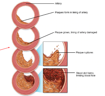
formed as a result of a deposition of material in the arterial walls between the intima and media, its an accumulation of lipid containing material, smooth muscle cells, fibrin and platelets,
What are the types of plaque ( the atherosclerotic process)
(Fatties find cute ultrasound images )
fatty streak- thin layer of lipid material on the intimal layer
fibrous plaque- lipids become covered by more material (collagen, elastic fiber deposits)
complicated lesion- a fibrous plaque that contains fibrous tissue, more collagen, calcium and cellular debris
ulcerative lesion- the deterioration of the normally smooth surface of the fibrous cap, this gives a higher tendency to shed debris causing emboli to travel distally
intraplaque hemorrhage: plaque bleeding from within
Write out the entire cerebrovascular anatomy, including the circle of willis
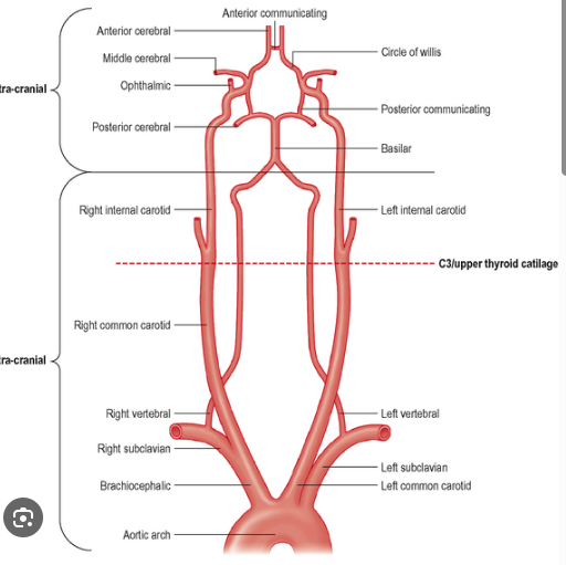
-remember innominate is the same as brachiocephalic
What is the boundary layer separation
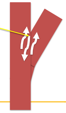
at branch points where blood flow can have both forward and backward velocity components
the effective velocity is zero
What is Reynolds number, what is it associated with
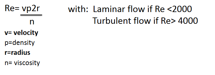
its calculates the ratio that helps predict if when fluid patterns will be turbulent
shows that turbulence depends on the size of the vessel and the velocity of the blood flow
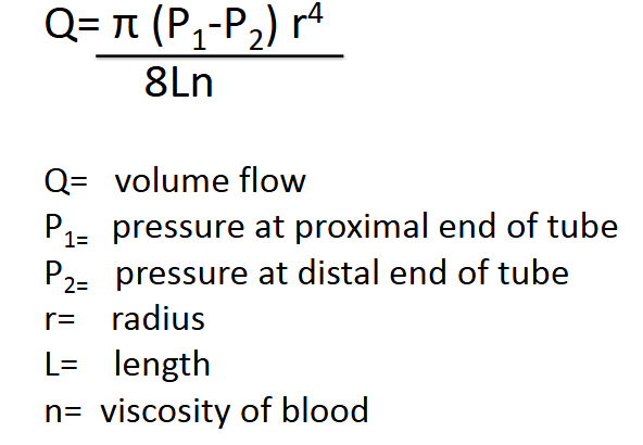
What is Poiseuille’s law and equation, what does it determine
its used to understand how vessel narrowing (stenosis) or dilation significantly affects blood flow.
discovered that a small change in radius results in a large change in volume flow ( 50% decrease in radius leads to a 95% decrease in volume flow)
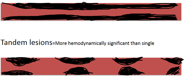
What’s the difference between stenosis and tandem lesions
tandem lesions is when there are two or more stenotic lesions occurring in a serios, they had a more pronounced effect on blood pressure and flow then a stenosis does
stenosis- single long lesion, less hemodynamically significant.

explain this waveform, where is it seen
tardus parvus: indicates a severe upstream artery stenosis or occlusion, has a slow and delayed upstroke and a low amplitude velocity.
its caused by a narrowed vessel forcing blood flow to arrive slowly and at a reduced velocity ( usually at renal, carotid, and hepatic arteries)
if a low resistive waveform becomes abnormal its ____ if a high resistive waveform becomes abnormal its ___
damped
monophasic
What does it mean if a venous waveform becomes pulsatile
indicates elevated right atrial pressure or systemic venous congestion
usually seen in conditions like right heart failure
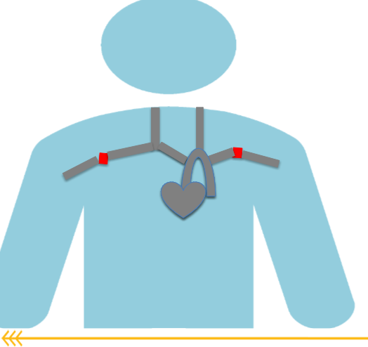
What is the thoracic outlet, what pathology comes from this
the space created by the scalene muscle, clavicle and first rib.
Subclavian artery, Subclavian vein and Brachial nerve travel
through this space
TOS (thoracic outlet syndrome) : occurs with abduction of the patients arm. with abduction the Sub A, Sub V, and brachial nerve can become compressed the order of compression is the nerve first, vein then the artery
symptoms: Nerve only- tingling and numbness
Vein and nerve- addition of pain and swelling
Vein nerve and artery- addition of paleness and loss of pulse
use can do two exams: segmental exam: using the PPG of the digits while patient performs dynamic maneuvers
Duplex exam: evaluating the distal subclavian artery while patient performs dynamic maneuvers
TOS maneuvers
90 degrees to the side ( arms in a T)
180 degrees from sides ( arms in a I)
Military stance ( arms pulled back at sides with elbows bent)
adsons- arms at 90 degrees with head turned toward arm, then away from the arm being tested
symptomatic position: the position the patient feels symptoms.
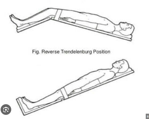
What position is Trendelenburg and reverse Trendelenburg
Trendelenburg: head lowered with legs in the arm
Reverse Trendelenburg: head up with legs down ( shown in image)
reverse Trendelenburg is better for venous exams to get blood flowing to your legs better.
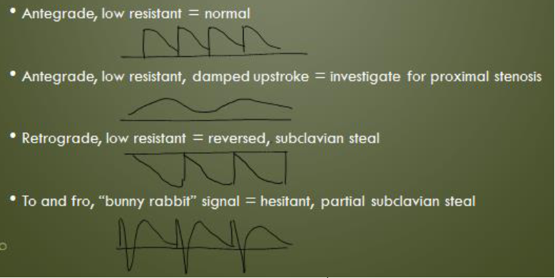
What is subclavian steel, when does it occur
occurs when a significant stenosis is in the subclavian artery proximal to the vertebral artery
this decreases the pressure in the subclavian artery causing the vertebral artery to change direction (retrograde)
for diagnosis you need a hemodynamically significant stenosis in subclavian, damped waveforms distally
a greater than 20 mmHg difference between right and left brachial BP
hesitant or reversed flow in the vertebral artery
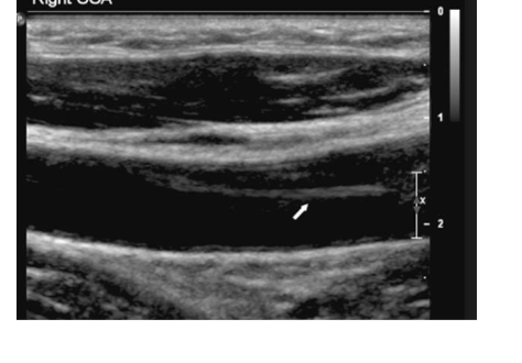
What causes arterial dissection what is it? what symptoms may a patient have with it.
the intimal layer tears and separates from the arterial wall
common causes: genetic diseases like marfans or ehlers- danlos
atherosclerosis
FMD
neck trauma
HTN and smoking increase risk
Patients may be asymptomatic or describe neck pain, coughing or dizziness
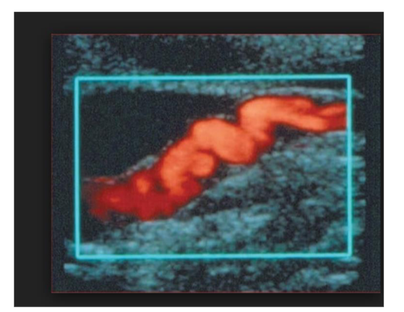
Whats FMD, fibromuscular dysplasia, what does it mostly affect
more likely in young women (25-50)
usually occurs in the ICA, renal artery, brachial, iliac and femoral
FMD is an overgrowth of the smooth muscle cells and fibrous tissue in muscle walls
patients usually have HTN due to decreased renal artery blood flow
has a string of pearls appearance, occurs mid to distal vessel, at least 1 cm from bifur. creates tandem stenosis effect within vessel. Common differential with vessel tortuosity.
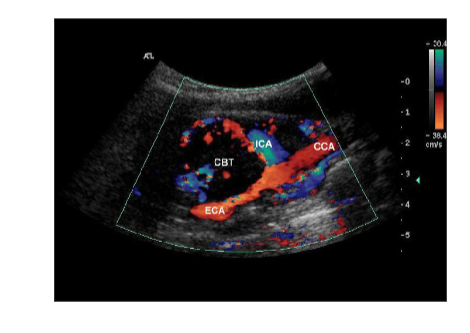
What causes a carotid body tumor (CBT) what is it?
its a tiny structure that controls PH, blood gas and BP, located at the carotid bifurcation
presents with palpable neck mass and headache, if untreated can cause stenosis, occlusion or rupture
On ultrasound it will bow out at the ICA and ECA bifurication
What is Moya Moya (MMD) who does it usually effect
rare progressive arterial occlusion of arteries in the basal ganglia section of the brain ( has puff and smoke appearance on angio)
not really seen on ultrasound but if u have a stenosis at the end of ICA and or the initial segment of the ACA and/or MCA
Usually affects children or young adults with a TIA or CVA as the first symptom
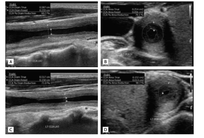
What is Takayasu’s arteritis, how does it look on ultrasound
rare vasculitis that affects large arteries ( aorta and branches)
this inflammation can cause narrowed or blocked vessels or aneurysms
more common in females, also known as pulseless disease due to lack of palpable extremity pulses
A >___ mmHg difference between left and right brachial systolic blood pressure is indicative of a hemodynamically significant stenosis on the ___
BP side
>20 mmHg
lower
this is because within a stenosis velocity increases and blood pressure decreases
If there is a hemodynamically significant stenosis or occlusion within a vessel, the blood pressures will decrease ____ to the stenosis or occlusion
distal
What are the 6 P’s of acute arterial occlusion
pain
pallor
Pulselessness
Paresthesia
Paralysis
Polar sensation ( cold) - caused by embolism, thrombus formation or trauma
What is Raynauds?
which the digital arteries become vasospastic
– Vasospasm (“vessel contraction”)
– Arteries close in response to cold, vibration, or emotional stress
– Often occurs bilaterally
– Can involve fingers and/or toes
To test for it you usually do a cold sensitivity training, PPG tracings obtained from the digits before and after immersion in ice water for up to 3 mins.
immediately after the immersion the amplitude of the waveform will decrease.
its abnormal if the amplitude of the waveform doesn’t return to normal in 5 mins.
In Raynauds the tracing will have a double peak with stimulation
Symptoms include skin changes (such as paleness, bluish discoloration, or dark red discoloration), numbness and pain
what is angioplasty and stent, where is it used and why
When a stenosis is found, physicians can use a catheter based procedure to insert a balloon to open the artery and reduce the plaque in the artery (angioplasty)
Stents enter artery compressed Then are opened with a balloon, Balloon is then deflated and stent remains
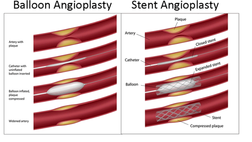
what’s the difference between Angioplasty/Stent and a Bypass Graft
When an artery becomes occluded, catheter based procedures are no longer possible, physician must then treat arterial insufficiency with bypass graft
– Graft a creation of a new arterial route for the patient
Bypass grafts connect patent artery proximal to occlusion to patent artery distal to arterial occlusion
If there’s a PSV of less then <45 cm/sec that indicates possible graft failure
what are the common UEA Bypass Grafts
Carotid to Subclavian (to revascularize patient with proximal subclavian occlusions)
Brachial artery to radial or ulnar artery (to revascularize patients with brachial artery or proximal radial or ulnar artery occlusions)
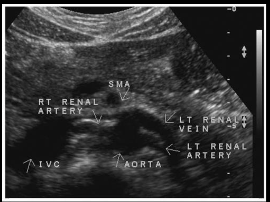
What pathology comes from this
Nutcracker syndrome, SMA compresses the left renal vein against the aorta. This can lead to left renal vein thrombosis and left renal vascular congestion. This will also cause scrotal varicocele in men
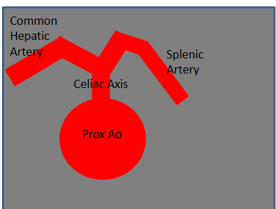
What is seagulls sign
Branches into 3 Vessels
– Common Hepatic Artery
– Splenic Artery
– Left Gastric Artery
• Typically only Common Hepatic Artery and Splenic Artery visualized on ultrasound
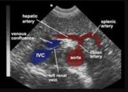
What is SMA resistance dependent on
depends upon
pre prandial/ post prandial state,
normally:
- High resistant pre-prandial ( before eating)
- Low resistant post- prandial ( after eating)
The SMA velocities will be elevated if its not pre-prandial
What does the RI (resistive index) show? what are the normal values
Resistive Index: calculated from cortical and medullary arteries at upper, mid and lower poles bilaterally
RI = (PSV-EDV ) / PSV
Normal <0.6
Borderline Increased Resistance 0.6-0.7
Increased Resistance >0.7
As an organ kidneys should normally have lower resistance, therefore increased resistance indicates likely parenchymal disease
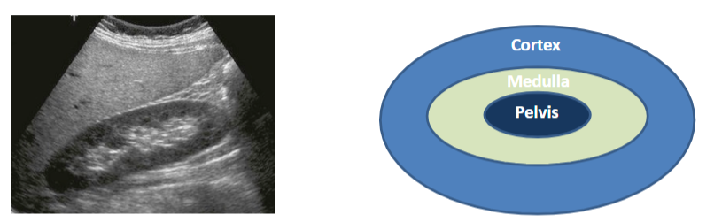
explain the difference between the cortex medulla and pelvis
Cortex- less echogenic than medulla, contains tubules for filtration
Medulla -is more echogenic than cortex, collection chamber
Pelvis- hypoechoic when urine is present
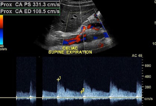
What is MALs explain the maneuvers and what happens
Occurs when the Celiac artery is compressed by the median arcuate ligament
usually in younger females ( around 40)
Accompanies abdominal pain and weight loss
if PSV in Celiac axis PSV >200cm/sec then you evaluate for MALS
Inspiration= relief of compression on celiac axis by median arcuate ligament as diaphragm moves inferiorly
– Celiac PSV then return to normal range
• Expiration= celiac artery compressed by median arcuate ligament as diaphragm moves superiorly
– Celiac PSV increase above normal criteria range
Renal Exam indications
Hypertension, new onset, or nonresponsive to medication
• FMD (Fibromuscular Dysplasia)
• Follow up known stenosis or intervention (angioplasty or stent )
• Renal artery aneurysm
• Epigastric or Flank bruit
• Renal transplant evaluation
• Decreasing kidney function
Why is Hypertension an indication for a renal artery exam
when atherosclerosis in renal artery increases, blood flow to kidneys decreases, body responds by raising blood pressure. Patients with new onset HTN will be routinely referred for renal artery duplex to ensure bilaterally patent renal arteries
Whats the RAR
Renal Aortic Ratio (RAR) = Renal PSV/ Prox Aorta PSV
Both the Renal Artery PSV and RAR Criteria must be met for a Renal Artery to be >60% stenotic
>180cm/s and the RAR is >3.5
What are the regular diameters for the aorta and celiac arteries
Distal Aorta: Normal <3.0 cm
– Ectatic = a 20 % increase from proximal segment
– Aneurysmal > 3.0 cm considered aneurysmal and or 50% increase from proximal segment
– Critical = 5.0 cm alert physician of findings
– Surgical Intervention = 5.5 cm
• Common Iliac: Normal < 1.2 cm
– Ectatic= 1.3 cm
– Aneurysmal= 1.5 cm
– Critical = 2 cm
– Surgical Intervention= 3.5 cm
What is blue toe syndrome, what causes it
If thrombus accumulates in the aneurysmal sac, small pieces can break free and travel
distally
• Blockage of distal foot digital vessels causes blue toe syndrome
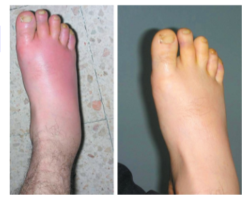
what is Dependent Rubor
Leg will appear red when in gravity dependent (lower than heart position)n and pale when nondependent (on same level as heart)
• Occurs when dermal arterioles and capillaries no longer constrict in the presence of increased hydrostatic pressure
What is popliteal entrapment
commonly affects male athletes, Popliteal artery becomes compressed by the medial head of the gastrocnemius muscle and tendon completely stopping blood flow to distal lower extremity
– Compression typically occurs after exercise and presents as pain with exercise
normal ABIs at rest and can have normal LEA Duplex findings
Often includes Duplex imaging of PTA, PER and ATA to obtain Spectral waveform while patient performs dynamic maneuvers of plantar flexion and dorsi flexion.
What is cystic adventitial disease
Cysts form in the adventitia of the arterial wall limiting blood flow. Usually seen in the popliteal artery. Symptoms mimic claudication in that pain occurs
with walking and subsides with rest, however on ultrasound no atherosclerosis (plaque) visualized
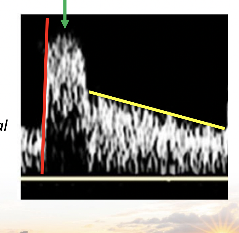
Explain the parts of this waveform
Inflow: in red, shows if theres a more proximal problem if its delayed.
Outflow: in yellow, shows if theres a more distal problem, like distal occlusion, AVF, or vasodilation
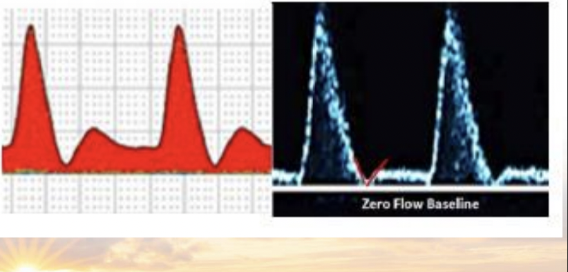
Describe an intermediate resistive waveform
Multiphasic but doesn’t go below the baseline, this suggests vasodilation and can be the result of exercise, increased temp, or a severe arterial obstruction.
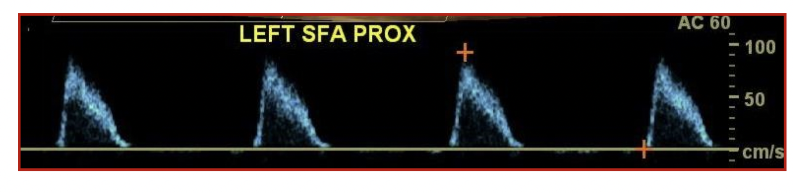
Resting superficial femoral artery waveforms from a 78 y.o. diabetic patient, what can you tell from it?
Abnormal, obstruction to outflow. You can tell by the no diastolic flow reversal, ( the outflow component). frequency seen in patients with multilevel calcific lesions
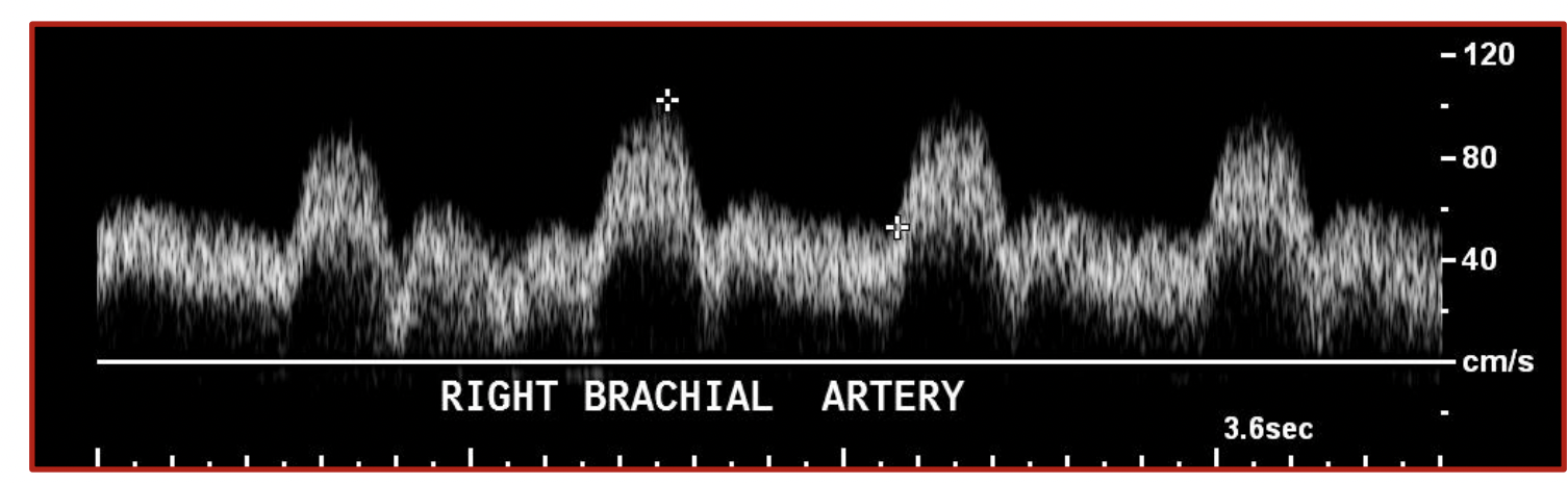
What does this waveform suggest
Abnormal, distal flow demand. There’s too much diastolic flow for the brachial artery, This almost looks like a low resistive pattern, suggesting a distal flow demand.
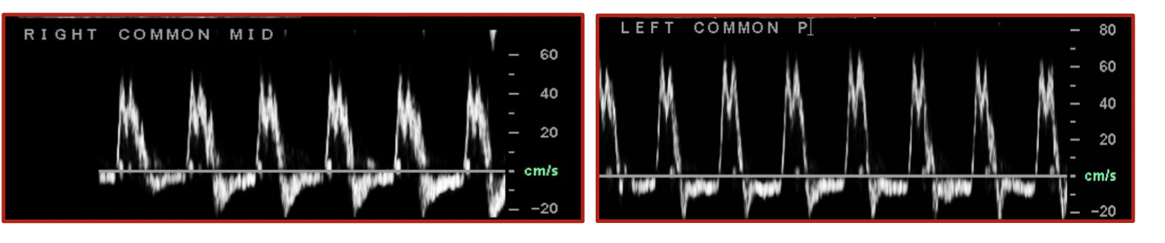
What does this waveform suggest
Aortic insufficiency/regurgitation you can tell because aortic insufficiency results in reversed flow throughout diastole and a bisferiens waveform, ( 2 distinct peak with the height of the second peak being equal to or higher than the first one)
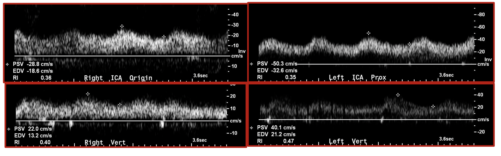
When would you expect this waveform?
If the patient is on ECMO, which provides support for failing heart and/or lungs by oxygenating blood outside of the body. So it gives largely continuous flow.
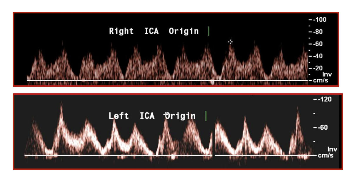
You see this waveform on a patient with a preop carotid duplex scan, how would you describe it.
its expected if the patient has an intra-aortic balloon pump, this is an atypical waveform of both the ICA and vert arteries, called a double hump.
make sure to measure velocities on the first hump
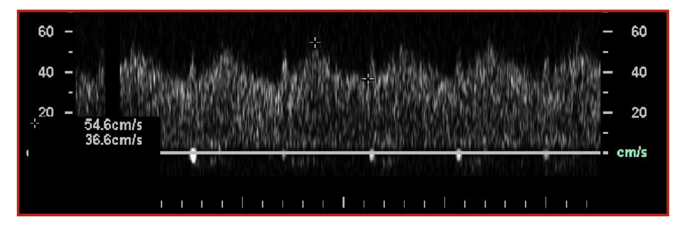
What is this waveform? its in the vertebral artery
this is abnormal suggestive of subclavian, pre-steal. you can see the mid-systolic flow deceleration.
also known as bunny sign
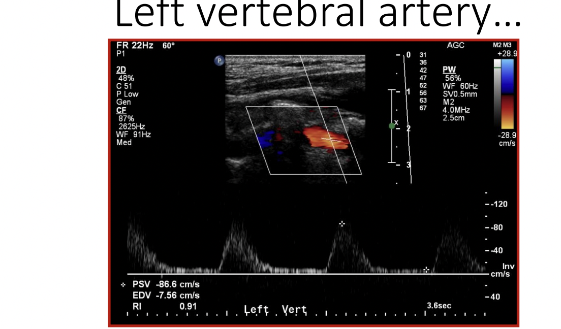
How would you describe this waveform
Complete subclavian steel, no diastolic flow, and its retrograde.
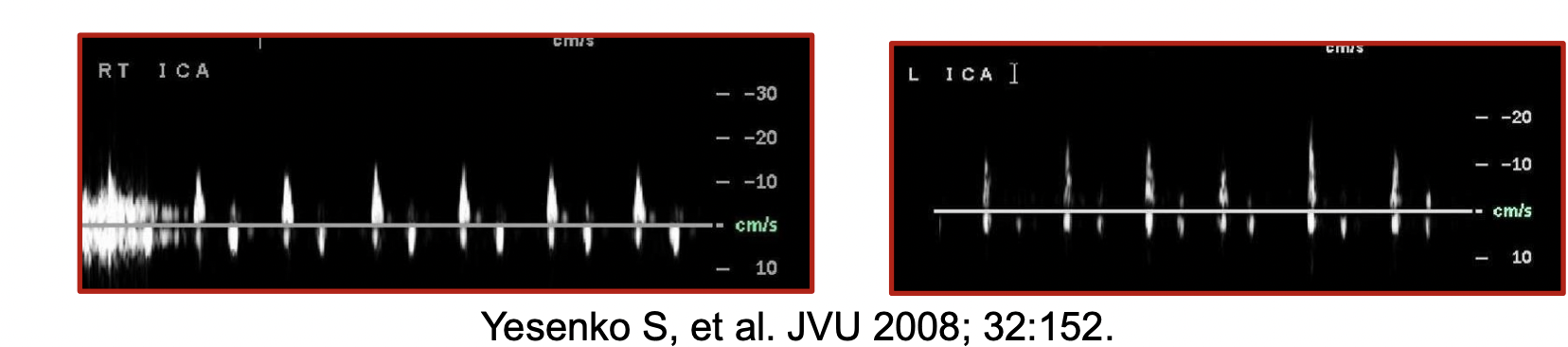
When would you see this waveform in the ICA
In someone with cerebral circulatory arrest, you’ll see these spikes intracranially in cases of brain death.
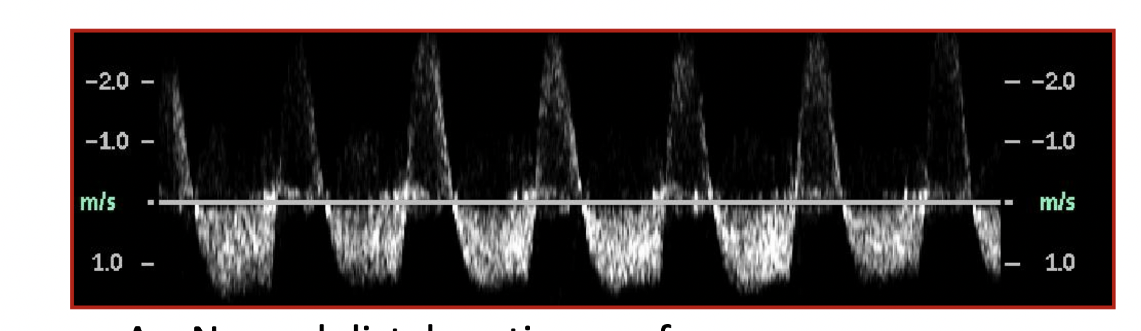
The following waveforms were recorded during an abdominal duplex exam in a patient who had undergone abdominal aortic surgery, what does it show?
two possible options, 1. arterial psuedoaneurysm (has the bidirectional to and fro flow). 2. type 2 aortic endoleak, you can tell by its retorgrade flow into the aneurysm sac.
usually a psuedoaneurysm will have more tubulence.

How would you explain this waveform
Hypertrophic obstructive cardiomyopathy, this results in waveforms with a rapid upstroke and a rapid descent, has a spike and dome appearance.
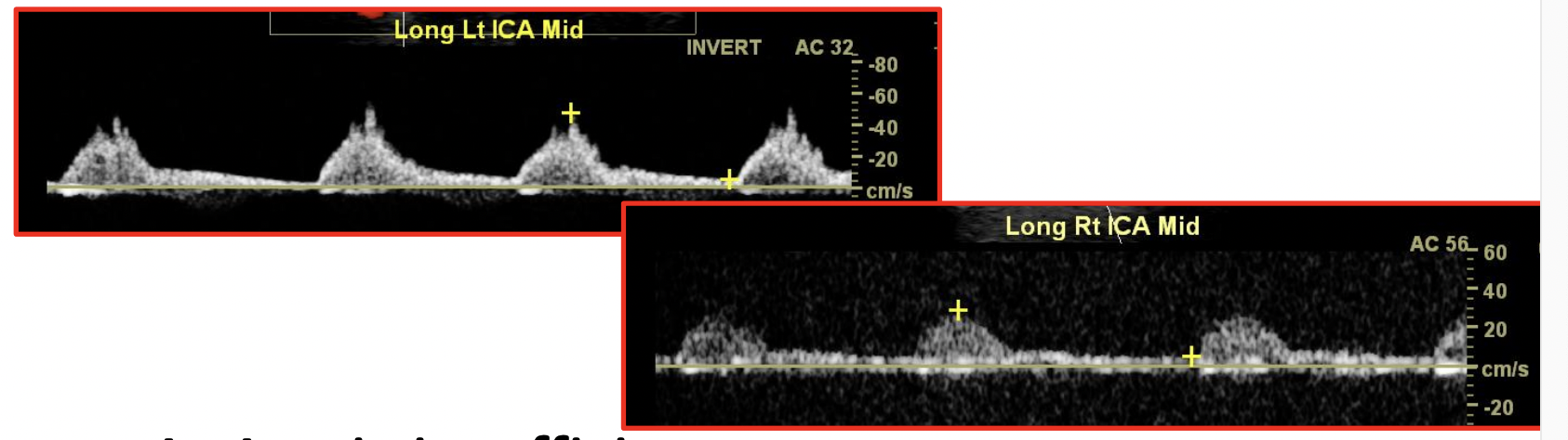
How would you explain this waveform
This is an example of an aortic stenosis, you can tell by the delayed systolic upstroke and rounded waveform appearance ( dampened)
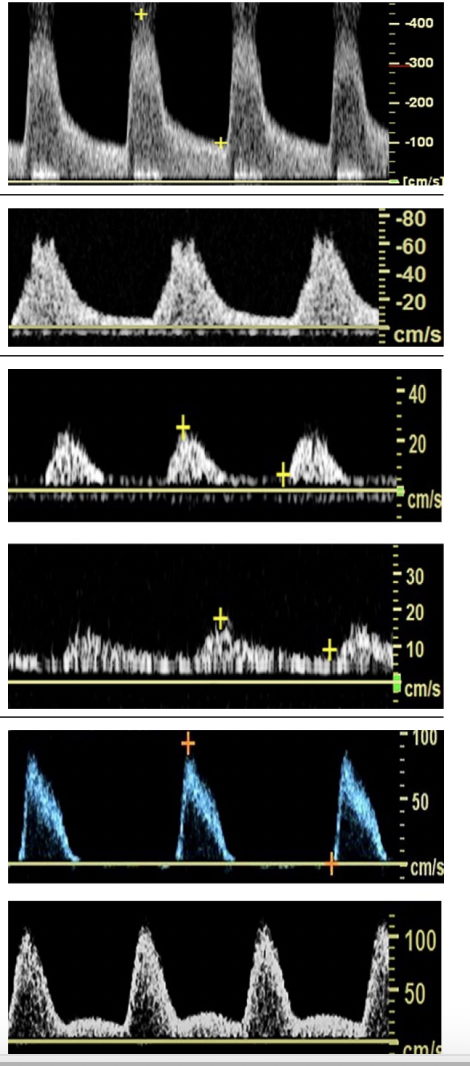
Draw out these waveforms and state where they are in response to a stenosis, which is prox at and distal to a stenosis
(5,6) image 5 and 6 are proximal to an occlusion, you can tell by the absent/abnormal outflow (diastolic) component
(1) Image 1 is at a stenosis, >75% you’ll get at least a fourfold velocity ratio increase compared to the normal proximal segment.
(2,3,4) image 2 3 and 4 show distal to an occlusion, waveform is lower velocity, dampened and mono phasic.
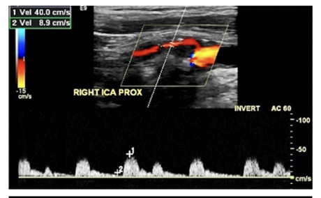
What does string sign mean?
The ICA is near occlusion, it shows theres a compromised ICA stenosis that often extends intracranially, a high-velocity jet may not be present and the waveform often demonstrates decreased velocities.
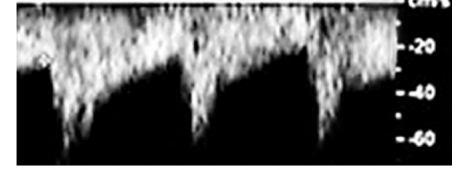
if there was a CCA occlusion and the ECA was being used as a collateral how would the waveform look like
Reversed flow, so theres collateral circulation. there would be a delayed systolic acceleration and an increased diastolic flow to compensate for the lack of flow in the CCA.
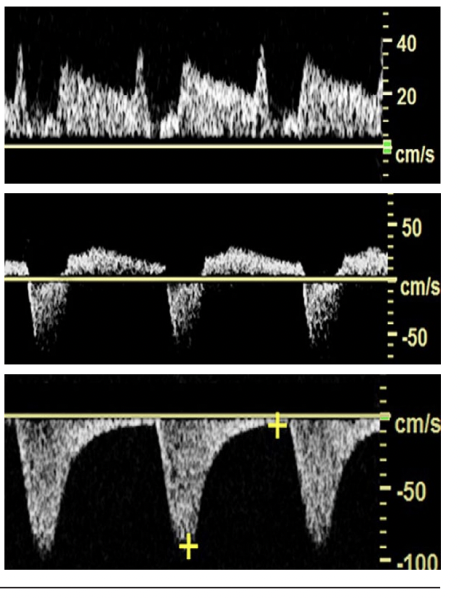
What do these waveforms represent
1) vertebral artery pre-steal: early in development of a subclavian steal, has bunny sign
2) Vertebral artery partial steal: alternating forward and reverse flow directions, seen as an incomplete steal
3) vertebral artery complete steal: waveform shows a complete steal with reverse flow direction
How does inspiration and expiration affect the venous waveforms ( upper extremities and lower extremities)
Opposite in lower and upper extremities
Inspiration: As diaphragm descends, decreases Intrathoracic pressure and increases intraabdominal
pressure
– Decreased venous return from the lower extremities
– Increased venous return from the upper extremities
• Expiration
– As diaphragm ascends, increases Intrathoracic pressure and decreases intraabdominal pressure
– Increased venous return from the lower extremities
– Decreased venous return from the upper extremities
What is Paget Schroetter Syndrome
form of upper extremity DVT characterized by blood clots in the subclavian and axillary veins, usually caused by repetitive motion,
Men more affected than women
• Incidence higher in patient’s dominate arm
may also be due to clavicle impingement due to associated anatomic abnormality of cervical rib
symptoms include: arm swelling, pain and heaviness, stiffness and visible veins
What are the symptoms for SVCS
Compression of the Superior Vena Cava
Symptoms include: Slow and progressive
– Swelling of face and arms
– Heaviness of upper extremities
– Formation of collateral veins that may be visible on surface
– Coughing or difficulty breathing
– Lightheadedness or confusion
Whats the diameter criteria for a UEV vein mapping
Diameter greater than 0.4 cm for arterial bypass graft or 0.25 for dialysis access
Whats the difference between AVF and AVG/DAG
Fistula (Arteriovenous fistula = AVF)= patient’s native artery and vein
Graft (Arteriovenous graft= AVG or Dialysis Access Graft= DAG) = patients native artery connected to synthetic material and then patient's native
deep vein
What’s the preference of access placement for dialysis?
Nondominant forearm cephalic vein fistula
Dominant forearm cephalic vein fistula
Nondominant or dominant upper arm cephalic vein fistula
Nondominant or dominant upper arm basilic vein transposition fistula
Forearm loop graft
Upper arm straight graft
Upper arm loop graft (ax artery to ax vein)
Lower extremity loop graft (CFA to CFV)
They prefer forearm first because the closer the fistula is to the heart the greater risk of developing heart failure
What does dialysis do?
Cleans excess fluid and waste material from the blood through a semi-permeable membrane
– Known as a “dialyzer”
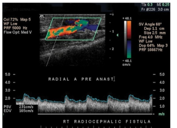
In a fistula, the arterial inflow will be, the diastolic flow will be___, and the PSV will be ___.
low resistive, increased, increased
In a fistula the venous outflow will be____, the velocities will be ___, will likely demonstrate ____ just distal to anastomosis.
arterialized (low resistive), increased, turbulence
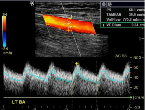
whats the criteria for the volume flow (TAMV) in a fistula
Obtain volume flow at mid access (TAMV)
• Determine volume flow at straight part of mid access
• Low <600 ml/min
• Adequate 600- 800 ml/min
• High >800 ml/min
too low <460- 500mL/min): Increased risk of fistula clotting (thrombosis).
Too High Flow (>1,500-2000 mL/min):
Cardiac Overload: High-output heart failure, as excessive blood is shunted back to the heart.
Vascular Complications: Increased risk of aneurysmal dilation (weakening of the vein wall) and venous stenosis.
what happens in a Dialysis Access Fistula Steal
Distal arterial blood flow is reversed into the access instead of supplying the distal extremity (often hand)
• Symptoms include pain, coolness and paleness in the extremity (hand) - can occur after exercise (claudication)
Radial and ulnar artery steal: Alternating or completely reversed flow seen in the radial and ulnar arteries upon spectral Doppler evaluation
if there is a steal in a fistula what happens?
an arterial bypass graft is placed from proximal to the arterial anastomosis of the fistula to distal to the anastomosis to shunt blood to the distal extremity
Distal Revascularization with Interval ligation = DRIL
- Ligation of native arterial connection between distal extremity and fistula
– Ligation portion not always completed
• Depends on the severity of the steal and the surgeons practice philosophy
The IVC is ___ to the spine and to the patients ___
anterior, right
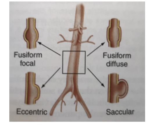
what are the different types of aneurysms
Fusiform: (focal or diffuse) a concentric enlargement
Saccular: enlargement in a shape of a sac
Eccentric: pouch-like bulge on one side of an artery wall, rather than a uniform swelling of the entire vessel
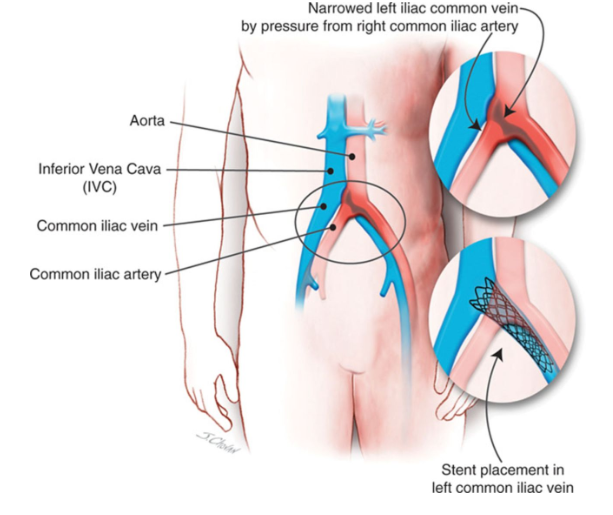
What is May Thurners syndrome
Compression of Left Common Iliac Vein by Right Common Iliac Artery against the lumbar vertebrae with or without DVT
– Causes stasis of the LCIV, often results in DVT
Typically diagnosed in women in 20s to 40s after prolonged immobilization or pregnancy
youll see High velocities venous flow or Doppler silence in LCIV Continuous waveforms distally in Left LLE vein (including EIV and CFV)
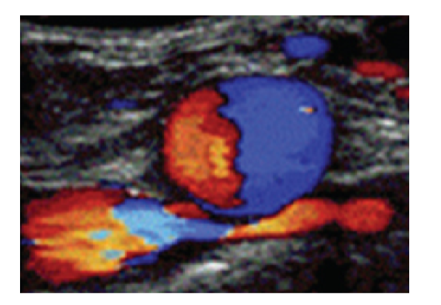
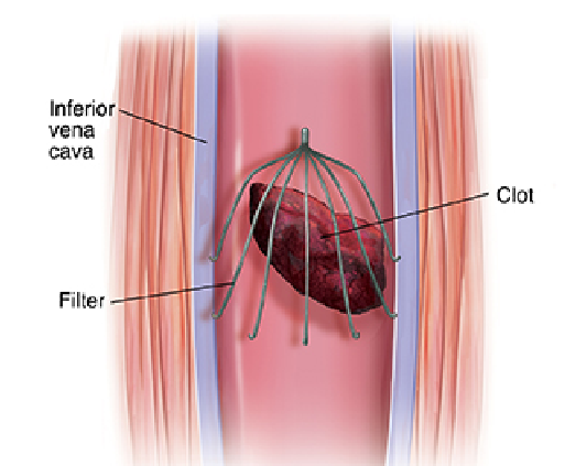
What is an IVC filter
Small device placed within proximal to mid abdominal IVC ( just inferior to the renal veins) to catch thromboembolic particles before reaching the lungs and becoming a pulmonary emboli
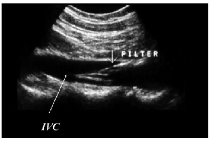
Why do we scan the ophthalmic artery in a TCD
We scan the ophthalmic to look for potential collaterals, the waveform should be red ( toward) and high resistive, if collateral are present it will not have these qualities. This shows external-to-internal carotid artery (ECA-to-ICA) collateralization
this should be shallow at 40-60mm depth
what are the depths for TCDs? (MCA, TICA, ACA, MCA/ACA bifur, Distal ICA, VA, BA)
MCA: 60 mm
TICA: 55-65
ACA: 60-80
PCA: 60-70
MCA/ACA bifur: 55-65
Distal ICA: 60-80
VA: 60-90
BA:70-120
What’s vasospasm, whats the criteria for it?
Vasospasm: a complication of subarachnoid hemorrhage related to a rupture of an intracranial aneurysm or head trauma. The sudden, temporary tightening or constriction of an artery, causing the vessel to narrow and significantly reducing blood flow to tissues.
Hyperdynamic flow: greater than or equal to 80 ratio less than 3
Mild: 120-149 ratio greater or equal to 3.
Moderate: 150-199 ratio greater than 3.
Severe: greater or equal to 200, ratio greater or equal to 6
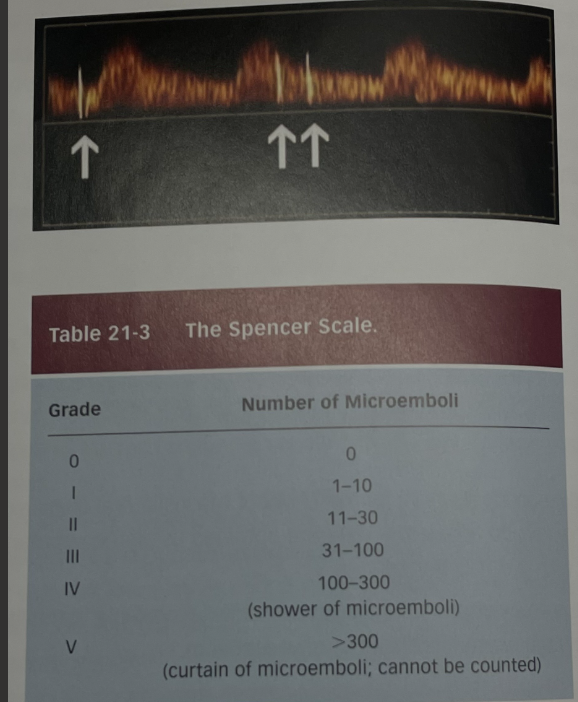
How would we use TCD for a patient with a PFO
We monitor the MCA’s and insert a contrast agent into an arm vein during a resting state. If no microbubbles are detected monitoring continues as the patient does the Valsalva maneuver.
Microbubbles are also referred to as HITS, criteria is above.
What waveforms would you see in someone with brain death ( TCD wise)
in the development of cerebral circulatory arrest you will see 4 stages.
1: increasing pulsatility with end diastole reaching zero
Oscillating flow, with forward and reverse flow are equal
Some patients may reach the stage where flow patterns turn into reduced velocity spikes.
Some patients may have no flow. but this could be an absence of a temporal bone window so we repeat the study 30 mins apart.
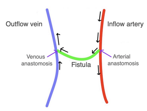
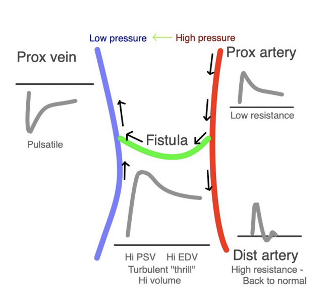
What waveforms will you see in someone with dialysis (inflow and outflow)
Proximal vein: pulsatile, below baseline
Inflow artery Proximal to anast: low resistive, high velocity with significant diastolic flow
Distal artery: bac
k to normal, high resistive
Fistula: low resistive, high PSV and high EDV
Why do we scan the radial and ulnar veins in a fistula
to evaluate for steal syndrome, scan the DISTAL ulnar and radial arteries to look for reversal of flow ( below baseline).
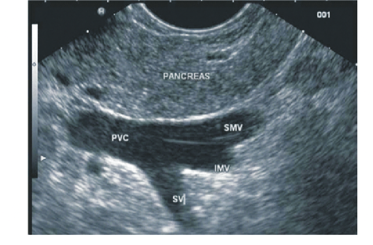
What is the portosplenic confluence
confluence of the portal vein, SMV and splenic vein
What will u see in PTN?
Caput Medusae: dilated tortuous vessels around the umbilicus caused by portal HTN.
Splenomegaly: an enlarged spleen, this is due to the backlog of flow into the portal system
Cavernous transformation: the formation of varices from the increased portal pressure. Accessory veins dilate at the porta hepatis.
What is normal MPV diameter
less than 1.3, an AP larger than 1.3 is indicative of portal hypertension
in someone with CIrrhosis or other chronic liver disease what would we see on ultrasound.
Cirrhosis is usually what causes portal hypertension, on ultrasound you will see flow reversal in the portal vein and will see portosystemic collaterals. you may visualize abdominal varices by the spleen and stomach. a Umbilical vein may also be present.
Caput medusae- refers to the dilated, tortuous vessels around the umbilicus caused by portal HTN.
What is Budd Chiari syndrome
Hepatic vein obstruction, usually caused by thrombosis but can also be caused by tumor infiltration or stenosis.
common causes of thrombosis are oral contraceptives and myleoproliferative processes.
What are the indications and things we’ll see on ultrasound on someone with acute PV thrombosis
usually due to cirrhosis and portal hypertension.
if acute it will present with abdominal pain, distention, diarrea, GI bleeding and lactic acidosis
absent or minimal portal vein flow, with vessels dilated distal to thrombus
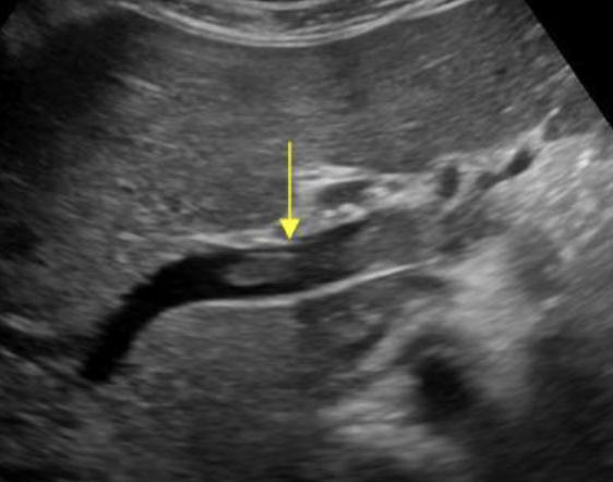
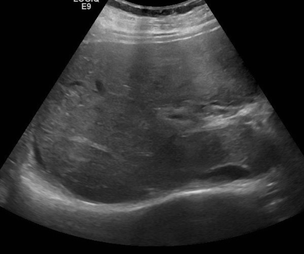
What are the indications and things we’ll see on ultrasound on someone with chronic PV thrombosis
patients may be asymptomatic or present with varices or hypersplenism.
PV will become fibrous and contracted, you will see multiple collateral vessels surrounding normal territory of PV
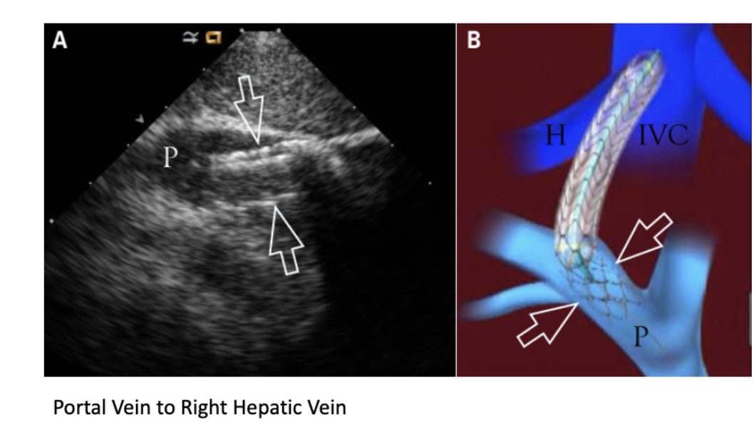
What is TIPs, what will we see on ultrasound,
Transjugular Intrahepatic Portocaval Shunt, IJV is the insertion site for the portocaval shunt.
treatment for portal hypertension
shunt placed between Portal vein and Hepatic vein (typically right hepatic vein) held open with metal stents
• Portal blood flow is diverted to one of the major Hepatic veins
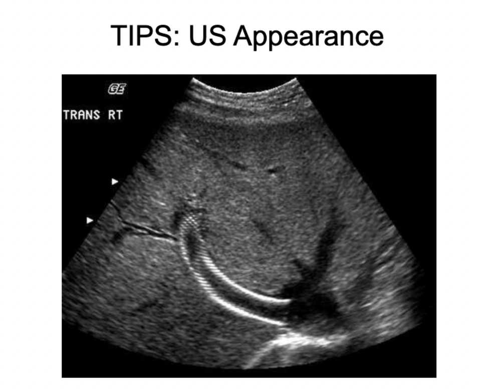
there will be pulsatile flow throughout the stent, PV will be hepatopedal at prox anastomosis and hepatofugal at the distal anastomosis ( hepatic veins)
-The hepatic vein anastomosis is the most common site for stenosis
What is the TIPS criteria
a velocity lower than 30cm/sec indicates impending shunt failure.
usually ranges between 90-190cm/sec
Common complications include a stenosis of stent, or the hepatic outflow vein, or complete thrombosis of stent or outflow vein
What is aortic coarctation
a congenital heart defect where a segment of the aorta is narrowed, restricting blood flow to the lower body
causes high blood pressure in the arms/head and low pressure in the legs, with symptoms like weak pulses, cold feet, and feeding difficulties
this is why we do a limited ABI on peds patients
What does an allens test do
determines if the hand can rely soley on the ulnar artery if the radial artery is blocked or removed.
The ulnar artery supplies most of blood flow to the hand.
the connection between the superficial arch and deep palmar arch allows blood to reach all fingers even if one primary artery is blocked
Superficial palmar arch is formed by ulnar artery and superficial branch of radial artery. Deep palmar arch is formed by radial artery and deep branch of ulnar artery
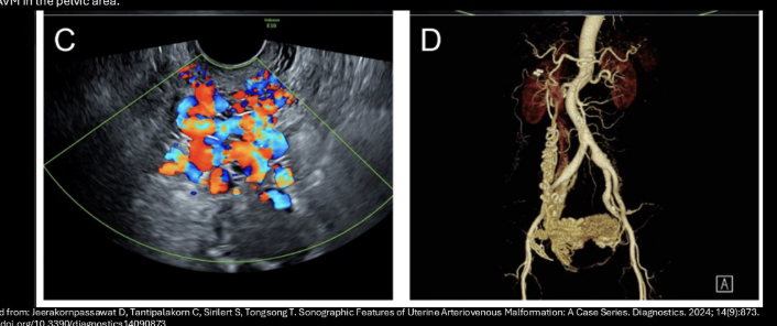
What is an AVM
Congenital connection between the arterioles and venules (lack of capillary bed)
will have multiple connections and surrounding venous collaterals and dilated veins
Seen anywhere but most common include the brain. kidney, uterus/pelvis
What is a segmental exam, what is the criteria
4 calfs are placed HT (high thigh) AK ( above knee) BK ( below knee) and ankle levels. CW probe is placed at dpa or PTA. same criteria as ABI <0.9 is abnormal
The proximal thigh cuff pressure should be 30-40mmHg higher than the brachial pressure.
What is helical flow, where do you see it
where blood moves in a spiral or vortex pattern rather than laminar flow, this can be seen in bifurications (normal) or in the portal vein after liver transplant
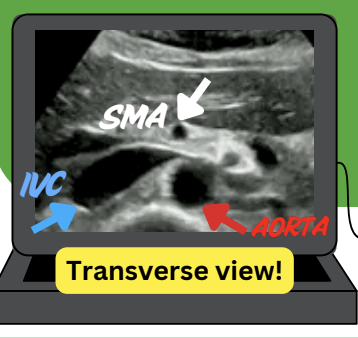
Post-prandially what happens to the SMA, what is the criteria for a SMA stenosis
it will exhibit a low-resistive waveform due to increased blood flow to the intestines
>275 cm/sec
Where do you see the IMA?
Go to the bifurcation and angle superiorly, it’s just 1.3 cm from the aortic bifurication, travels slightly to the left and will be high resistive during fasting and low resistive after fasting.
What are the types of endoleaks
Will be seen in EVAR. Endovascular aortic repair.
Type 1: leaks at the Proximal or distal graft attachment sites. Flow will be high-velocity, pulsatile
Type 2: Retrograde flow into the anuerysm sac from the side branches of the aorta such as the lumbar or inferior mesenteric arteries. ( youll see bi-directional flow)
Type 3: Caused by a defect in the graft either due to a fabric tear or disconnection (one of the most dangerous)
Type 4: Graft porosity, diffuse oozing through graft material ( not really a problem anymore due to advances in graft construction)
Type 5: Sac enlargement without visible leak ( inc by 0.5cm or more)