Looks like no one added any tags here yet for you.
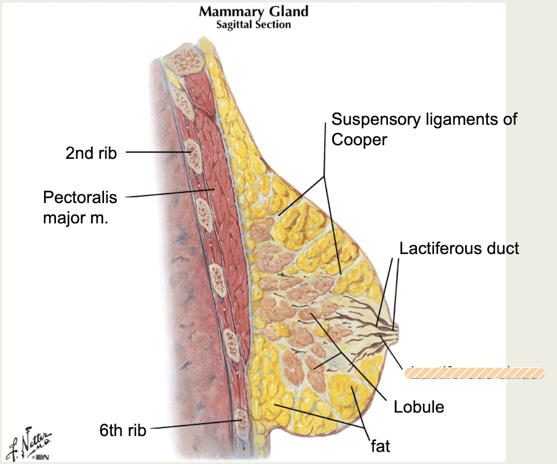
female breast
-subcutaneous tissue
-Glandular and supporting fibrous connective tissue embedded in a fatty matrix
-Contains lobes, lobules, lactiferous ducts, blood vessels, lymphatics and nerves
-Modified sweat gland
female breast
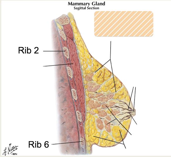
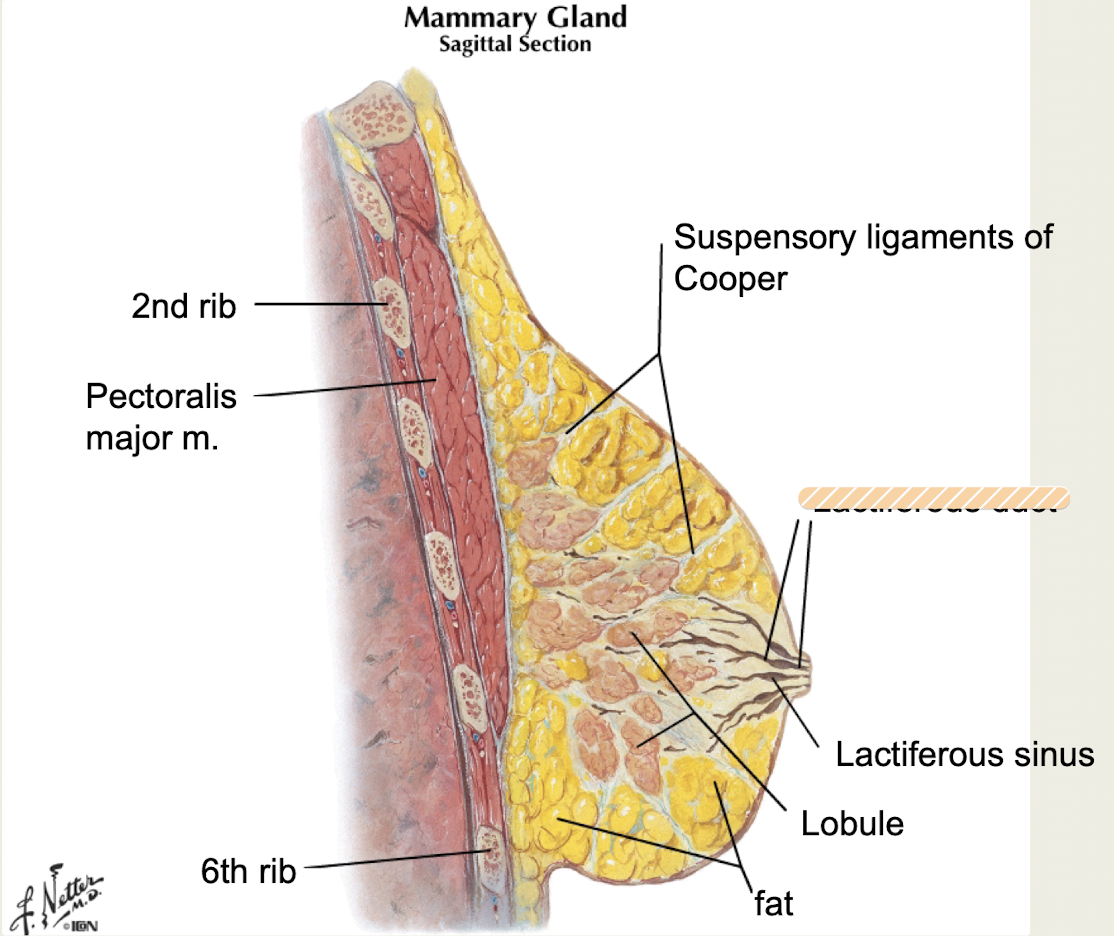
-overlies pectoralis major/minor, parts of serratus anterior and external oblique mm
-transversely from lateral border of sternum to midaxillary line
-vertically base located from ribs 2-6
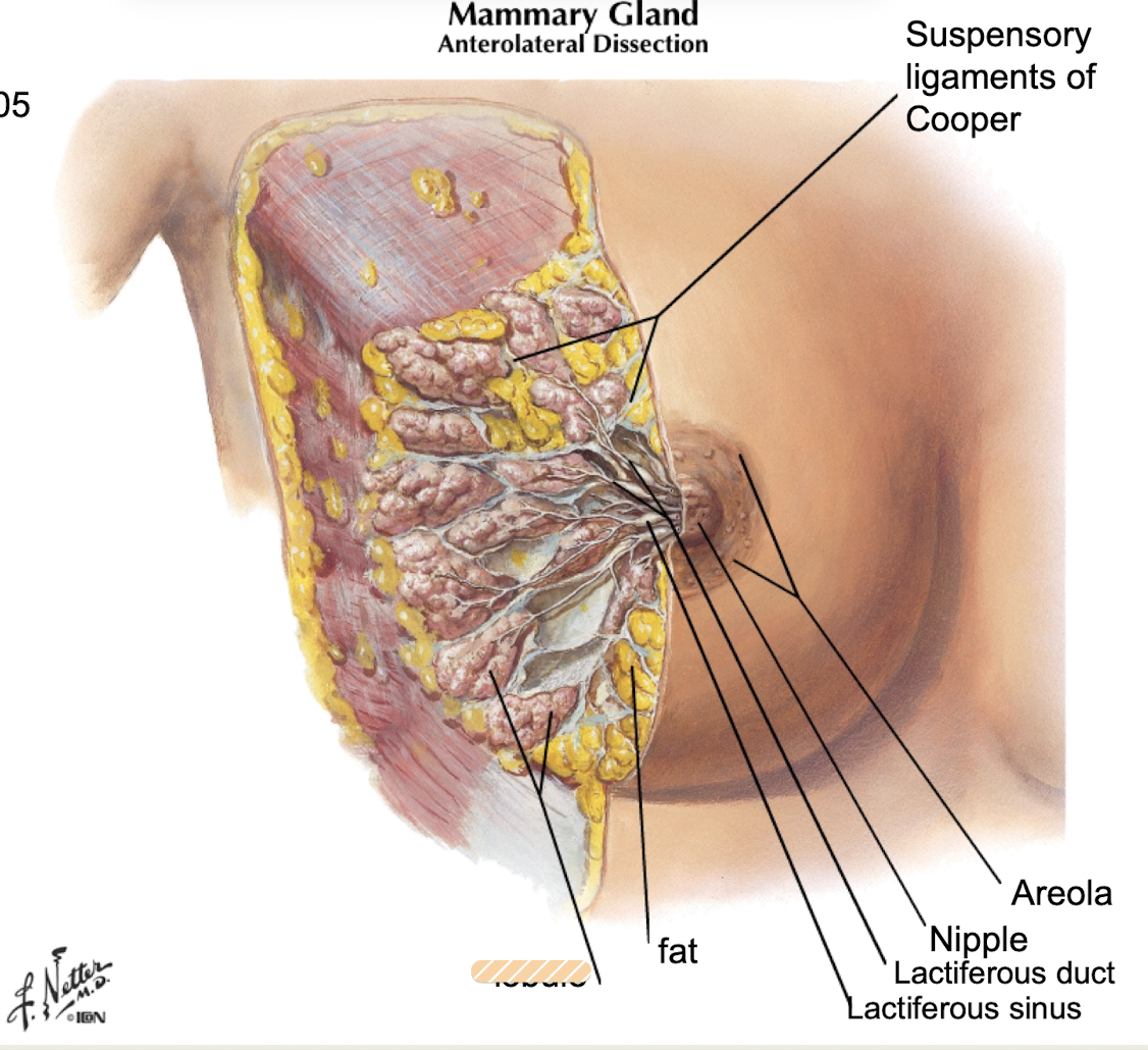
Suspensory ligaments of Cooper
fibrous connective tissue skin ligaments that attach breast tissue to dermis and provide support
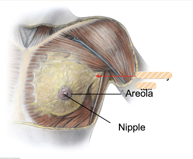
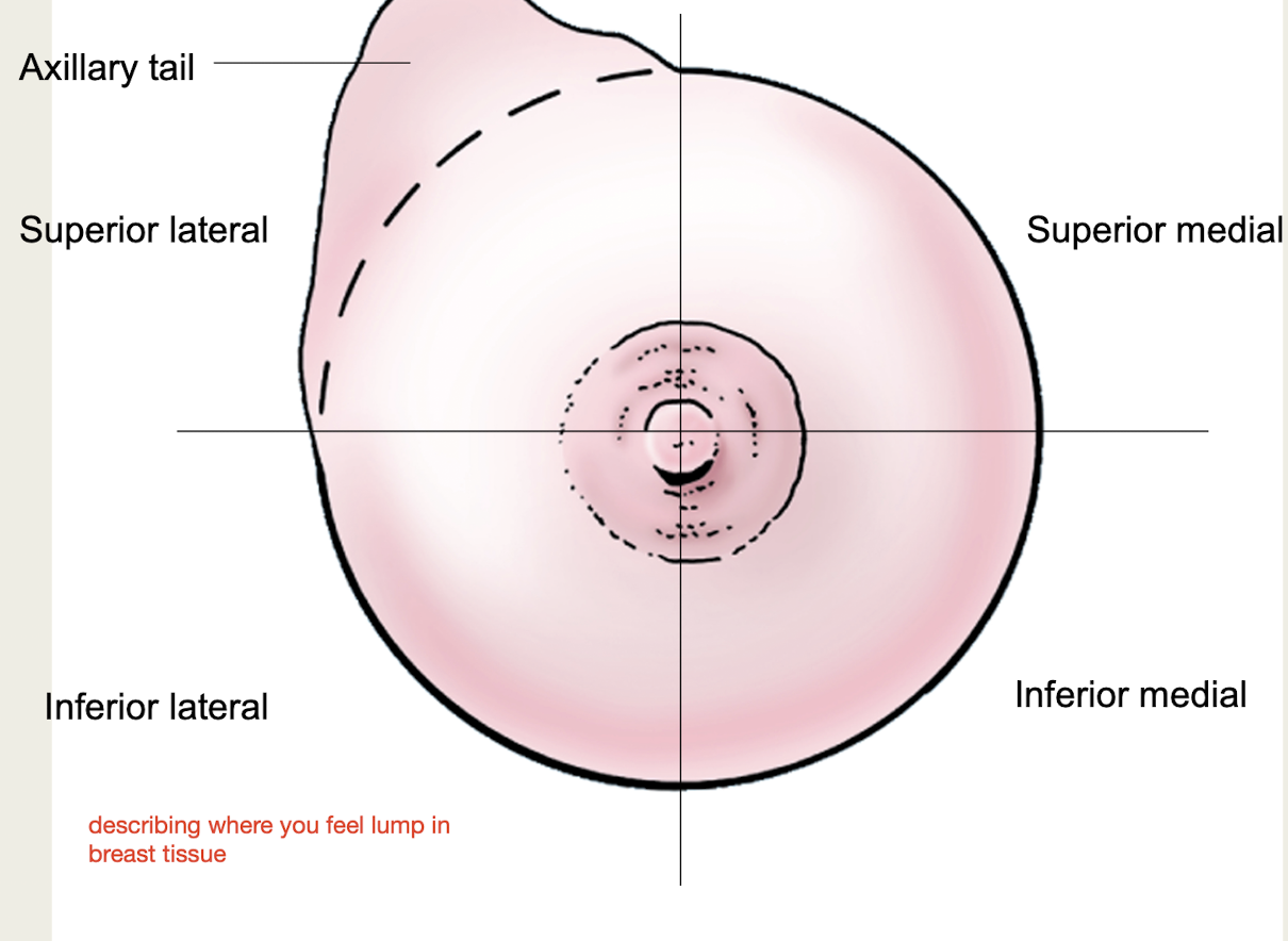
Axillary tail/process (of Spence)
part of breast that extends over pectoralis major muscle to the axilla
Lobes
15-25 compound alveolar glands in breast
Lobules
clusters of alveoli in each lobe of breasts where milk is produced
Ducts
carry milk from glandular tissue in breasts
Lactiferous ducts
largest ducts, drain lobules
Lactiferous sinus
deep to areola, dilated region of lactiferous duct
Stroma of breast
Adipose tissue and connective tissue that surrounds the lobules and ducts
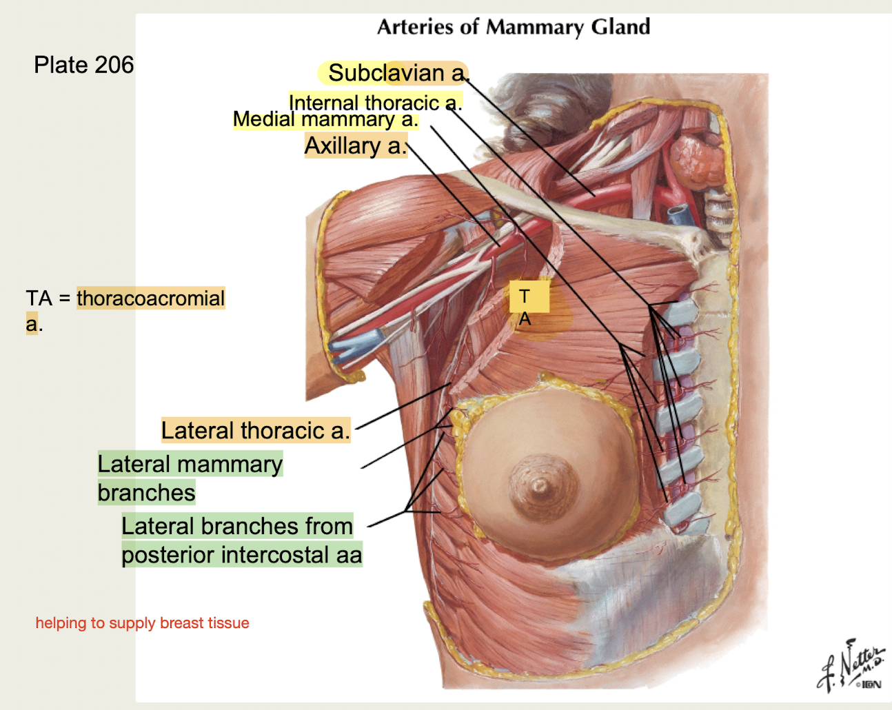
Breast vasculature
1.Subclavian a → internal thoracic a → medial mammary branches
2.Subclavian a → axillary a → lateral thoracic and thoracoacromial aa
3.Thoracic aorta → posterior intercostal aa → lateral mammary branches
Supply breast tissue
Breast Quadrants
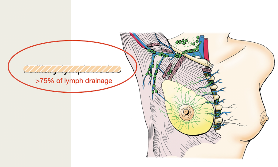
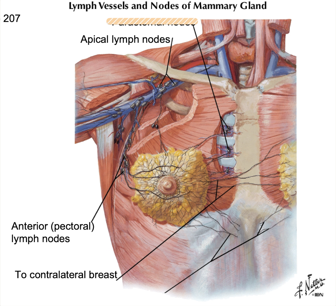
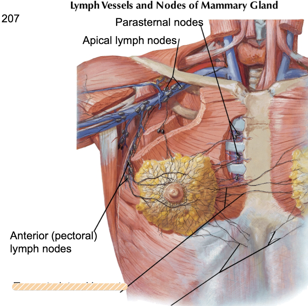
Axillary lymph nodes
75% of lymph drainage in breast
-Primary route for metastasis of breast carcinoma
Parasternal lymph nodes
Medial quadrants of breast drain into _________ along internal thoracic blood vessels
Contralateral lymph nodes
Medial quadrants of breast drain into _________ by crossing midline
Abdominal lymph nodes
Lower quadrants of breast drain into _____
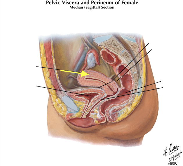
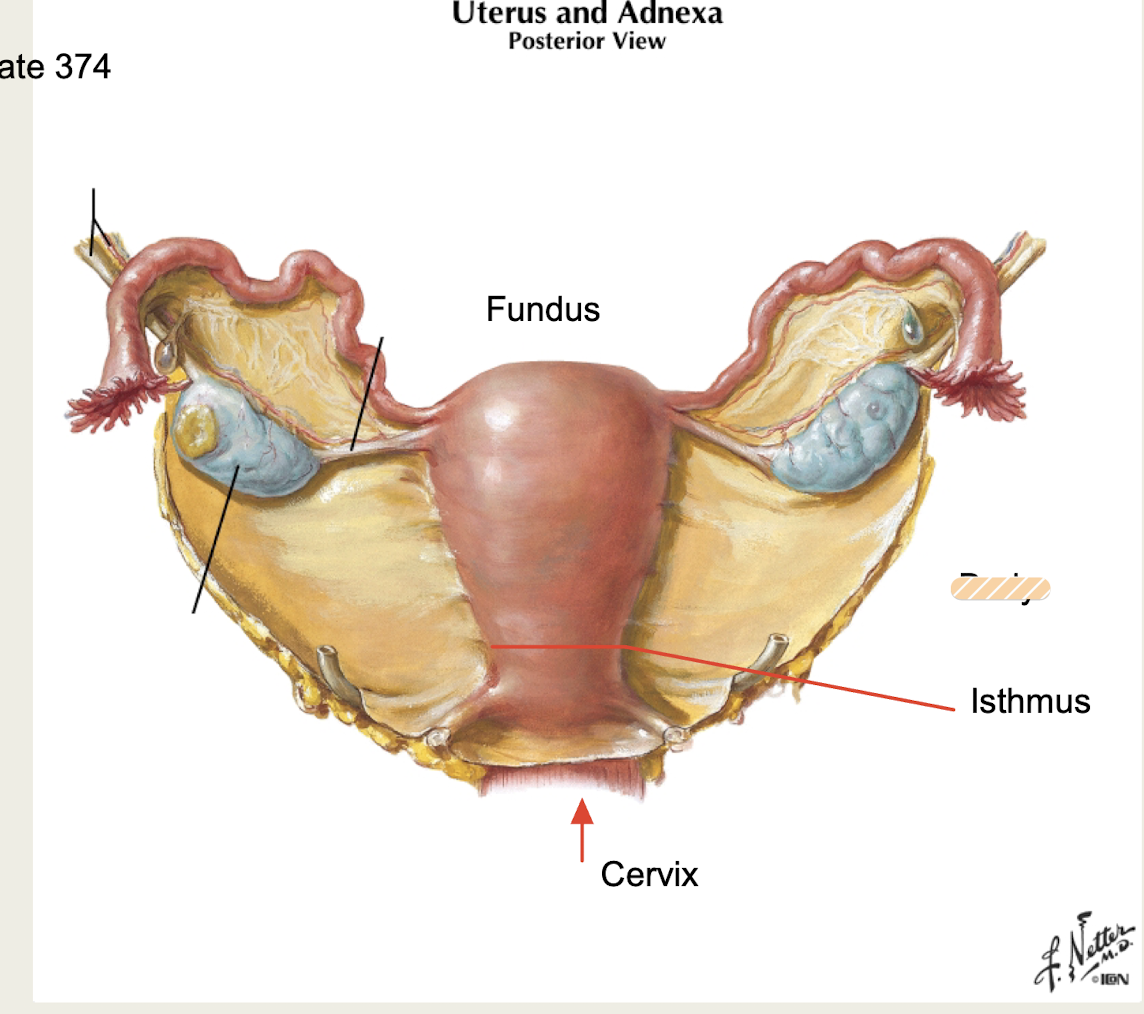
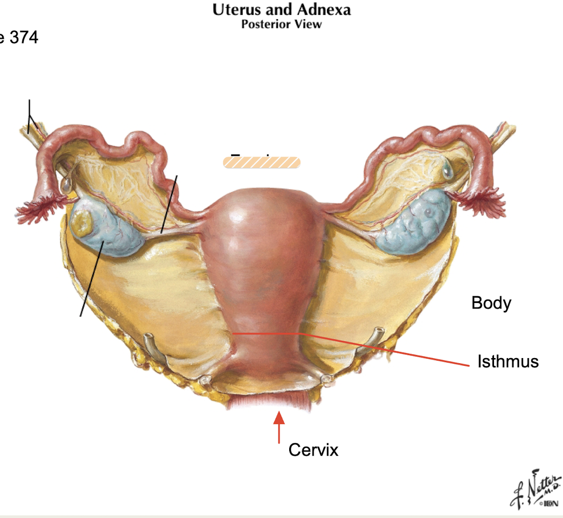
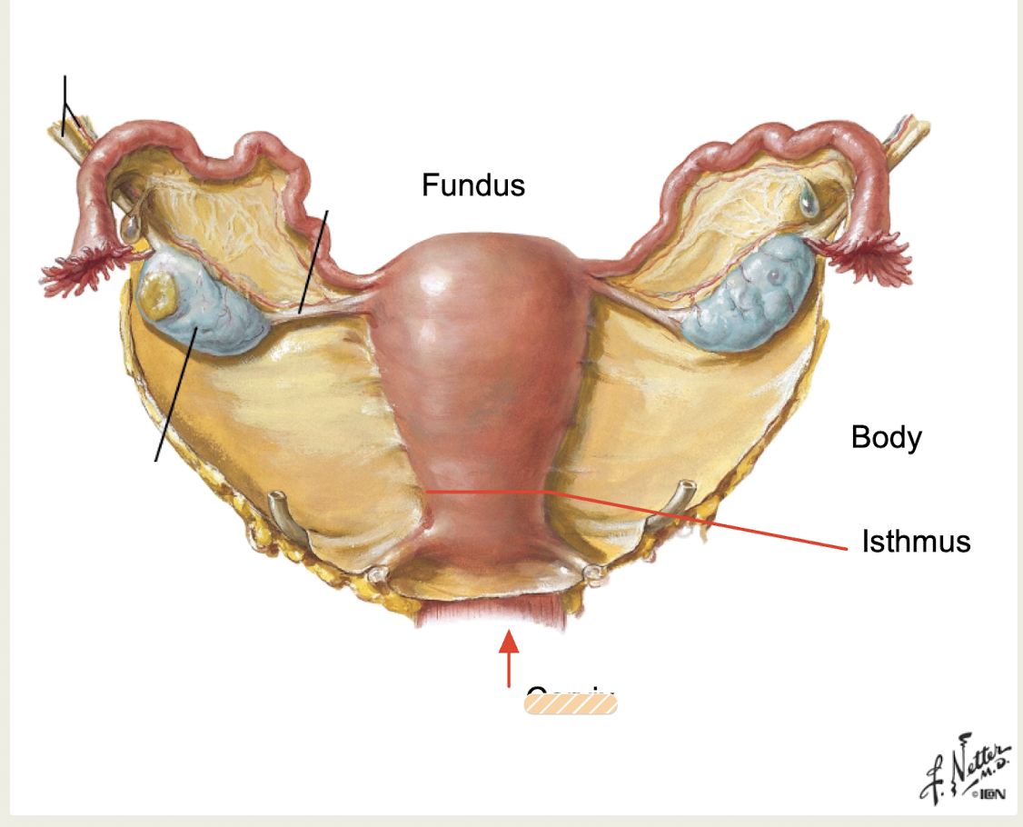
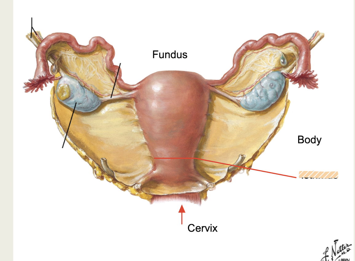
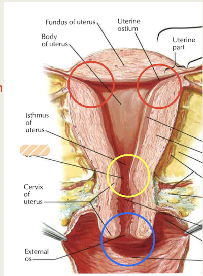
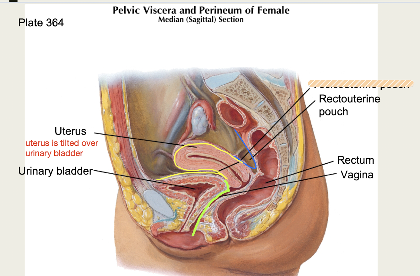
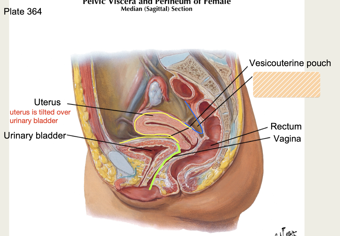
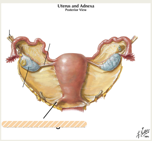
Uterus
-Pelvic hollow muscular organ; anterior to rectum, posterior to bladder
-Typically tilted over fundus of urinary bladder; covered superiorly by peritoneum
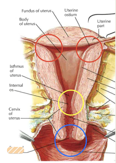
Body
main part of uterus
Fundus
rounded top of uterus
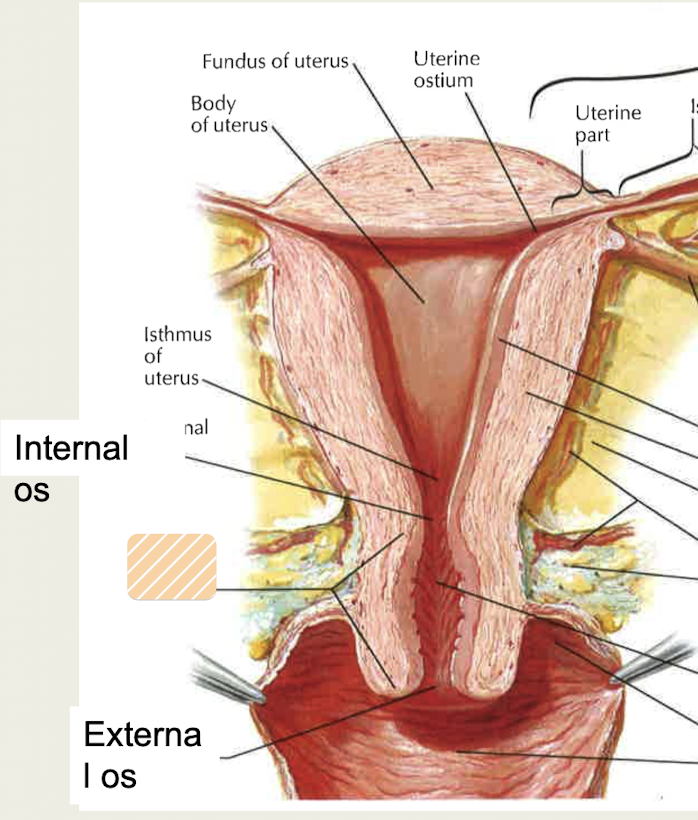
Cervix
narrow neck of uterus, projects into vagina
Isthmus
narrowed region of uterus between body and cervix
External os
cervix of uterus; opening into vagina; visible on speculum exam
Internal os
cervix of uterus; opening into uterus
Perimetrium
visceral peritoneum, external layer of uterus
Myometrium
smooth muscle, middle layer of uterus
Endometrium
-inner layer of uterus, shed with menses; highly vascular tissue where embryo implants
-contains stratum functionalis and stratum basalis layers
Stratum functionalis
-Layer of endometrium closer to lumen; simple columnar epithelium + underlying stroma
-Sloughed off with menstrual cycle
-Temporary tissue that regrows each cycle
Stratum basalis
-Permanent stromal tissue of endometrium + glandular tissue
-Source of regrowth for stratum functionalis
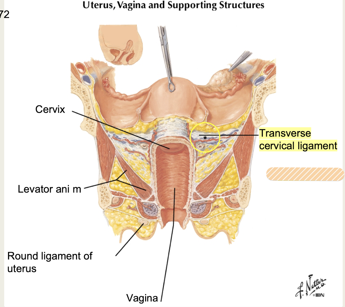
Cervix
Narrow inferior 1/3 of uterus that opens into vagina; contains internal and external os
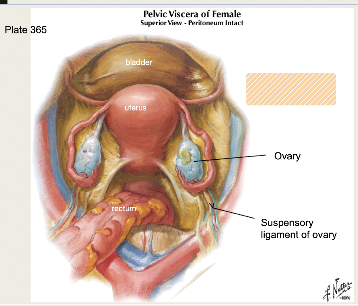
Broad ligament
Part of peritoneum of uterus; includes Mesometrium, Mesovarium, Masosalpinx
Mesometrium
Broad ligament (peritoneum) of uterus that provides lateral support and covers uterus
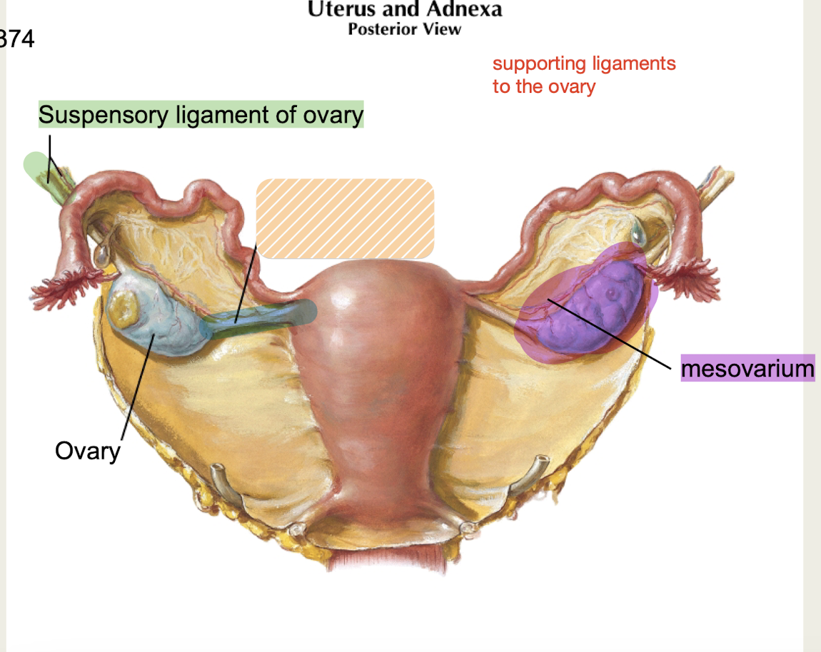
Mesovarium
ovarian support, part of broad ligament (peritoneum) of uterus that covers ovaries
Mesosalpinx
Broad ligament (peritoneum) of uterus that covers uterine tubes (fallopian tubes)
Vesicouterine pouch
Portion of broad ligament (peritoneum) of uterus found between bladder and uterus
Rectouterine pouch
Portion of broad ligament (peritoneum) of uterus found between uterus and rectum
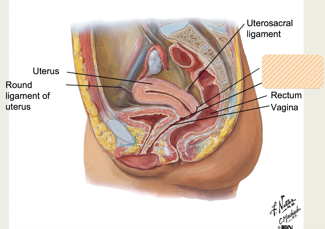
Round ligament
-Bilateral peritoneum attaches to superior lateral wall of uterus
-Runs through inguinal canal, attaches to labia majora
aka ligamentum teres
Uterine a/v
Which uterine blood vessels run in the transverse cervical ligament?
Transverse cervical ligaments
bilateral ligaments that attach cervix and superior portion of vagina to lateral wall
contain uterine artery and vein*
Uterosacral ligaments
bilateral ligaments that attach to posterior inferior portion of uterus and sacrum
provide posterior and inferior support
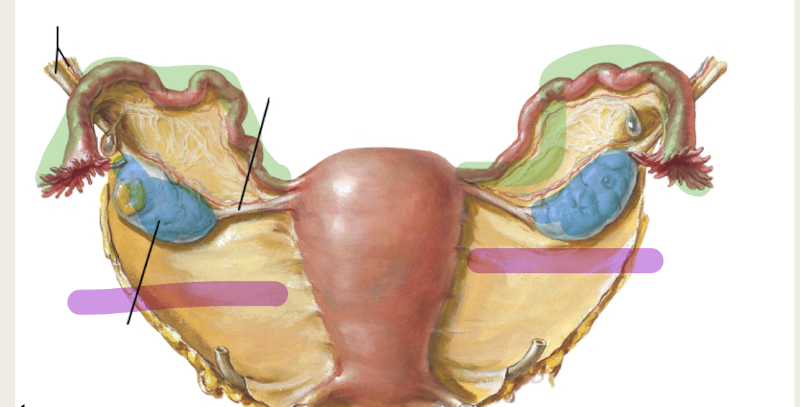
Adnexa
region adjacent to the uterus that includes ovary, uterine tube, broad ligament
Clinical significance: site of tumors, ovarian cysts, ectopic pregnancy
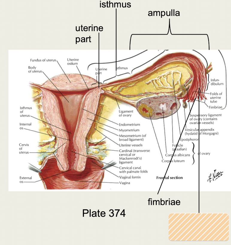
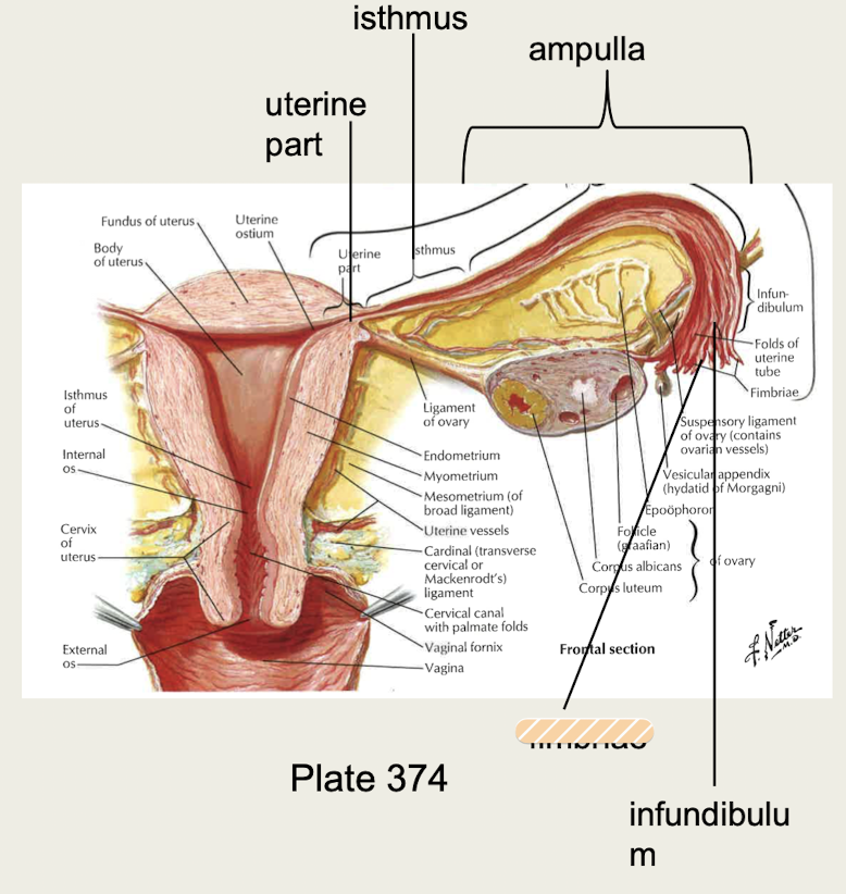
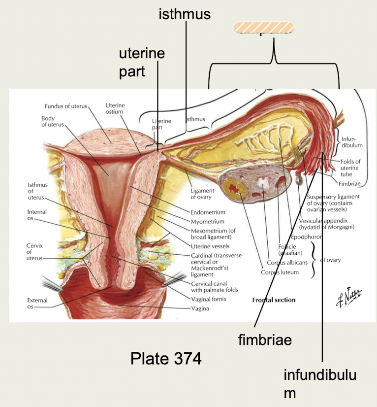
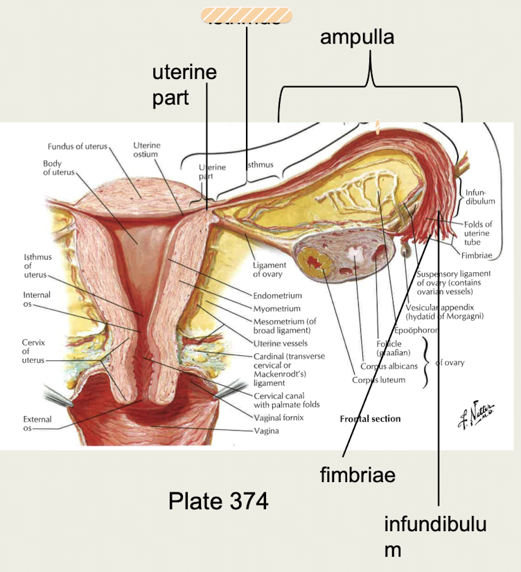
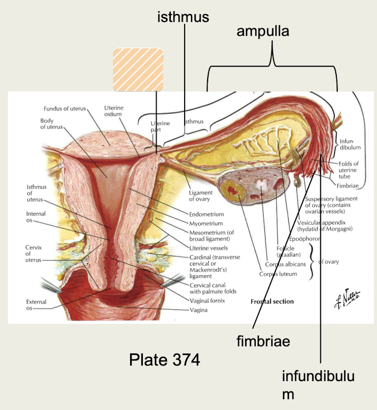
Uterine tube (Oviduct, Fallopian tube)
site of fertilization, receives oocyte after ovulation and transports oocyte from ovary to uterus
Infundibulum
funnel-shaped beginning part of uterine tubes
Fimbriae
Finger-like projections at end of uterine tubes
Ampulla
Main part of uterine tube where fertilization typically occurs
Isthmus
Narrowing of uterine tube
Uterine part
Section of uterine tube that travels through muscle of uterus
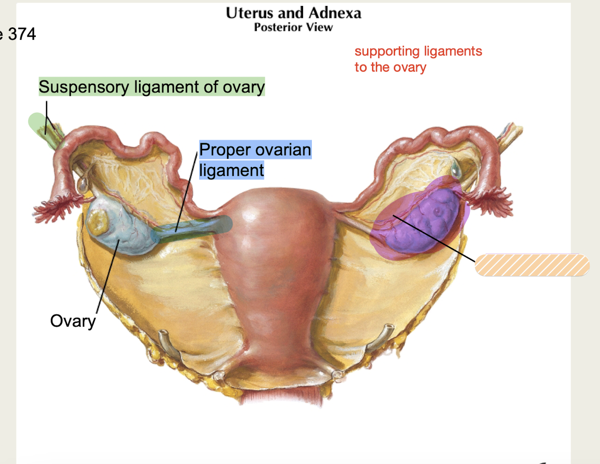
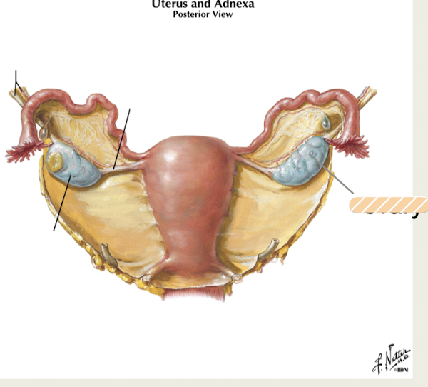
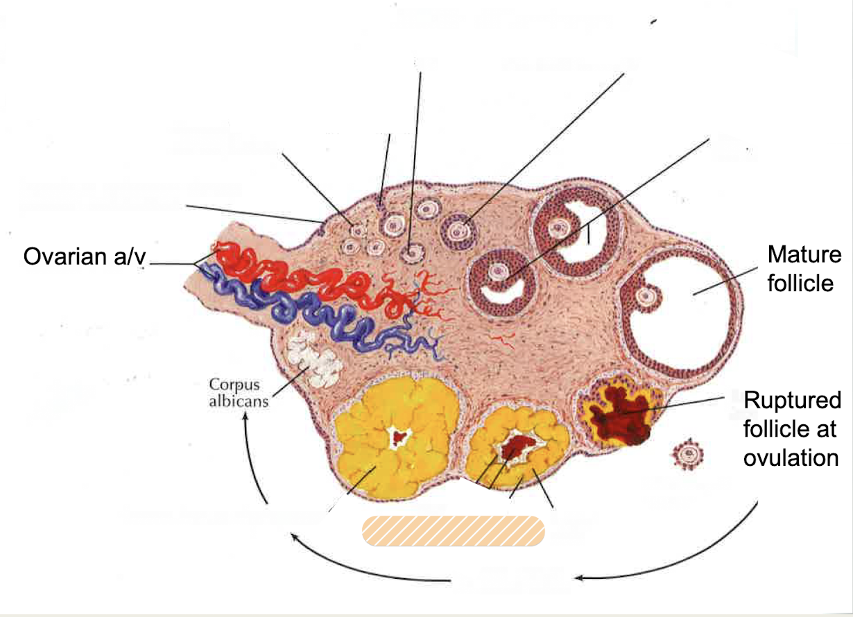
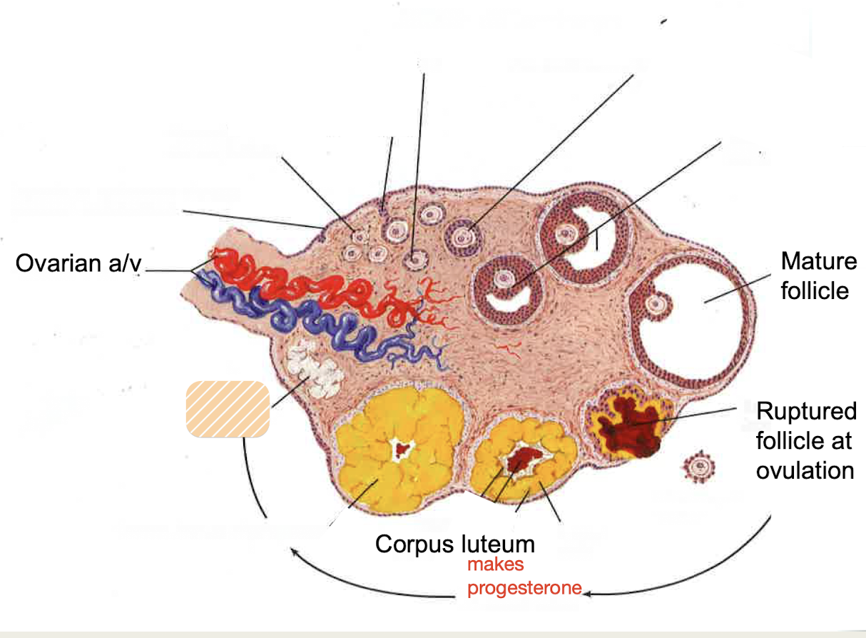
Ovary
female gonad, make hormones estrogen and progesterone (as well as androgens)
releases oocyte at ovulation
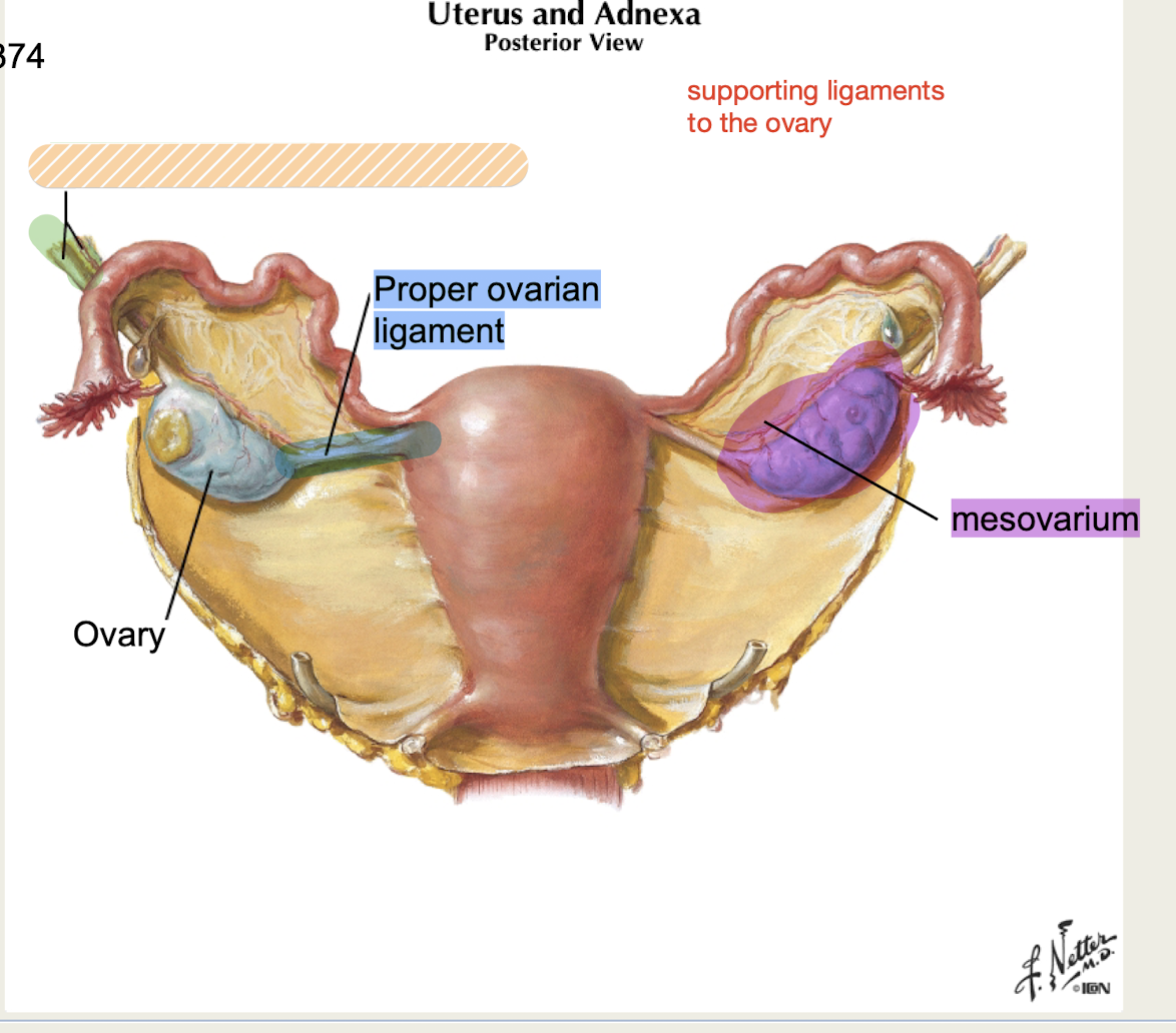
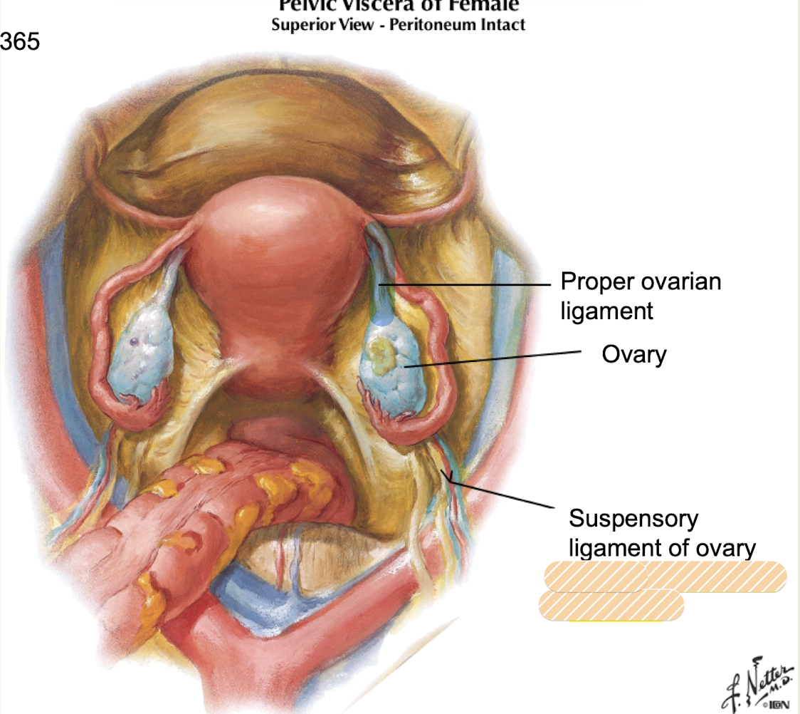
Suspensory ligament
ovarian support attached to lateral body wall
contains ovarian artery and vein*
Ovarian a/v
What blood vessels are contained within the suspensory ligament of ovary?
Proper ovarian ligament
ovarian support from ovary to uterus
Germinal epithelium
simple cuboidal epithelium on the surface of the ovary
Tunica albuginea
connective tissue capsule that surrounds the ovary
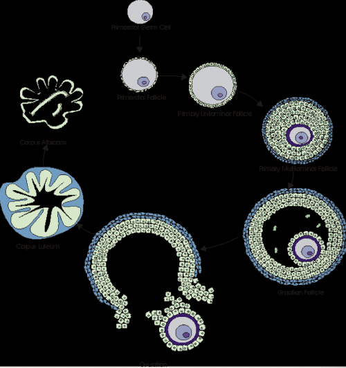
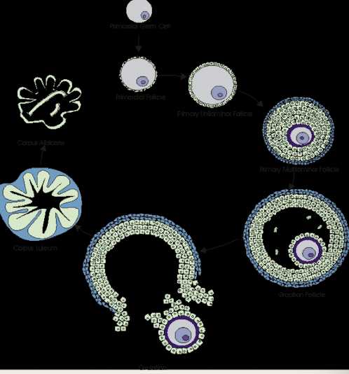
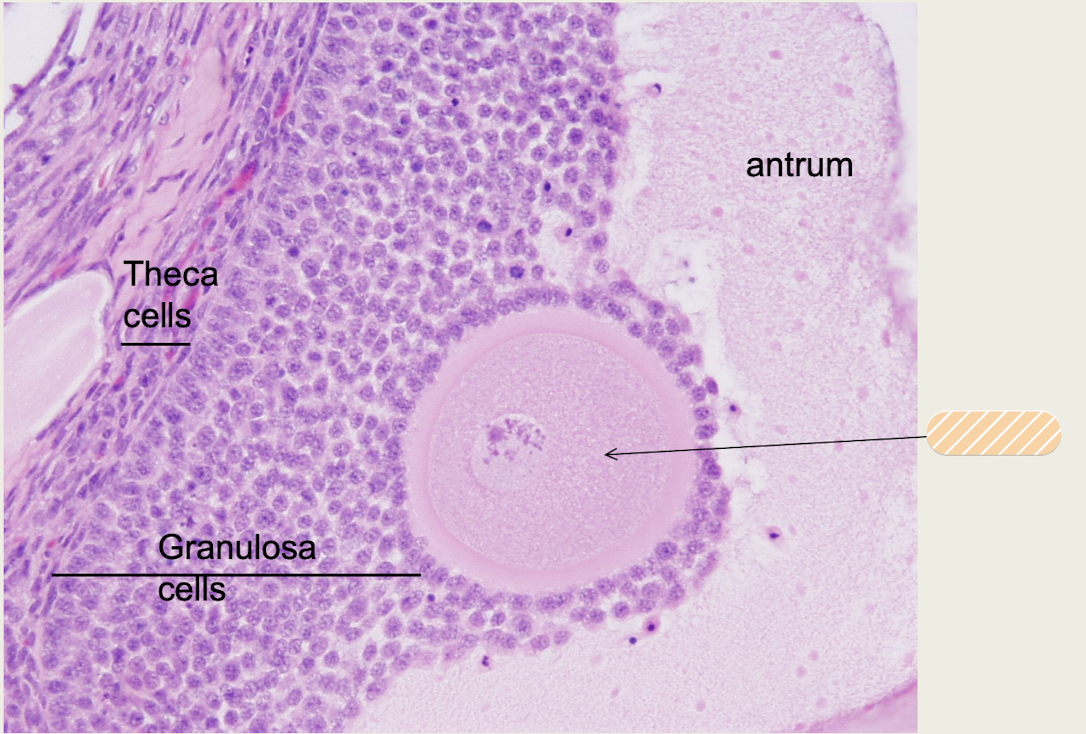
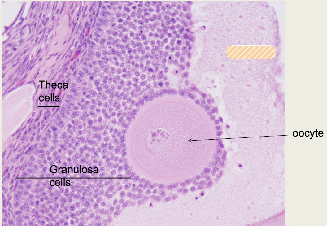
Follicle
cells that surround the oocyte in the ovary
Contains outer theca cells (make androgens) and inner granulosa cells (make estrogen)
Theca cells
outer follicle cells in the ovary that produce androgens
Granulosa cells
inner follicle cells in the ovary that produce estrogen
Oocyte
female haploid gamete
Graafian (mature) follicle
after follicle develops, typically contains antrum (fluid filled space) that will rupture
Antrum
fluid-filled space of a mature follicle
Ovulation
What is the term for when the follicle ruptures and oocyte is released?
Corpus luteum
remaining portion of follicle after ovulation (when follicle ruptures and oocyte released)
makes progesterone that maintains endometrium for first trimester of pregnancy until placenta takes over
*without fertilization, lifespan is 14 days, stops producing progesterone causing endometrial shedding
Corpus albicans
degenerated corpus luteum, forms in the absence of fertilization
Vagina
distensible musculomembranous tube and birth canal
canal for menstrual fluid, receives sperm
Anterior/Posterior/Lateral Fornix
What are the recesses around cervix?
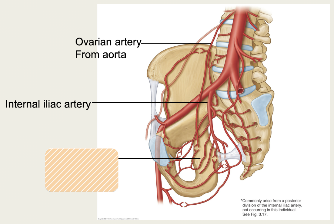
Ovarian artery
Which pelvic artery travels from abdominal aorta in the suspensory ligament of the ovary?
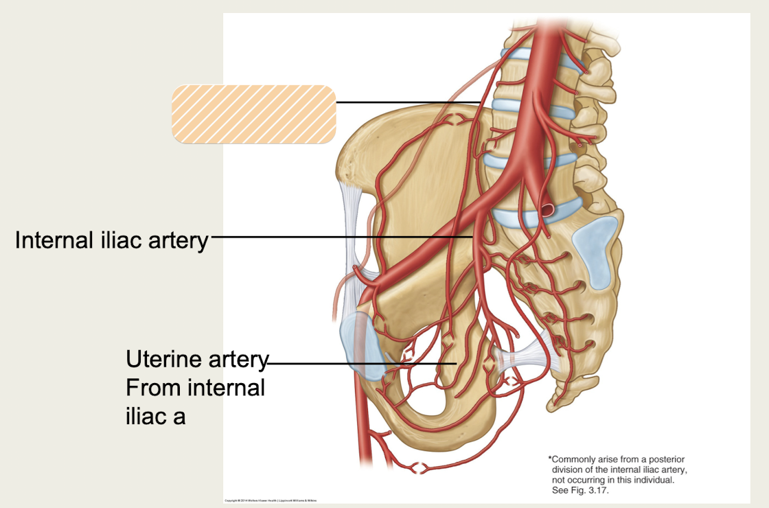
Uterine artery
Which pelvic artery travels from internal iliac artery in the transverse cervical ligaments of the uterus?
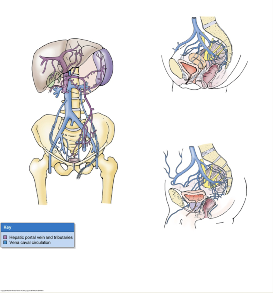
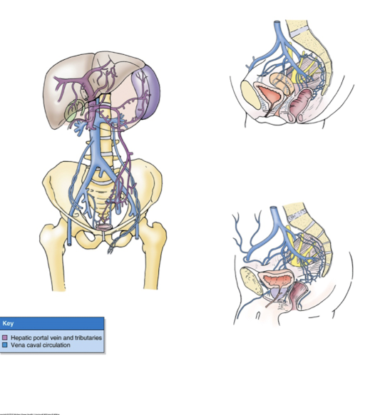
Lateral sacral veins
Which pelvic veins make anastamosis with internal vertebral venous plexus draining internal vertebral canal
*metastasis of prostate/ovarian cancer to vertebral or cranial sites
plexuses
Pelvic veins form _____ that drain into:
-Internal iliac vein
-Superior rectal vein
-Lateral sacral vein: forms anastamosis with internal vertebral venous plexus*
*metastasis of prostate/ovarian cancer to vertebral or cranial sites
Pelvic cavity
What portion of pelvis is within pelvic girdle and sacrum?
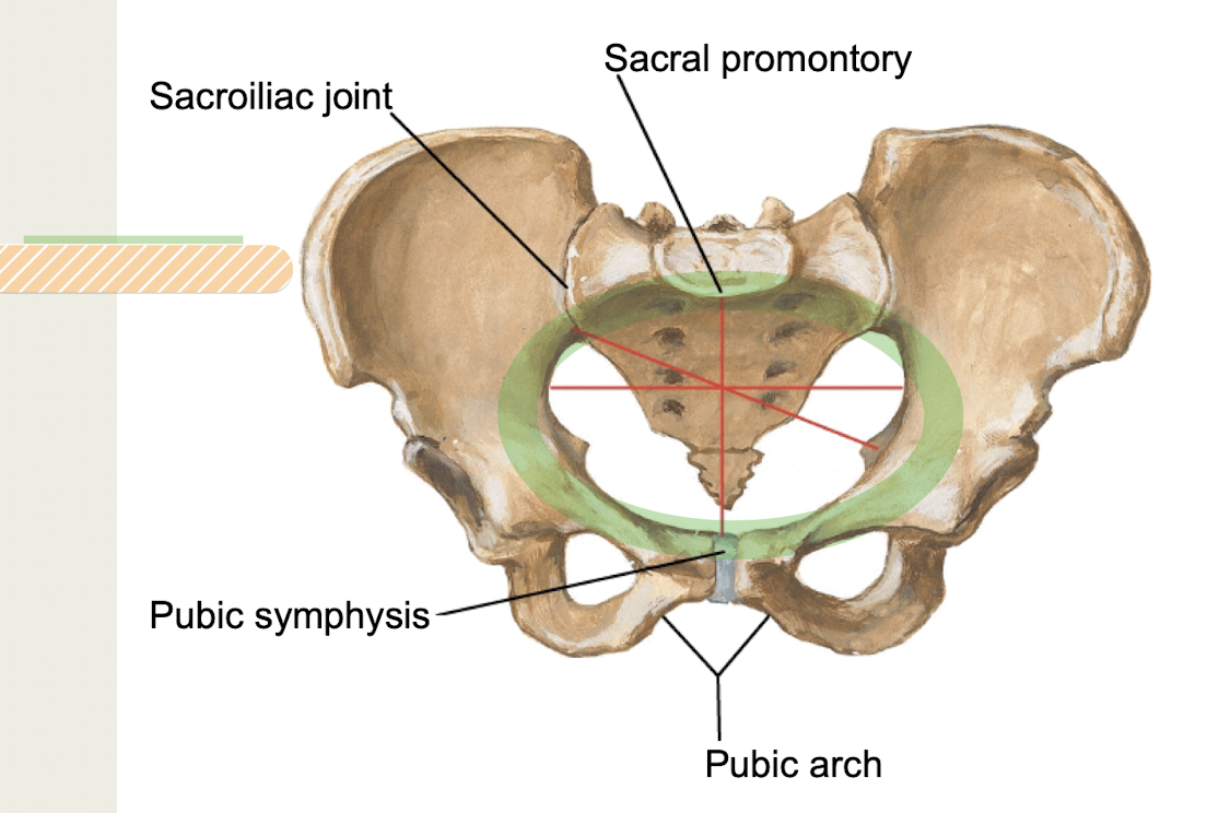
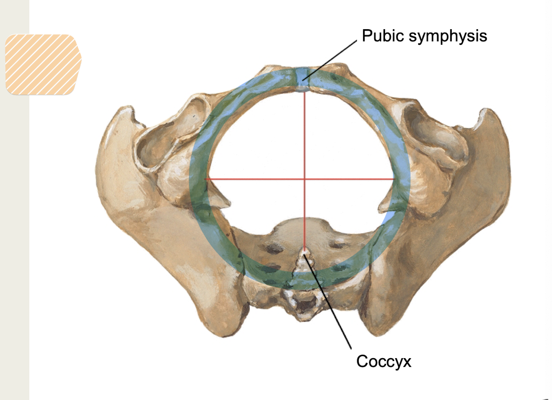
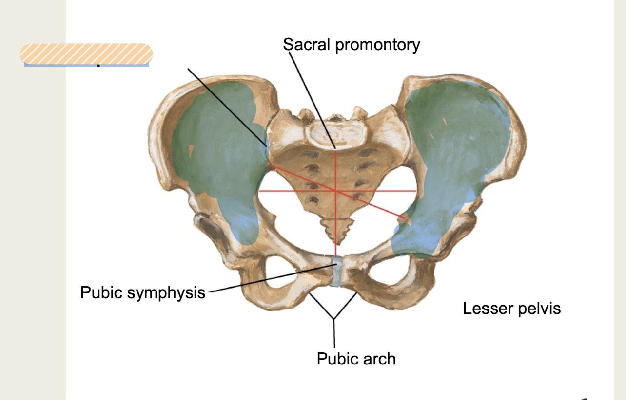
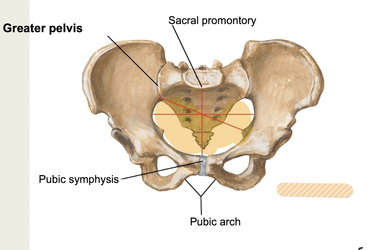
Pelvic brim
What portion of pelvis is between pubic symphysis and sacral promontory?
Pelvic inlet
separates greater and lesser pelvis
Pelvic outlet
inferior margin of pubic symphysis, ischial tuberosity, sacrotuberous ligament, tip of coccyx
Greater pelvis (pelvis major, false pelvis)
above pelvic brim, within iliac fossa, part of abdominal cavity, contains sigmoid colon*
Lesser pelvis (pelvis minor, true pelvis)
below pelvic brim, between pelvic inlet/outlet
contains pelvic viscera and true pelvic cavity*
Male pelvis
thick, heavy, prominent markings
heart-shaped pelvic inlet, small pelvic outlet; narrow pubic arch
coccyx angled, large acetabulum anteriorly
Iliac ala less flared, round obturator foramen
Female pelvis
thin, light, shallower and wider, oval or rounded pelvic inlet
large pelvic outlet, wide pubic arch
coccyx angled, small acetabulum inferiorly
iliac ala more flared, oval obturator foramen
Sacroiliac joint
Part fibrous / part synovial joint of pelvis
Lumbosacral joint
Part secondary cartilaginous / part synovial joint of pelvis
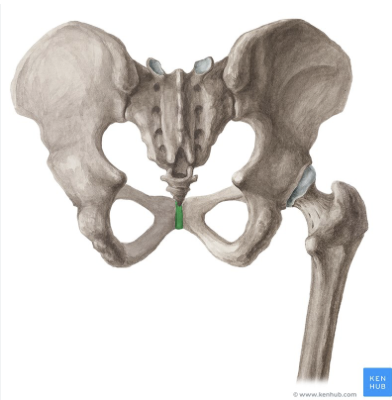
Pubic symphysis
secondary cartilaginous joint of pelvis
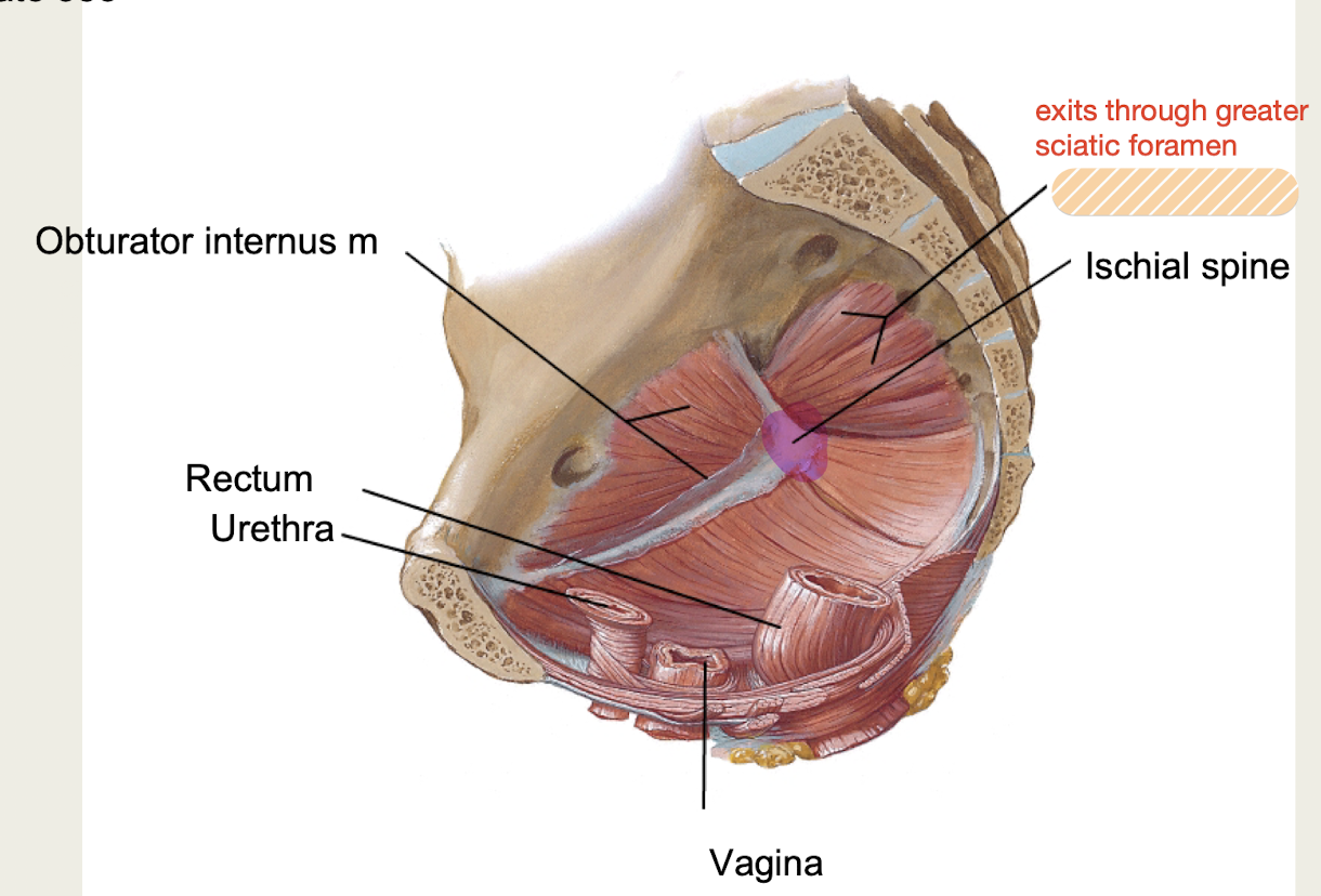
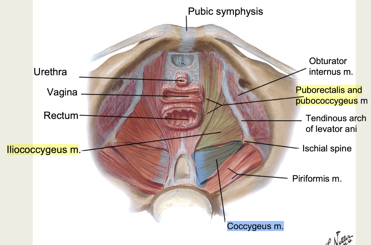
Obturator internus m
anterolateral pelvic wall, passes through lesser sciatic foramen
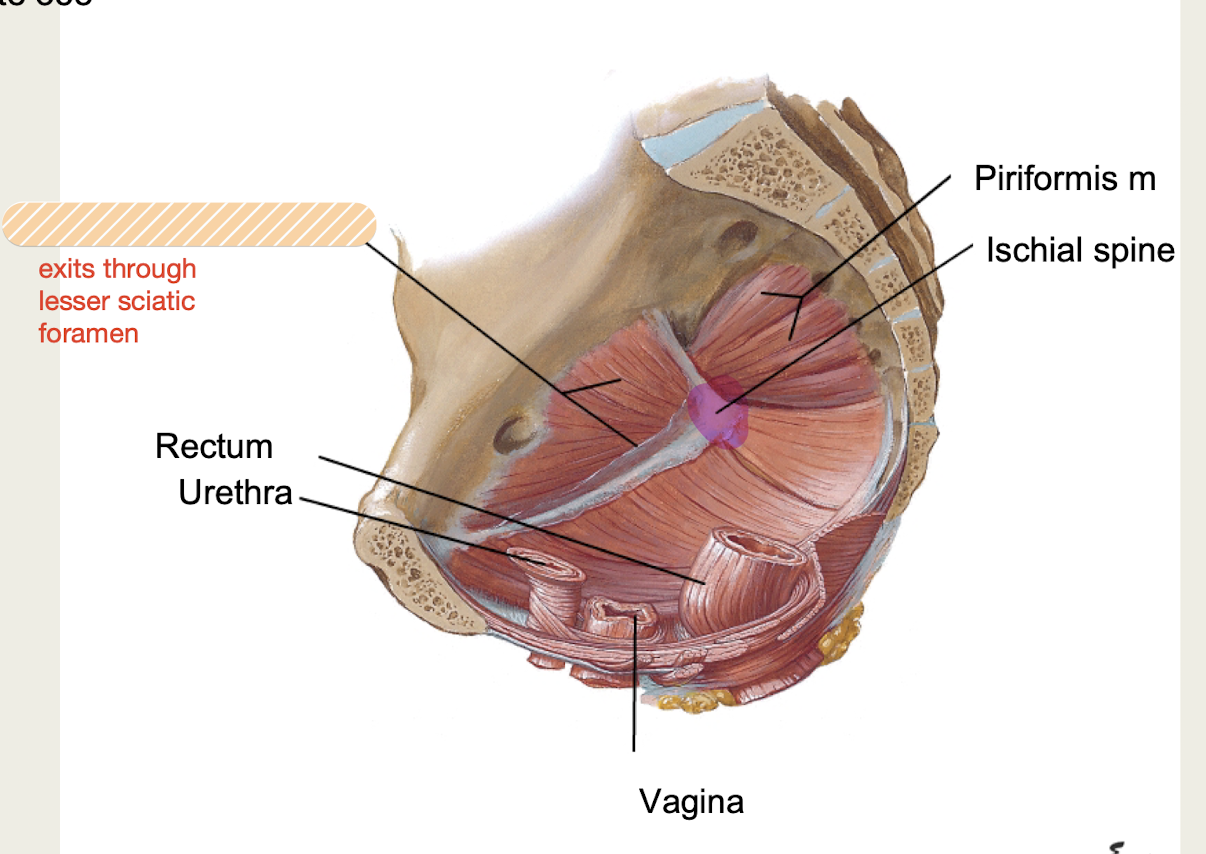
Piriformis m
posterolateral pelvic wall, passes through greater sciatic foramen
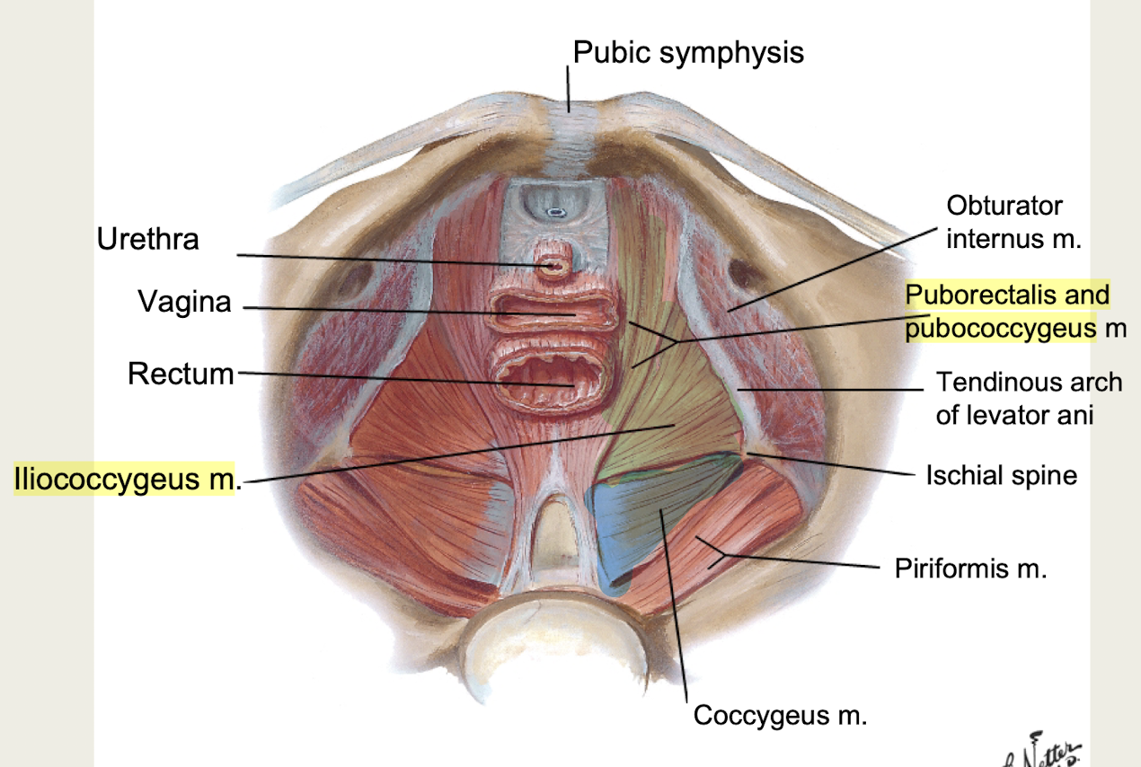
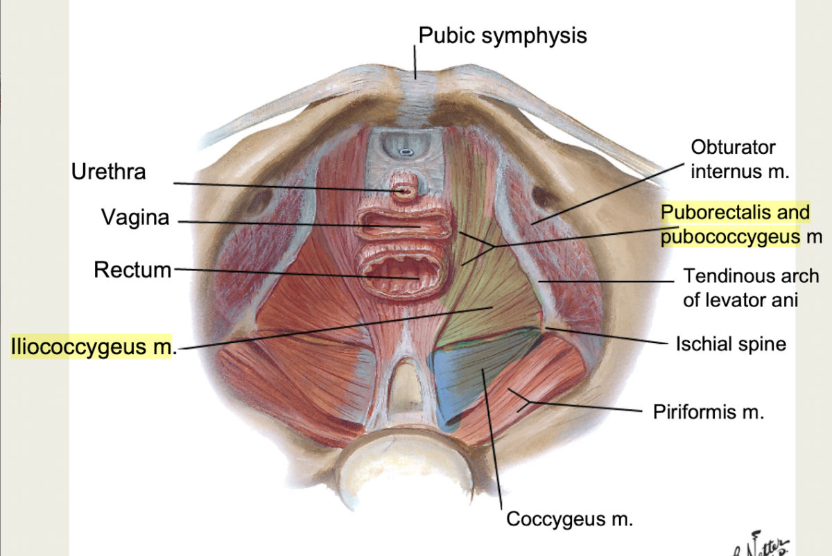
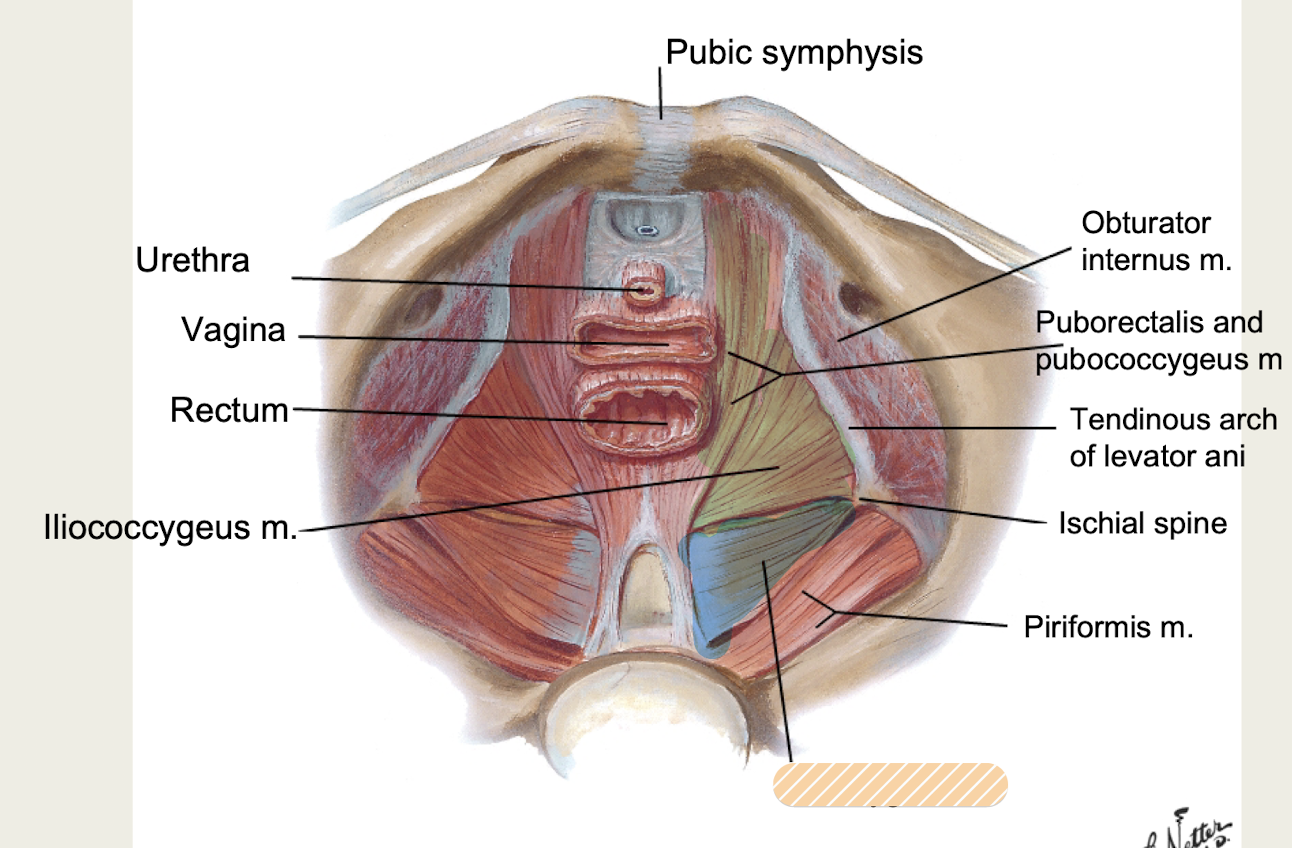
Pelvic diaphragm (floor)
funnel shaped, separates pelvic cavity from perineum
supports pelvic viscera, resists increases in intra-abdominal pressure*
Levator ani mm (Puborectalis, Pubococcygeus, Iliococcygeus) + Coccygeus m
Levator ani mm
innervated by ventral rami of S4
1. Puborectalis m
2. Pubococcygeus m
3. Iliococcygeus m
Muscles of Pelvic Diaphragm (floor)
S4 ventral rami
What is innervation to the levator ani muscles of pelvic diaphragm?
Coccygeus m
innervated by ventral rami of S4-S5
Muscle of Pelvic Diaphragm (floor)
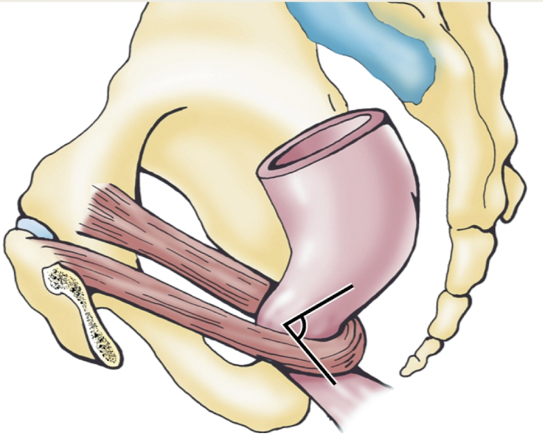
Puborectalis m
levator ani m (S4 ventral rami innervation) of pelvic diaphragm (floor)
attached laterally to pubic bone, makes a u-shaped sling around the rectum; helps to maintain fecal continence
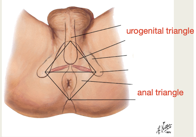
Perineum
diamond shaped area below pelvic diaphragm (floor), bounded by pelvic outlet
contains external genitalia (urogenital triangle + anal triangle)
Urogenital triangle
Boundaries: pubic symphysis, ischiopubic ramus, ischial tuberosities
Contents: urethra, external genitalia
Perineum of female pelvis
Anal triangle
Boundaries: coccyx, ischial tuberosity, sacrotuberous ligament
Contents: anus, anal canal
Perineum of female pelvis
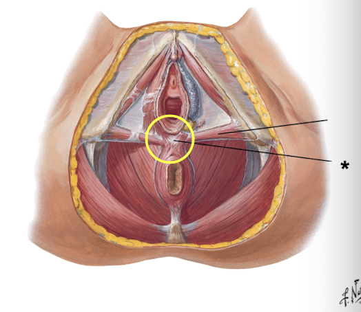
Perineal body
central tendon of perineum
posterior border of vagina, anterior border of external anal sphincter
attachment for urogenital diaphragm and external anal sphincter*
Episiotomy
surgical incision of perineum and vaginal wall, decreases trauma to pelvic diaphragm and perineum during delivery
easier to fix a cut muscle than torn muscle
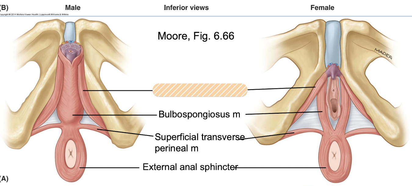
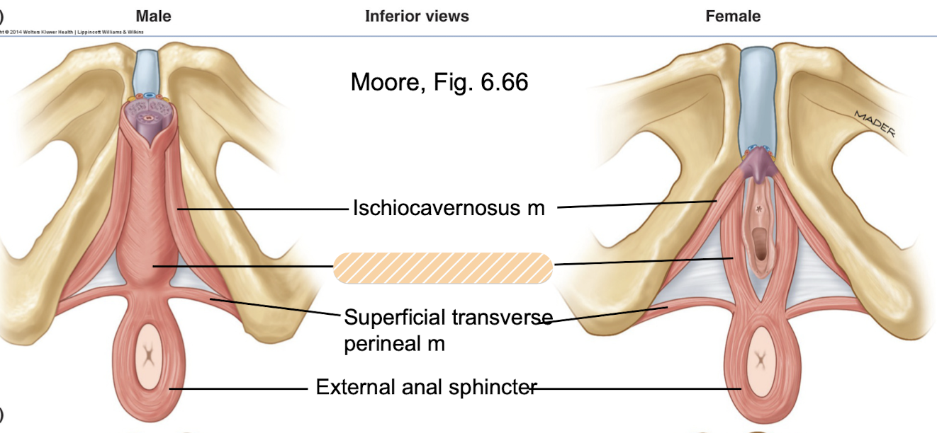
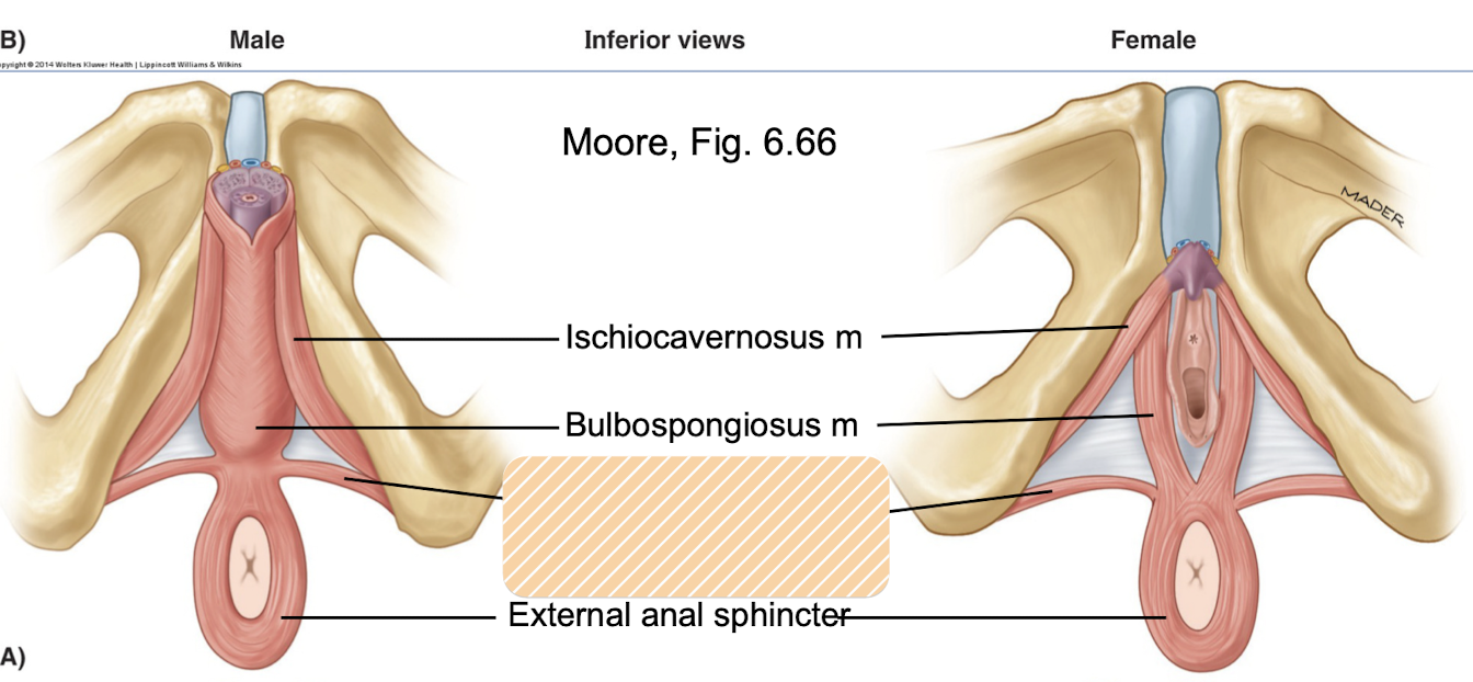
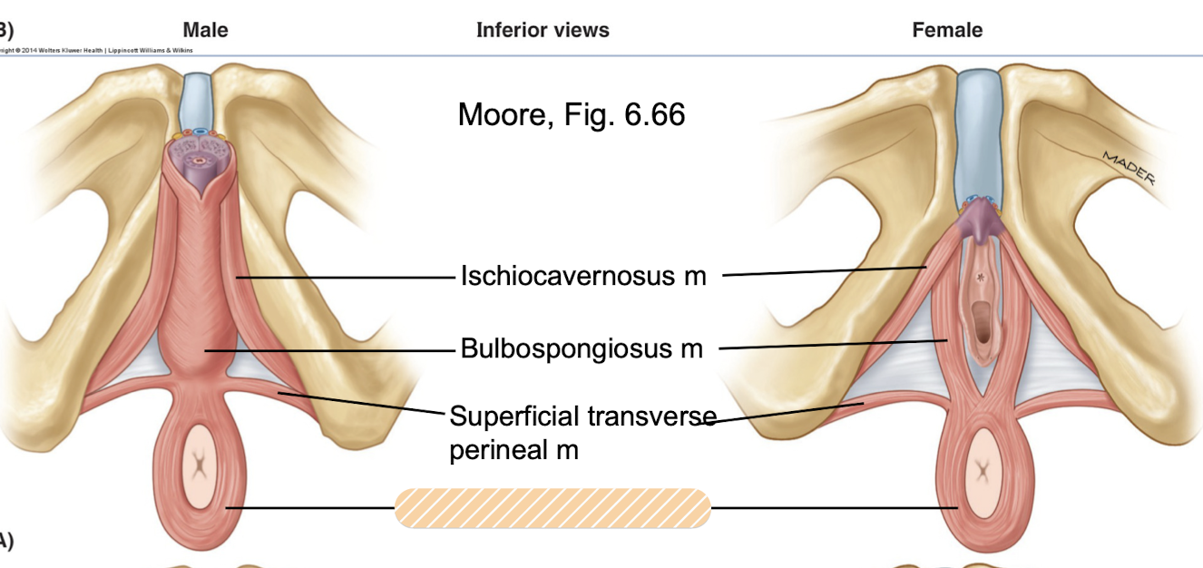
Perineal muscles
inferior to pelvic diaphragm, support pelvic diaphragm, support perineal body (except for ischiocavernosus m*), resists increases in intra-abdominal pressure
■ Ischiocavernosus m* - not attached to perineal body
■ Bulbospongiosus m
■ Superficial transverse perineal m
■ Deep transverse perineal m
■ External anal sphincter**
innervated by perineal nerve (pudendal nerve branch ventral rami S2-S4)
**external anal sphincter innervated by inferior rectal nerve (S2-S4)
Perineal n
What is innervation to the:
Ischiocavernosus m
Bulbospongiosus m
Superficial transverse perineal m
Deep transverse perineal m?
Perineal muscles; pudendal nerve branch ventral rami S2-S4
Ischiocavernosus m
does not attach to perineal body*, perineal nerve innervation
runs along ischial pubic ramus to ischial tuberosity, inserts along crus of penis or crus of clitoris, helps to maintain an erection
Perineal muscle
Bulbospongiosus m
origin on perineal body, perineal nerve innervation
surrounds bulb of penis/vagina, supports and stabilizes pelvic diaphragm/perineal body and resists increases in intraabdominal pressure, assists with erection
Perineal muscle
Superficial transverse perineal m
runs from ischial tuberosity to perineal body, perineal nerve innervation
supports and stabilizes pelvic diaphragm/perineal body and resists increases in intraabdominal pressure
Perineal muscle
Deep transverse perineal m
deeper muscle, runs from ischial pubic ramus to perineal body, perineal nerve innervation
supports and stabilizes pelvic diaphragm/perineal body and resists increases in intraabdominal pressure
Perineal muscle
External anal sphincter
anteriorly attached to perineal body, innervated by inferior rectal nerve (S2-S4)
surrounds anal canal, helps to maintain fecal continence (closing off the anus)
Perineal muscle
Ischiocavernosus m
Which perineal muscle does NOT attach to the perineal body?
runs along ischial pubic ramus to ischial tuberosity
Vulva/Pudendum
female external genitalia, present on surface
sensory and erectile tissue, directs flow of urine, prevents entry of foreign material to urogenital tract
Mons pubis
overlies pubic symphysis on female