Chapter 18: The Heart
5.0(1)
5.0(1)
Card Sorting
1/65
Earn XP
Description and Tags
Study Analytics
Name | Mastery | Learn | Test | Matching | Spaced |
|---|
No study sessions yet.
66 Terms
1
New cards
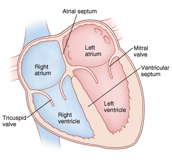
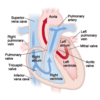
Chambers of the heart
right atrium, left atrium, right ventricle, left ventricle
2
New cards
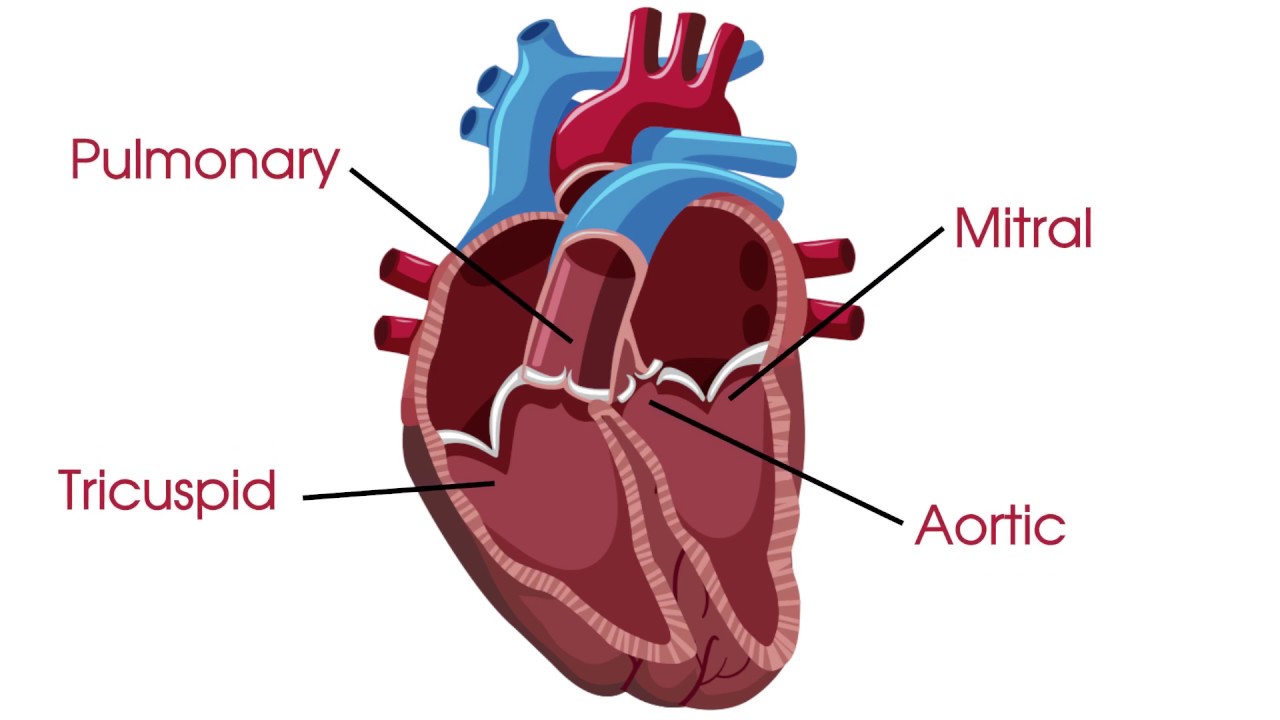
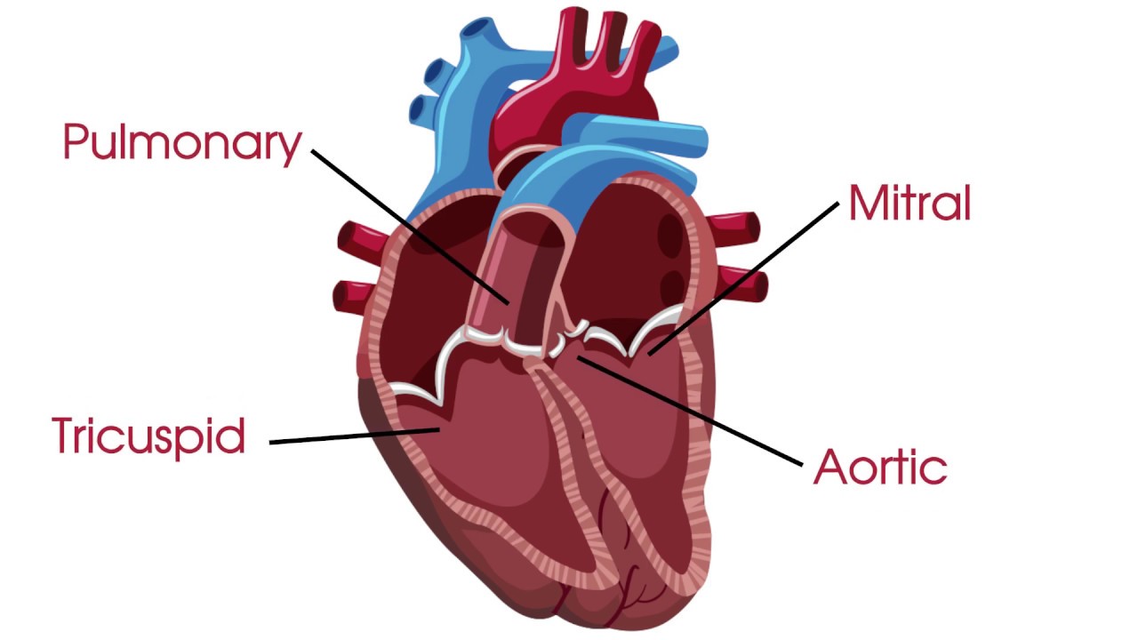
Valves of the heart
tricuspid valve, bicuspid valve, pulmonary valve, aortic valve
3
New cards
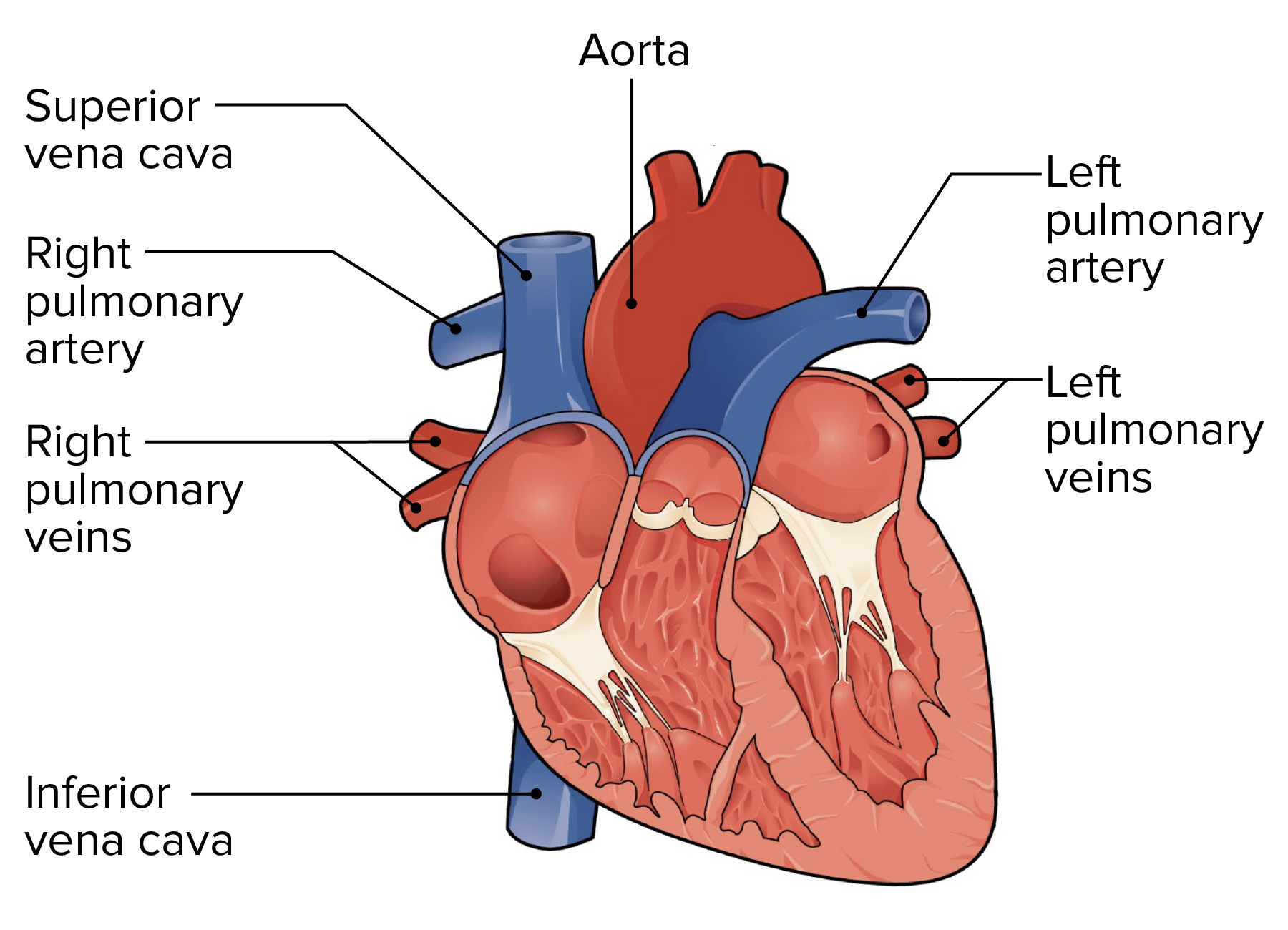
Great vessels of heart
pulmonary veins, pulmonary arteries, aorta, superior vena cava, inferior vena cava
4
New cards
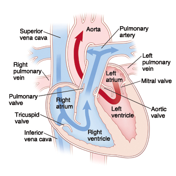
Deoxygenated structures
right atrium, right ventricle, tricuspid valve, pulmonary valve, pulmonary veins, pulmonary veins
5
New cards
Oxygenated structures
left atrium, left ventricle, bicuspid valve, aortic valve, pulmonary arteries, aorta
6
New cards
Systemic
left side, oxygenated, from heart -> body
7
New cards
Pulmonary
right side, deoxygenated, from heart -> lungs
8
New cards
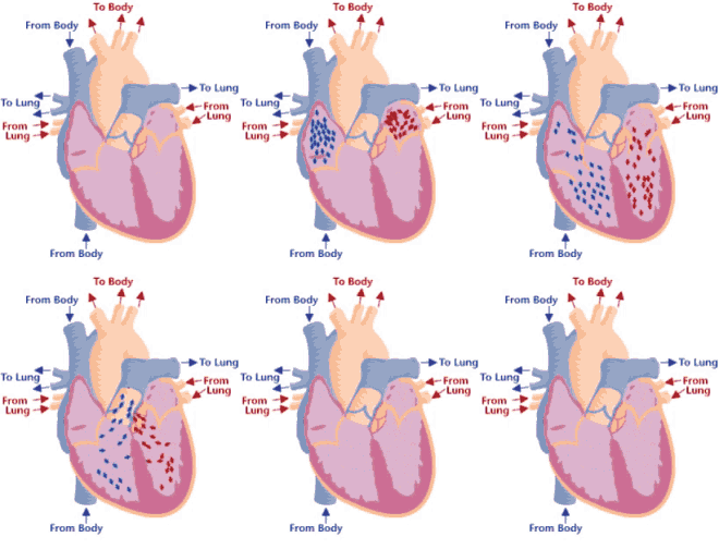
Step 1 of blood circulation
Deoxygenated blood pools in right atrium
9
New cards
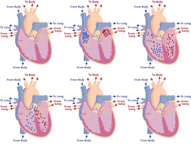
Step 2 of blood circulation
Right atrium fills and tricuspid valve opens
10
New cards
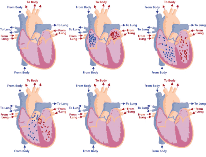
Step 3 of blood circulation
Blood is squeezed through pulmonary valve into pulmonary trunk
11
New cards
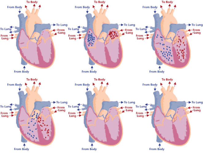
Step 4 of blood circulation
Deoxygenated blood continues through the left and right pulmonary arteries to the lungs
12
New cards
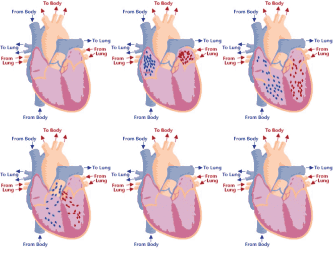
Step 5 of blood circulation
Lungs exchange CO2 for O2
13
New cards
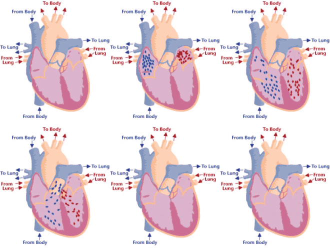
Step 6 of blood circulation
Now oxygenated blood collects in left atrium
14
New cards
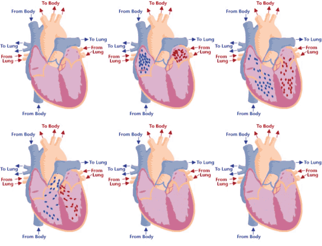
Step 7 of blood circulation
Left atrium fills and bicuspid valve opens, releasing into left ventricle
15
New cards
Step 8 of blood circulation
Pumped through aorta and into left and right pulmonary veins
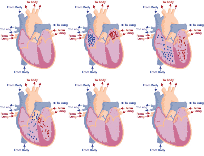
16
New cards
Step 9 of blood circulation
Travels through systemic circuit
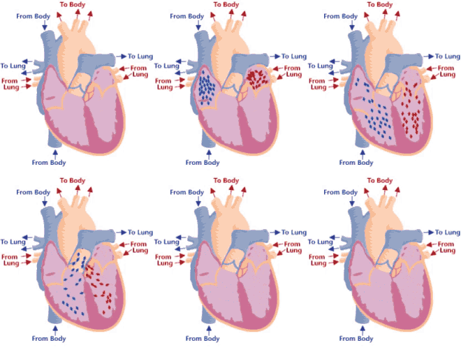
17
New cards
Step 10 of blood circulation
Returns via the superior vena cava and right atrium
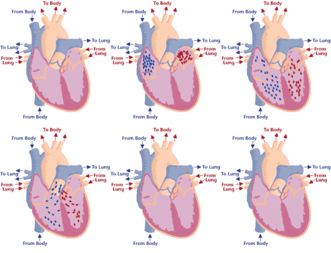
18
New cards
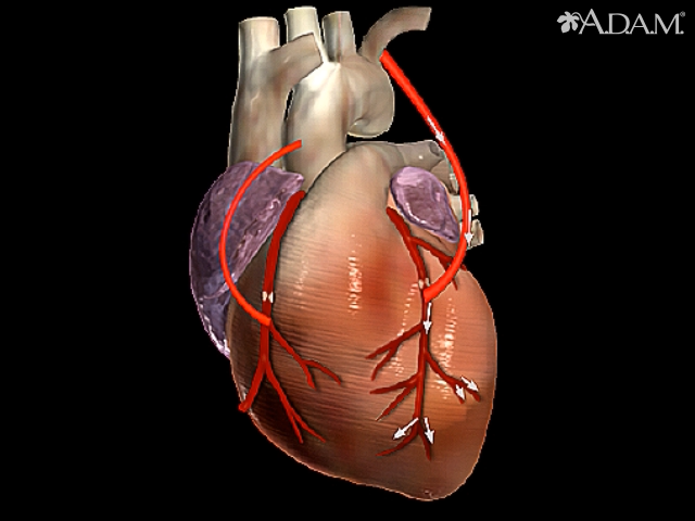
Coronary circuit
Supplies the heart with blood, coronary arteries bring blood to myocardium of the heart, form anastomoses
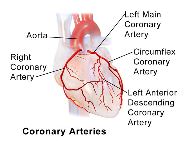
19
New cards
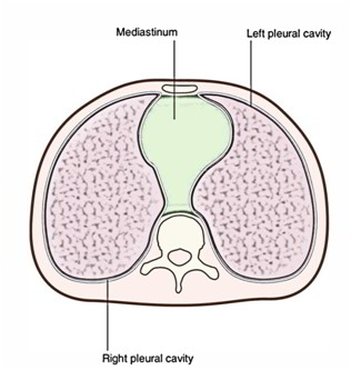
Mediastinum
cavity in center of chest between lungs (where the heart is located)
20
New cards
Anastomoses
connections between arteries
21
New cards
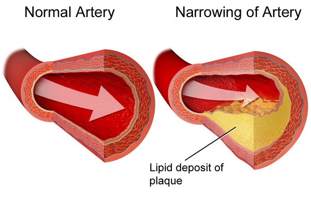
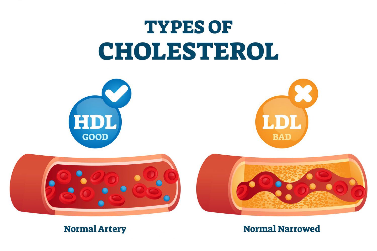
Atherosclerosis
narrowing of arteries due to fatty plaque
22
New cards
Foam Cell
localized to fatty deposits on blood vessel walls, where they ingest LDLs and become laden with lipids
23
New cards
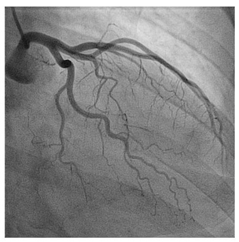
Angiogram
test injecting dye into vessels to see blockage
24
New cards
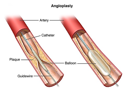
Angioplasty
inserting a balloon into artery to smash plaque against wall to open up vessel
25
New cards
Myocardial Infarction
heart attack, blockage of an artery to the heart due to atherosclerosis and blood clot
26
New cards
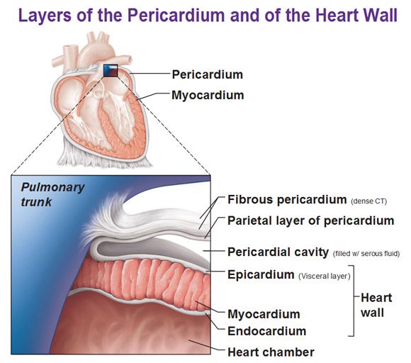
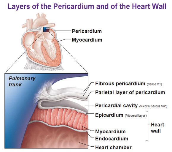
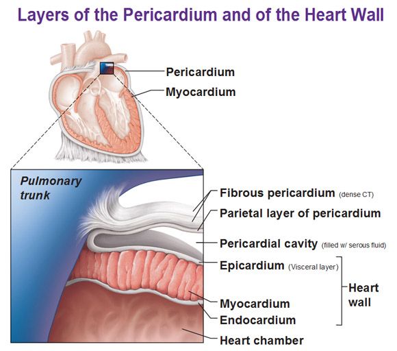
1st layer of the heart
Epicardium
27
New cards
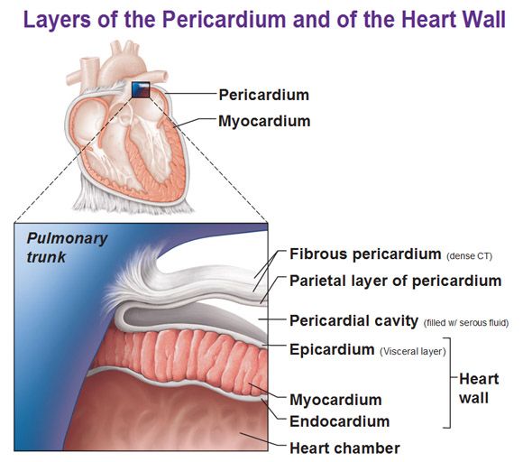
Epicardium
same as visceral pericardium, outermost layer composed of serous membrane
28
New cards
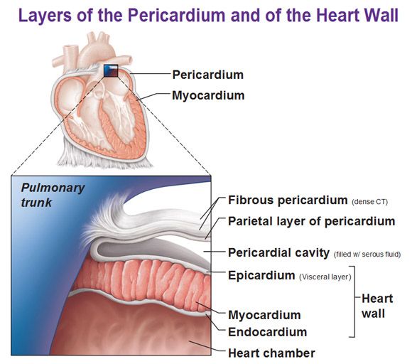
2nd layer of the heart
Myocardium
29
New cards
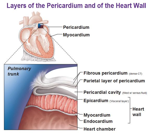
Myocardium
middle layer (thickest), cardiac muscle, contracts the heart
30
New cards
3rd layer of the heart
Endocardium
31
New cards
Endocardium
innermost layer, simple squamous epithelium, provides smooth lining for blood
32
New cards
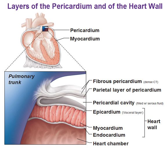
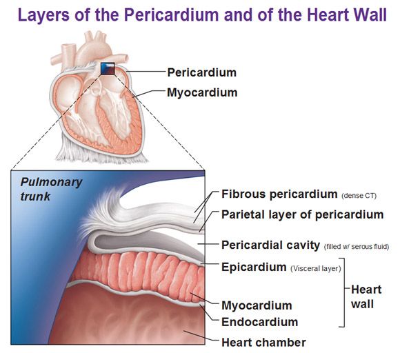
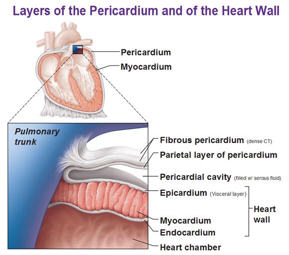
1st layer of the pericardium
Fibrous Pericardium
33
New cards
Fibrous Pericardium
outermost layer composed of dense connective tissue for protection
34
New cards
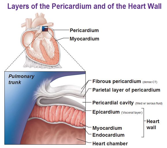
2nd layer of the pericardium
Parietal Pericardium
35
New cards
Parietal Pericardium
composed of serous membrane producing serous fluid to reduce friction
36
New cards
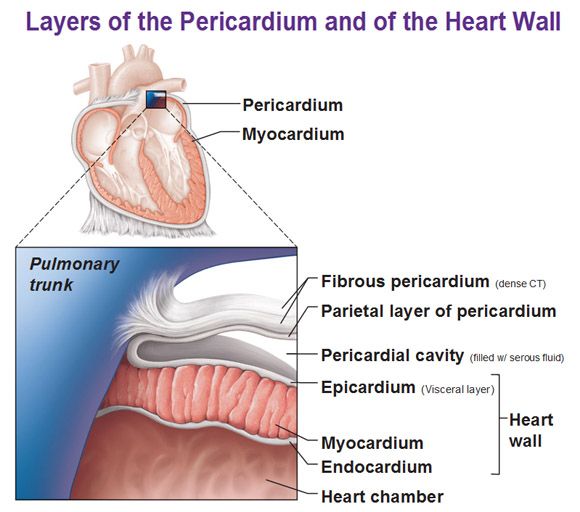
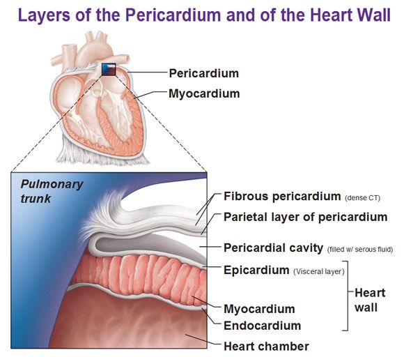
3rd layer of the pericardium
Visceral Pericardium (epicardium)
37
New cards
Visceral Pericardium (epicardium)
folded part of parietal pericardium, between parietal pericardium and visceral pericardium space called pericardial cavity for the serous fluid
38
New cards
Function of heart valves
one way flaps that close to prevent back-flow of blood
39
New cards
10
?
40
New cards
LDL
bad cholesterol
41
New cards
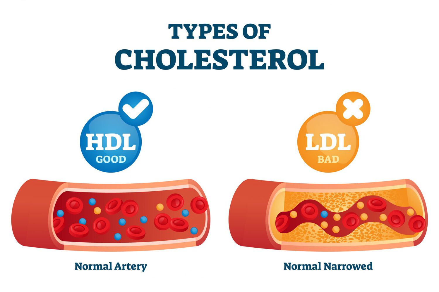
HDL
good cholesterol, scavenges LDL and brings it back to liver
42
New cards
Coronary bypass surgery
\-A vein from the leg is reattached around clotted coronary artery to maintain blood flow
\-Used when arteries are too clogged for angioplasty to work
\-Used when arteries are too clogged for angioplasty to work
43
New cards
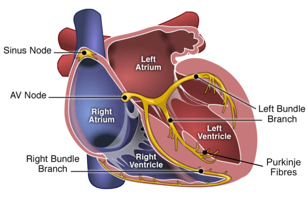
Heart (intrinsic) conduction system #1
SA (sinatrial) node
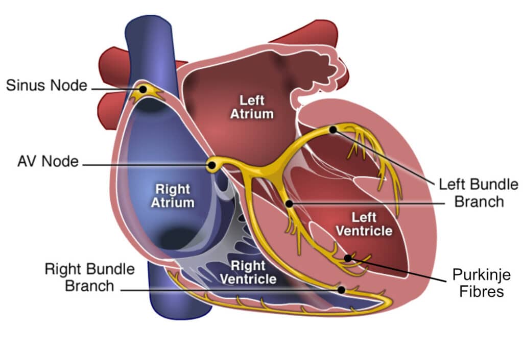
44
New cards
Heart (intrinsic) conduction system #2
AV (atrioventricular) node
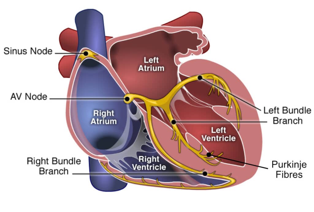
45
New cards
Heart (intrinsic) conduction system #3
AV bundle
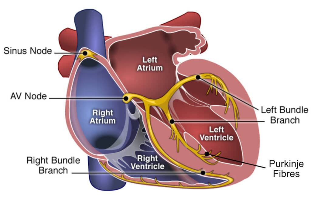
46
New cards
Heart (intrinsic) conduction system #4
Right and left bundle branches
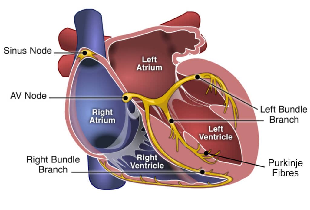
47
New cards
Heart (intrinsic) conduction system #5
Purkinje fibers
48
New cards
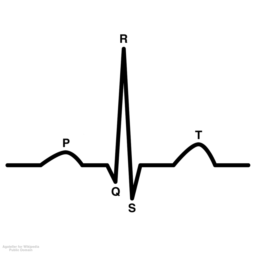
P wave
depolarization of atria which causes contraction of atria
49
New cards
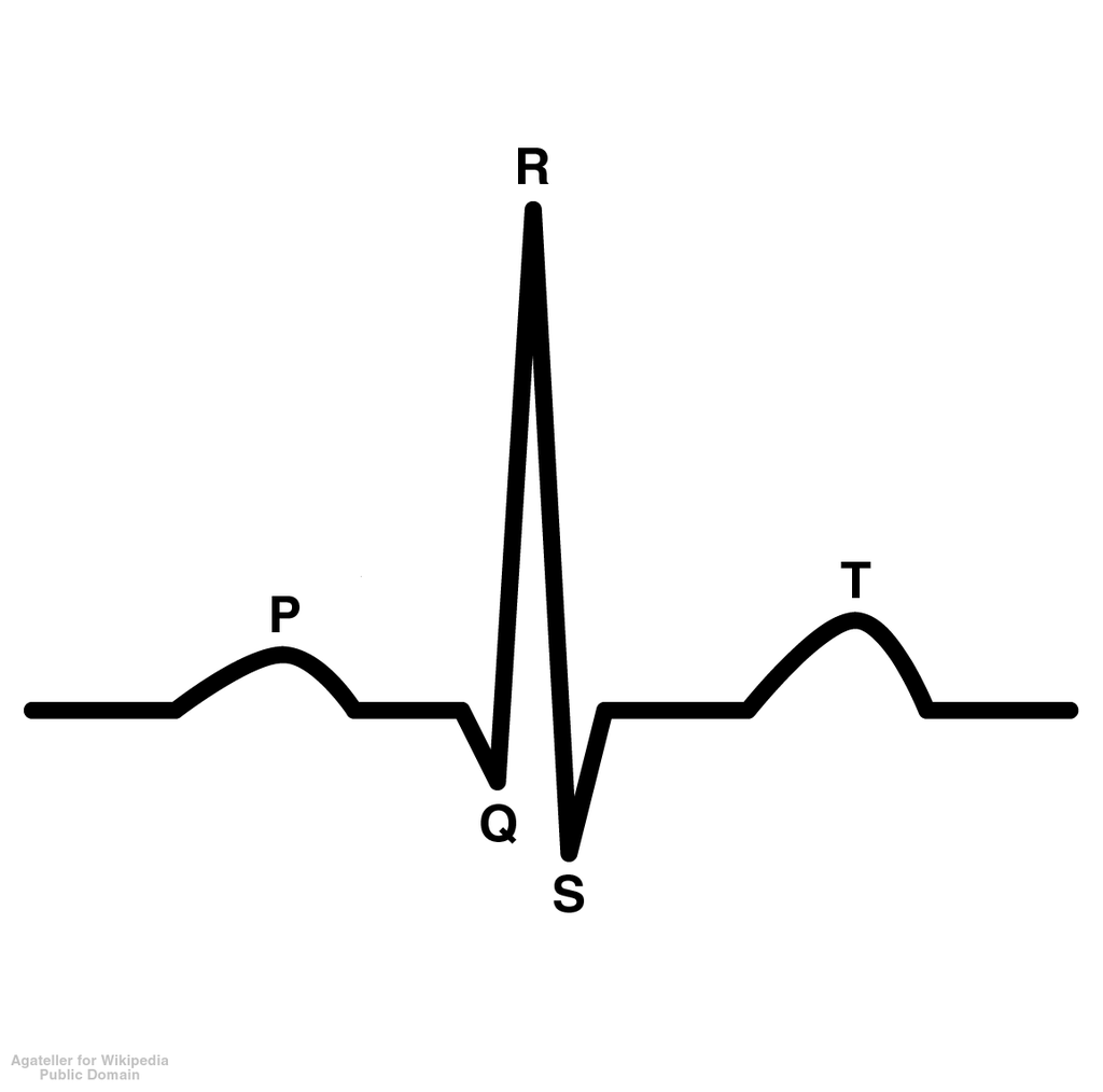
QRS wave
depolarization of ventricles; atrial repolarization is blocked
50
New cards
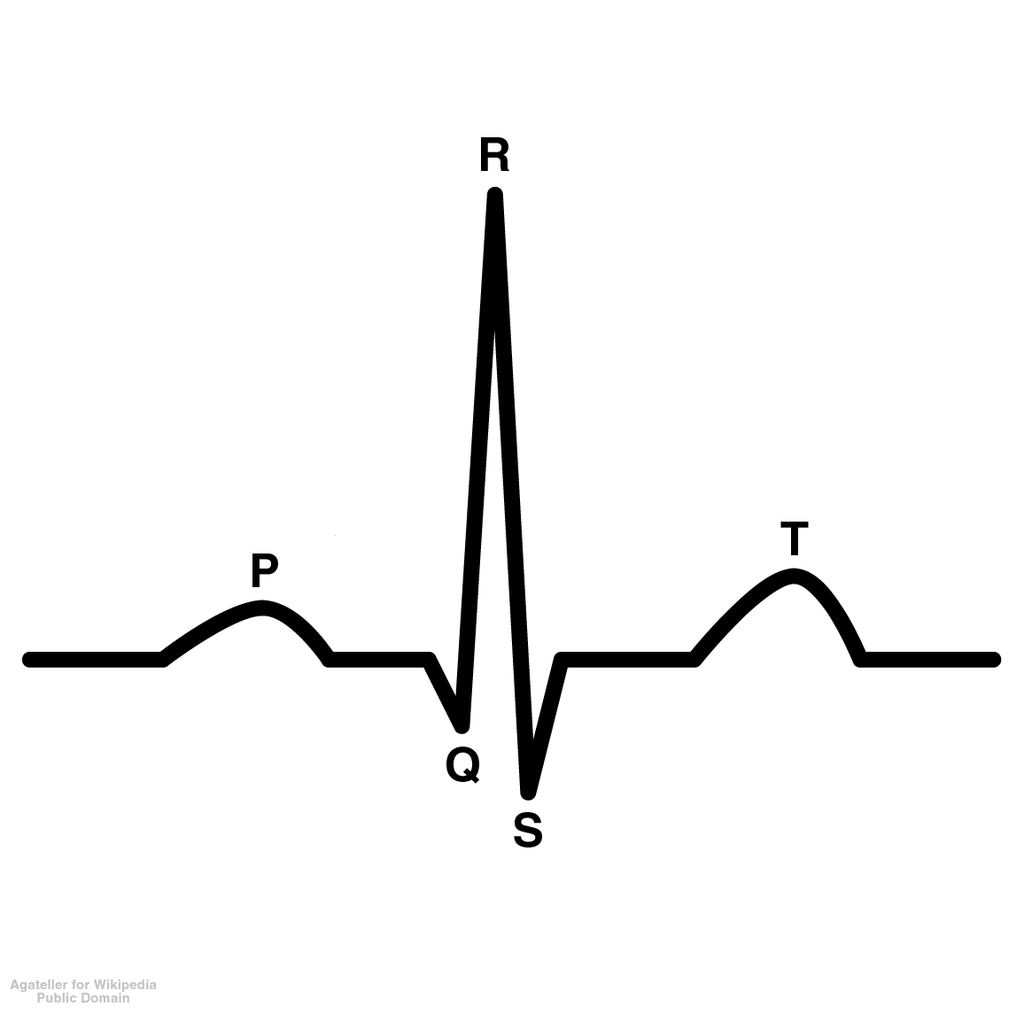
T wave
repolarization of ventricles
51
New cards
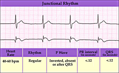
Arrhythmias
no p wave-> atria/SA node not working, irregular heart rate
52
New cards
Autorhythmic
heart produces its own pulses through electrochemical stimuli originating from a small group of cells in the wall of the right atrium, known as the sinoatrial node (or SA node)
53
New cards
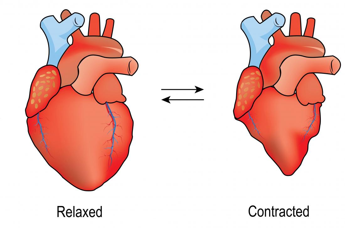
Systole
rate of contraction (depolarization), pressure when heart is squeezed
54
New cards
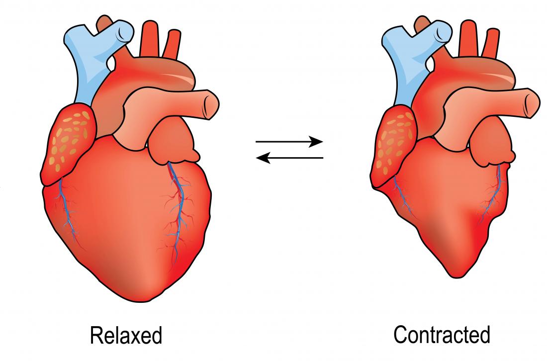
Diastole
rate of relaxation, heart relaxes
55
New cards
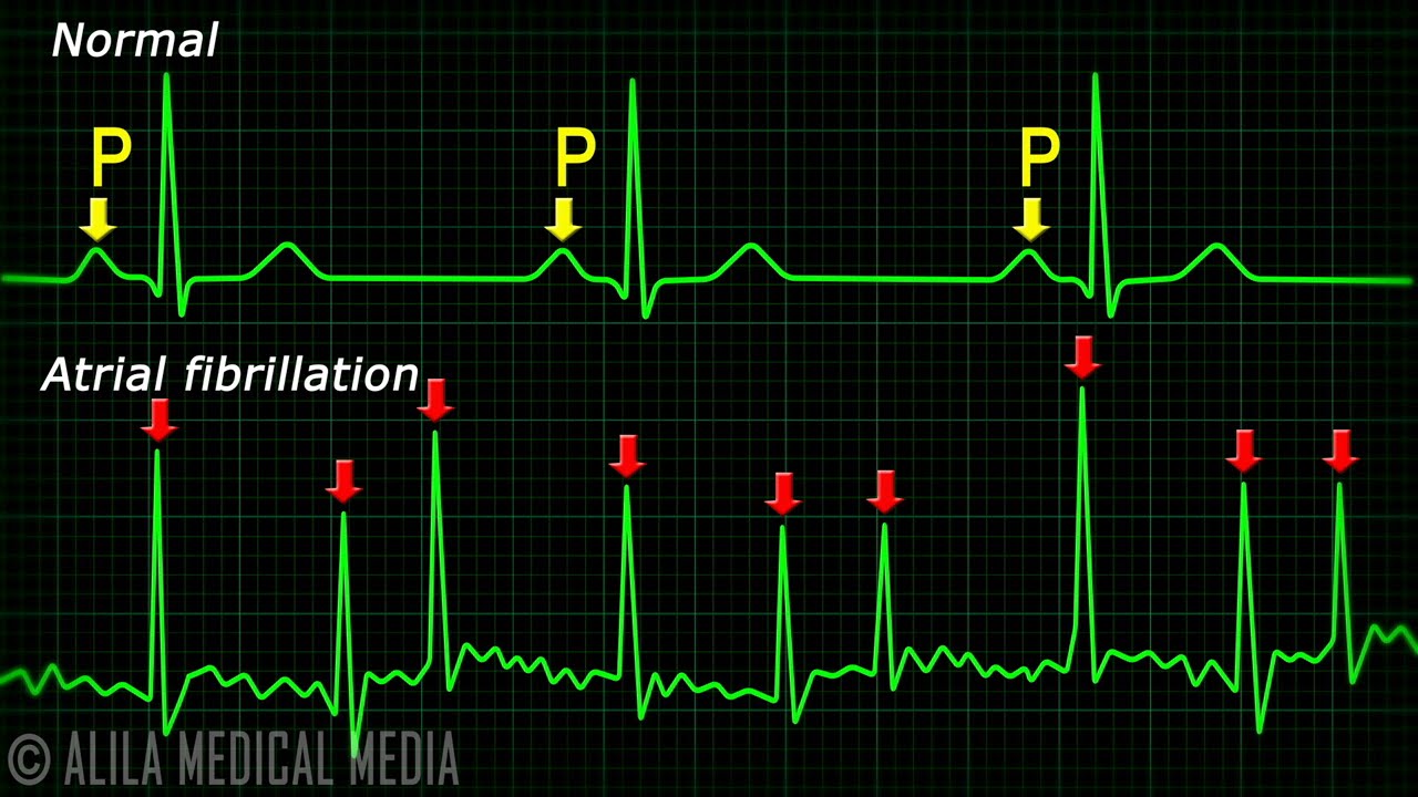
Fibrillation
waves undisguisable, chaotic rhythm, all cells have different electrical impulses and beat out of sync
56
New cards
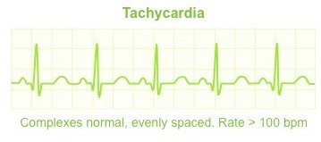
Tachycardia
heart beats too fast, bpm >100
57
New cards
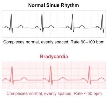
Bradycardia
heart beats too slow, bpm
58
New cards
Hypertension
high blood pressure > 140/90
59
New cards
16
?
60
New cards
Normal heart rate
60-100 bpm
61
New cards
Normal blood pressure
120/80
62
New cards
Systolic pressure
1st # in blood pressure, contraction of heart
63
New cards
Diastolic pressure
2nd # in blood pressure, relaxation of heart
64
New cards
Cardiac output
amount of blood pumped out by each ventricle in 1 minute, blood/min
65
New cards
? \* ?=blood/min
Heart rate \* stroke volume
66
New cards
70\*75=?
5250mL or 5.25L per minute