BIOL 2134 Anatomy Unit 4
1/213
There's no tags or description
Looks like no tags are added yet.
Name | Mastery | Learn | Test | Matching | Spaced | Call with Kai |
|---|
No analytics yet
Send a link to your students to track their progress
214 Terms
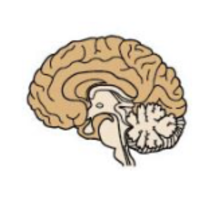
Cerebrum
Largest component of the human brain.
Cerebral cortex
Surface of the cerebrum; gray matter.
Gyrus
Ridge in the cerebral cortex.
Sulcus
Crease in the cerebral cortex.
Fissure
Deep sulcus.
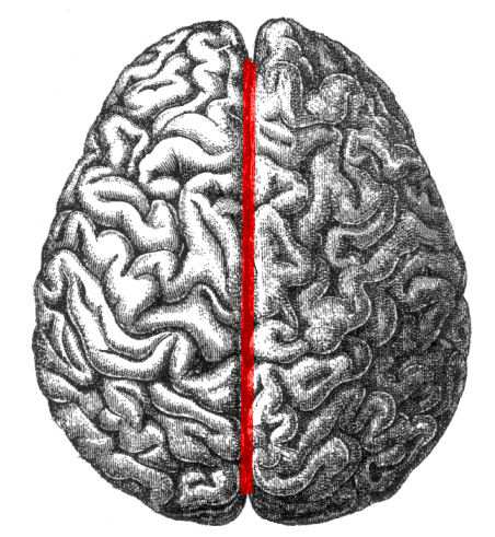
Longitudinal fissure
Separates the brain into left and right hemispheres.
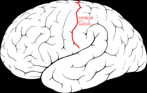
Central sulcus.
Pre-central Gyrus.
Post-central Gyrus.
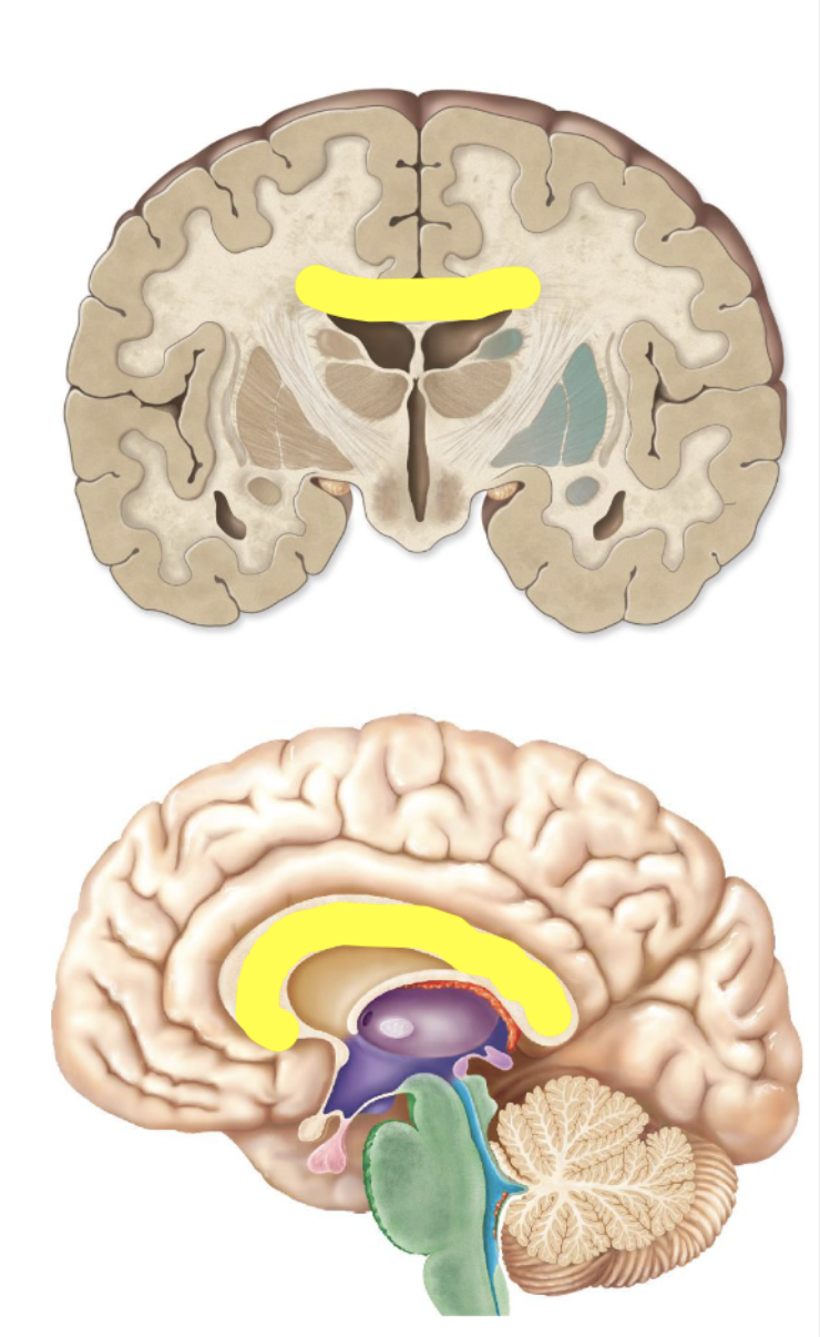
Corpus Callosum
Communication between left and right hemispheres of the cerebrum.
Is the Corpus Callosum made of white or grey matter?
White.
Physiology of the Cerebrum
Interpretation of sensation, voluntary movement, speech, memory, intelligence and other higher functions.
Functional Areas: Motor areas
Control voluntary motor functions.
Functional Areas: Sensory areas
Provide conscious awareness of sensation.
Functional Areas: Association areas
Integrate and store information.
What is an example of a Motor Area?
Frontal lobe.
Frontal lobe
Primary motor cortex, is the location of somas of neurons that initiate voluntary muscle movement.
Broca’s area
Located in the left lobe only, is the location of somas of neurons that produce speech.
What is an example of the Sensory Area?
Parietal Lobe.
Parietal Lobe
Primary somatosensory cortex, is the location of somas of neurons involved with conscious awareness of general somatic sensory information.
Pre-central Gyrus
The pre-central gyrus is in the frontal lobe and anterior to the central sulcus. It is also the location of the primary motor cortex.
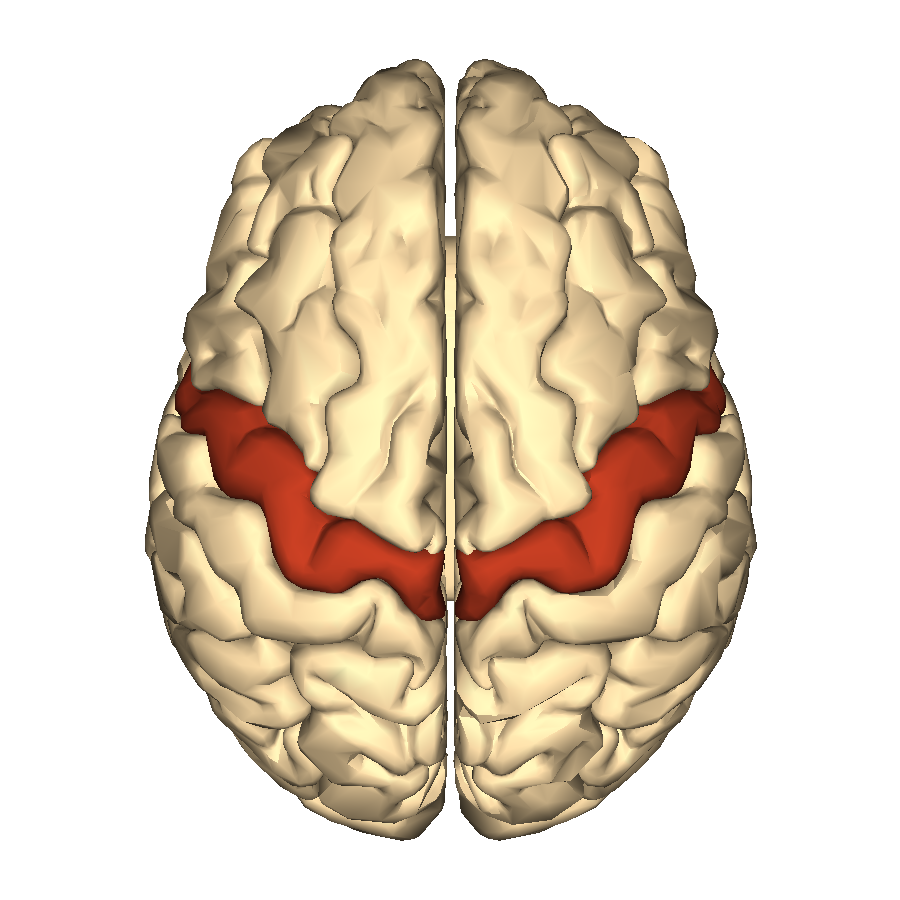
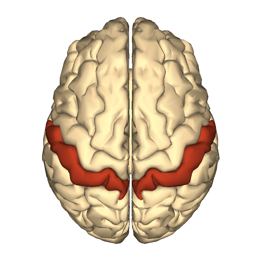
Post-central Gyrus
The post-central gyrus is in the parietal lobe and posterior to the central sulcus. It is also the location of the primary somatosensory cortex.
What is an example of the Sensory Area?
Insula Lobe.
Insula Lobe
Primary gustatory (taste) cortex, location of somas that interpret gustatory information.
Occipital Lobe
Primary visual cortex, location of somas that interpret visual information.
What is an example of a Sensory Area? (Temporal)
Temporal Lobe.
Temporal Lobe
Wernicke’s area (left only), location of somas involved with speech formation and comprehension.
Primary auditory and olfactory cortices
Location of somas that interpret auditory and olfactory information.
Are the motor and sensory cortices white matter or gray matter?
The motor and cerebral cortices are made up of gray matter – somas, dendrites, and synapses that are interpreting information and initiating action.
Left side of the body is controlled by the _______ primary motor cortex
Right.
Left side of the body is monitored by the ______ primary somatosensory cortex.
Right.
Broca’s Area
Related to speech. Motor. Allows for word expression. Left-sided only. Common target for stroke.
Wernicke’s Area
Related to speech. Sensory. Allows a word to be formed and understood. Left-sided only. Common target for stroke.
Words travel from gray matter in ______ to the cerebral white matter to ______ Area to the cerebral white matter to the muscles of the mouth.
Wernicke’s; Broca’s.
What is an example of Association Area? (Pre-frontal cortex)
Pre-frontal cortex.
Pre-frontal cortex
Complicated area, the site of learning, higher emotion, mortality, intelligence, and personality.
What is an example of Association cortex? (Pre-motor cortex)
Pre-motor Cortex.
Pre-motor Cortex
“Muscle memory”. Associations based on previous motor experiences.
What is an example of Association Area? (Parietal Lobe)
Parietal Lobe; somatosensory association area; associations based on previous sensory experiences.
What is an example of Association Area? (Occipital)
Occipital Lobe; visual association area; associations based on previous visual experiences.
What is an example of Association Area? (Temporal)
Temporal Lobe; auditory association area; stores memories of previous auditory experiences.

What part is green?
Temporal.

What part is red?
Frontal lobe.
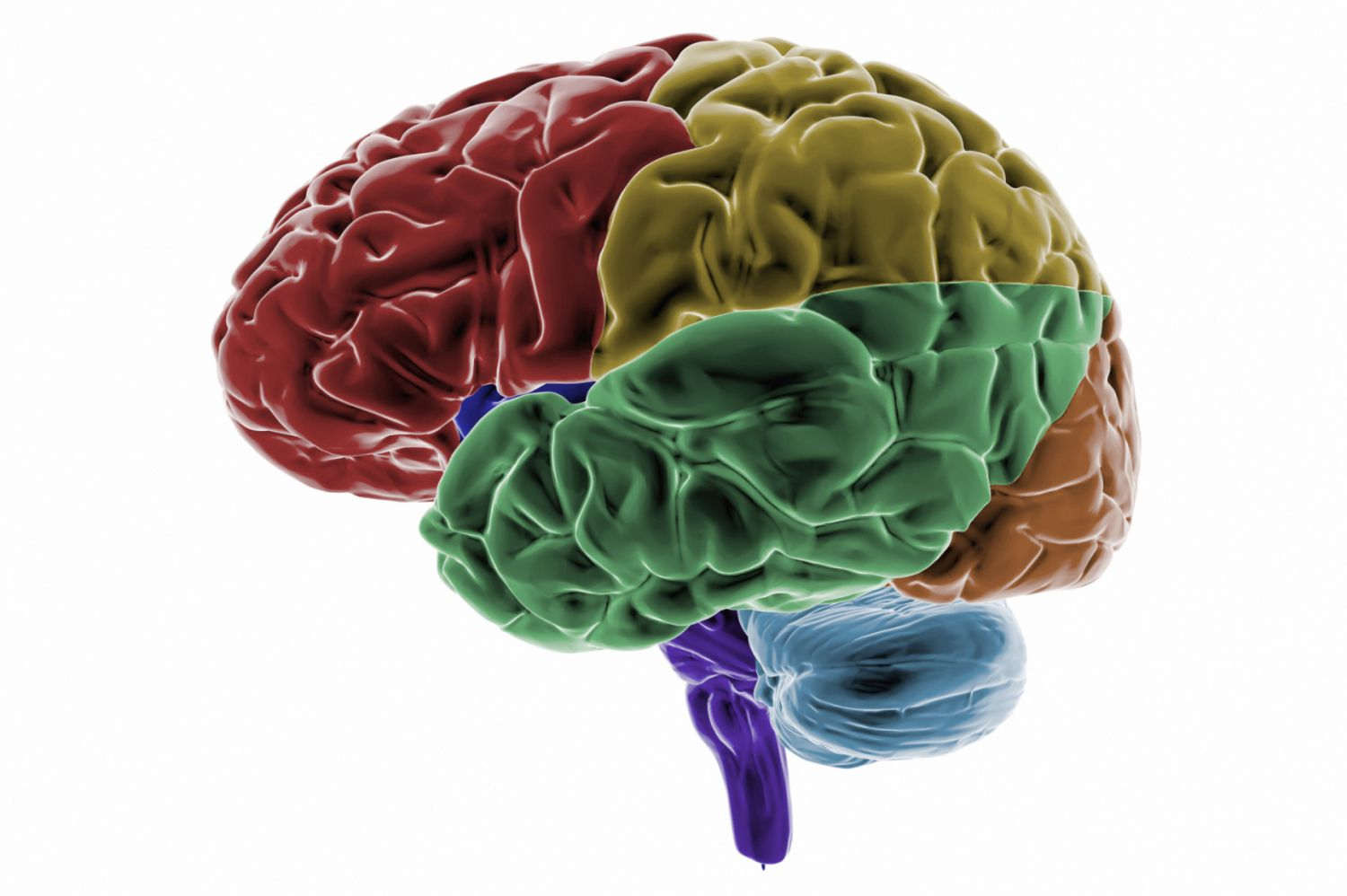
What part is yellow?
Pariteal Lobe.

What part is orange?
Occipital.
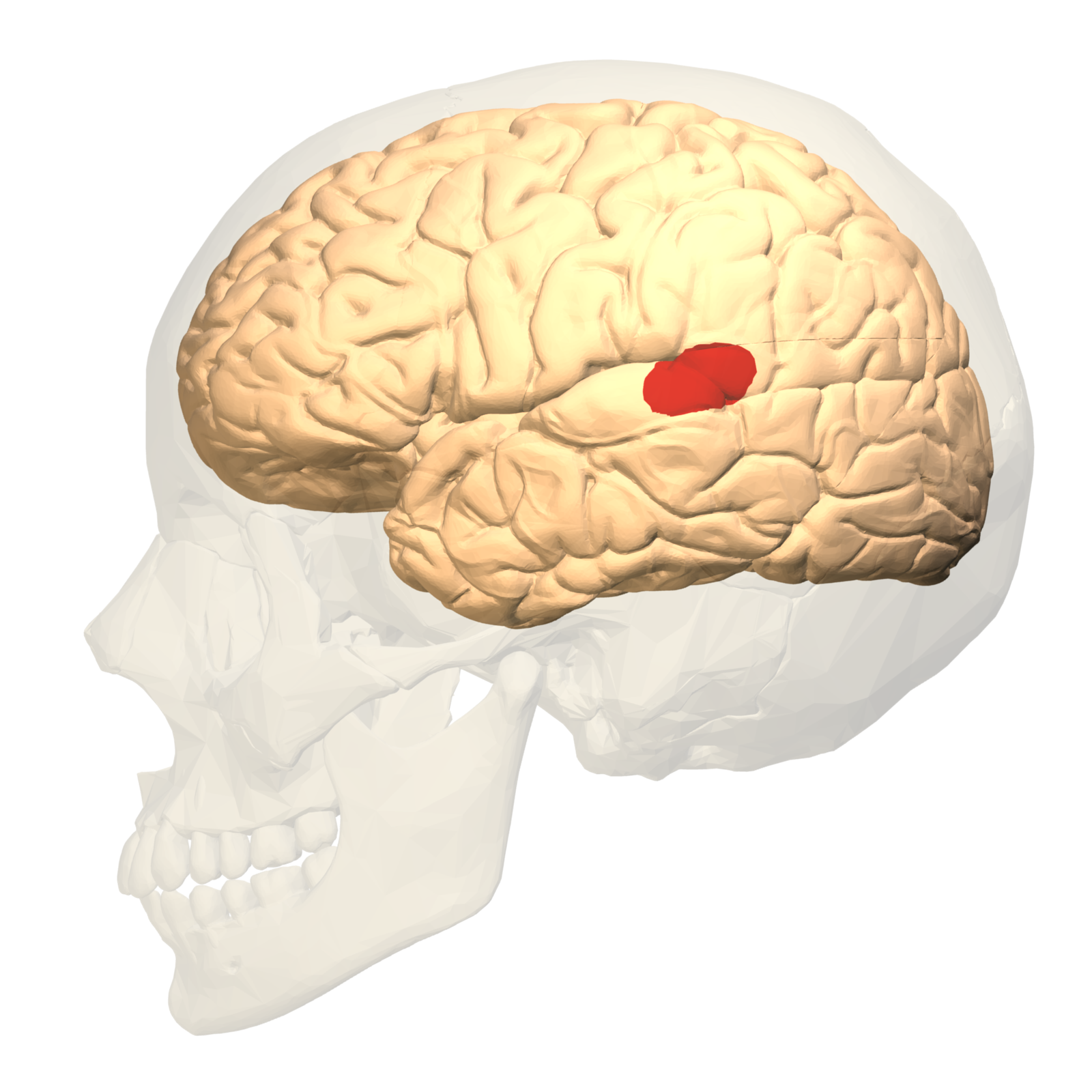
What is highlighted?
Wenicke’s area.
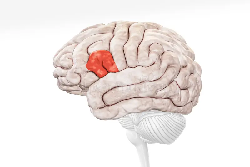
What is highlighted?
Broca’s area.
Gray matter
Somas, dendrites, and synapses; cortex; where awareness occurs and action is initiated.
White matter
Myelinated axons; deep to cortex; where information is carried somewhere else; called “tracts”.
Basal Nuclei
Areas of grey matter within the white matter tracts of the cerebrum; regulate motor information to produce smooth movements; affected by Parkinson’s Disease.
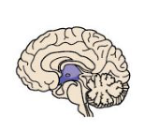
Diencephalon
Enclosed by cerebral hemispheres.
Thalamus
Relay sensory impulses to the primary somatosensory cortex in the cerebrum.
Hypothalamus
Controls the visceral nervous system. Controls the limbic system (“emotional-visceral brain”). Controls the endocrine system with the pituitary gland.
Hippocampus
Long-term memories and olfactory tracts.
Fornix
White matter that connects all gray matter of the limbic system.
Epithalamus
Contains the pineal gland and choroid plexus.
Pineal gland
Releases melatonin.
Choroid Plexus
Filters blood plasma to produce cerebrospinal fluid.
Brainstem
Axons traveling between the brain and the spinal cord; mostly white matter.
Nuclei
Areas of gray matter are involved with visceral motor system; areas of gret matter within the brainstem involved with the visceral motor activity,
Midbrain
Nuclei relay information about vision and hearing.
Pons
Nuclei control breathing and dreaming.
Medulla oblongata
Where motor neurons “cross-over”. Nuclei initiate visceral motor activity, like heart rate, blood pressure, swallowing, vomiting, etc. Merges seamlessly into spinal cord.
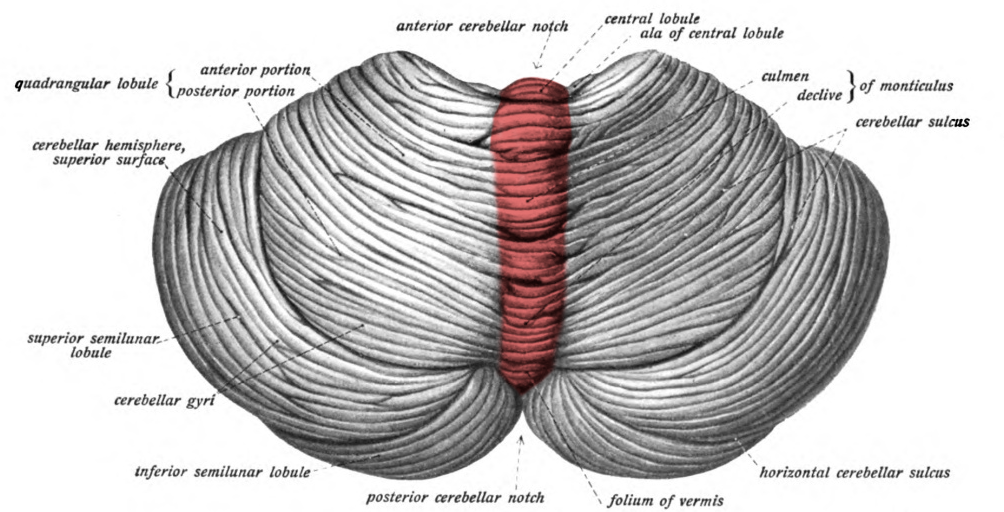
What is highlighted?
Vermis. (Looks like a worm).
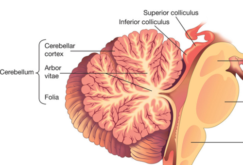
What is being shown?
Folia and arbor vitae. (They look like tree branches and leaves).
What are the functions of the Cerebellum?
“Fine tunes” skeletal muscle movement.
Controls balance.
Meninges
Specialized connective tissues that provide protection, physical stability, and shock absorption. Surround the brain and spinal cord.
Dura mater
Outermost and toughest layer of the meninges. Not attached to bone — forms epidural space.
Arachnoid mater
Middle layer of the meninges. Thinnest layer. Forms subarachnoid space where cerebrospinal fluid circulates.
Pia mater
The innermost layer of the meninges. Anchors blood vessels in place.
Transports nutrients and waste away from the CNS. Shock absorption.
Cerebrospinal Fluid (CSF).
Cerebrospinal fluid is made in the ______ of ventricles.
Chorioid plexus.
Enlarged chambers in the brain that contain cerebrospinal fluid.
Ventricles.
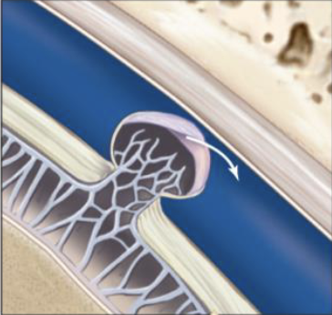
What is pictured?
Arachnoid Villi
______ lines the blood vessels of the central nervous system.
Astrocytes
Cerebrovascular accident (CVA)
Stroke.
One sided paralysis
Hemiplegia
Damage to speech center in left hemisphere.
Aphasia
Transient ischemic attack (TIA)
Temporary restriction of blood flow to the brain. Numbness, temporary paralysis, impaired speech.
General senses — Throughout body
Refers to temperature, pain, touch, pressure, vibration, and proprioception.
Special censes — Only in sense organs
Refers to vision, taste, hearing, balance, and smell.
Sensory receptors
The interface between the nervous system and the body.
Nociceptors
Detect the sensation of pain. Associated with free nerve endings and large receptor fields.
Thermoreceptors
Detect changes in temperature. Found in the dermis, skeletal muscles, liver, and hypothalamus.
Mechanoreceptors
Detect changes in pressure. (Touch, vibration, stretch)
Chemoreceptors
Detect change in the chemical composition of body fluids (specific molecules dissolved in fluid).
Mechanoreceptors — Tactile receptors
Provide sensations of touch, pressure, and vibrations.
Mechanoreceptors — Baroreceptors
Stretch receptors that monitor changes in the stretch of organs.
Mechanoreceptors — Proprioceptors
Monitor the position of joints, tension in the tendons and ligament, and the length of muscle fibers upon contraction. Controls the sense of body position.
What are the 5 special senses?
Vision
Gustation
Hearing
Equilibrium
Olfaction
Aqueous Fluid
Located in the anterior cavity, this liquid prevents collapse of the eye and is continuously replaced.
Flow of Aqueous
Ciliary body —> Pupil —> Scleral venous sinus —> Blood
Vitreous Fluid
Located in the posterior cavity, this gelatinous substance prevents collapse of the eye as well as supporting the overall shape of the eye and the position of the lens it is never replaced.
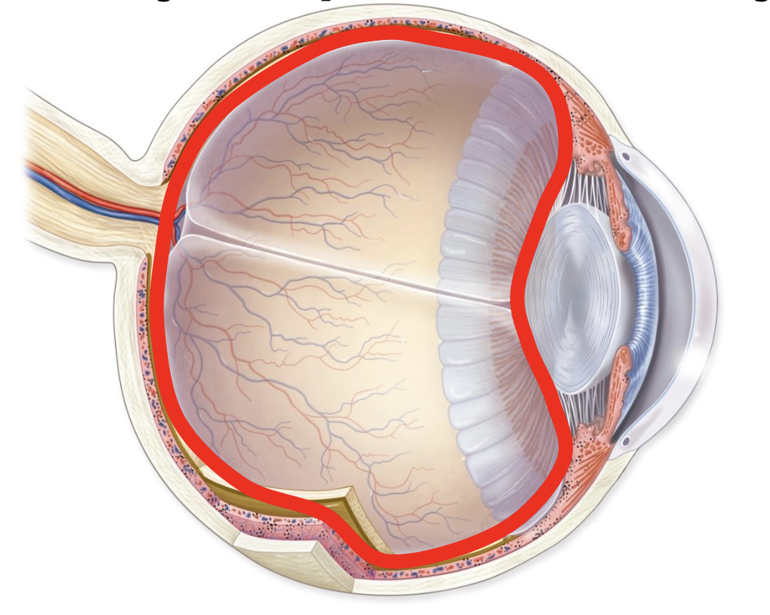
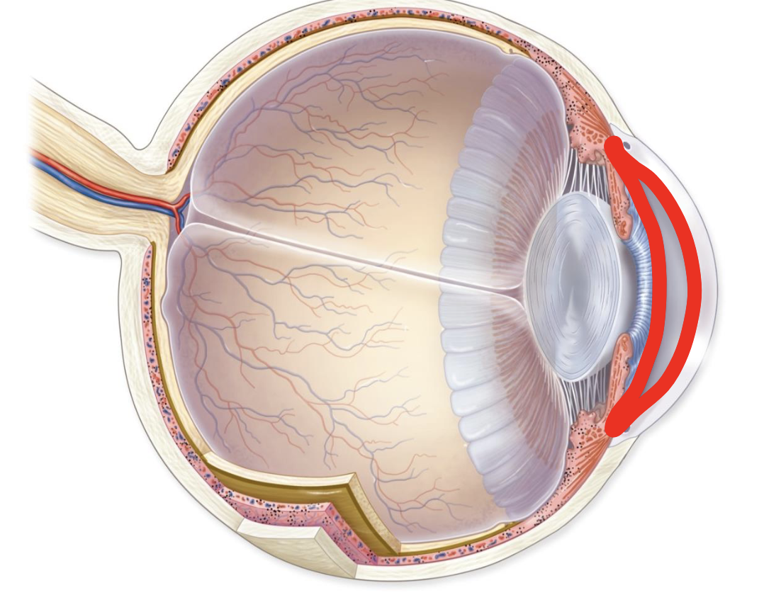
What is highlighted?
Posterior cavity
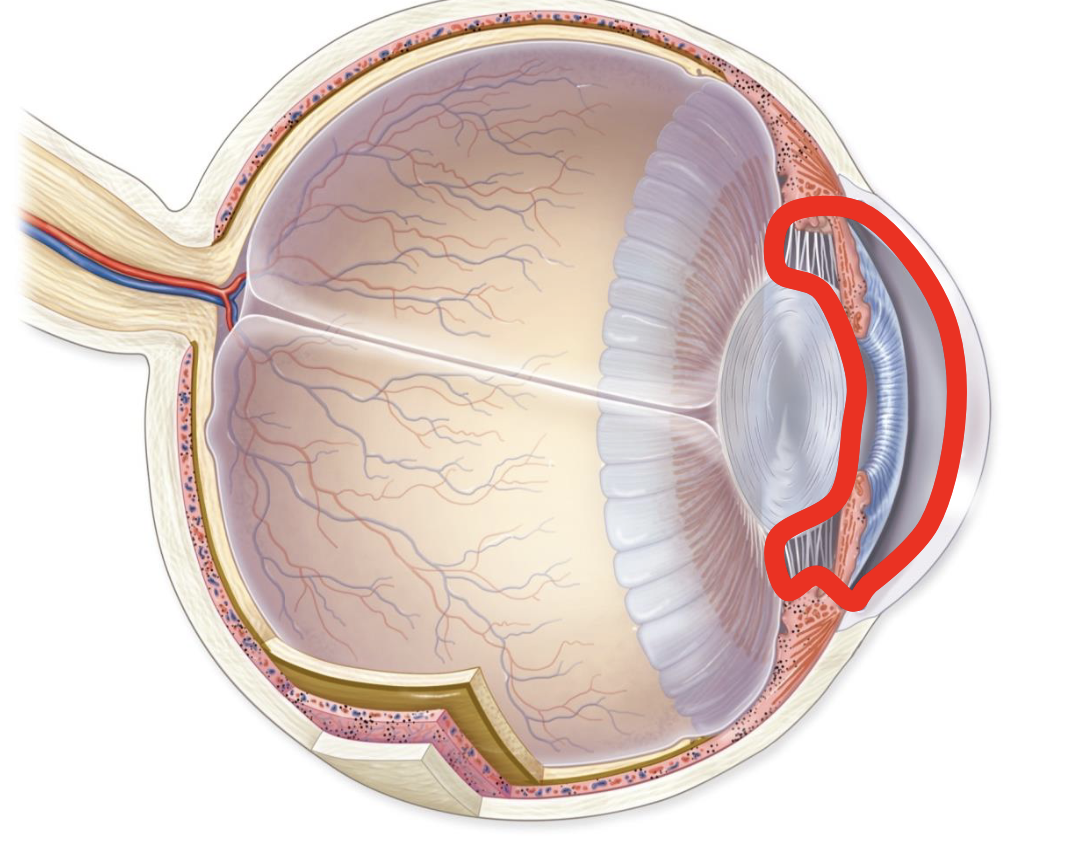
What is highlighted?
Anterior cavity
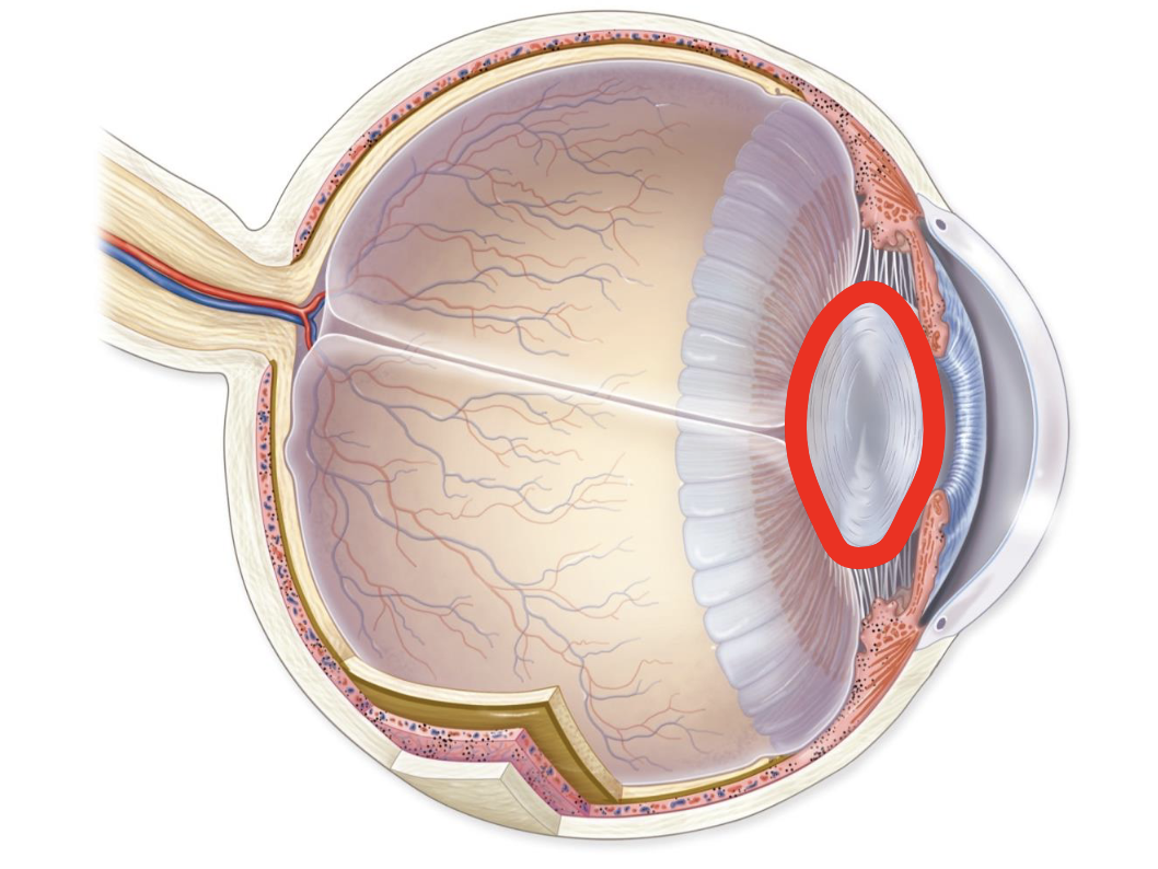
Identify the highlighted structure.
Lens.
What is highlighted?
Anterior chamber
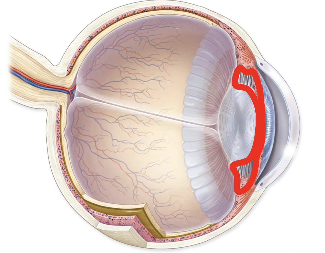
What is highlighted?
Posterior chamber
The Fibrous Tunic
Outermost layer for protection. Contains the sclera and cornea. Diseases of this layer are painful.
The Vascular Tunic
Middle layer for nutrition. Contains the iris, ciliary body, choroid. Disease of this layer are inflammatory.