Digestive System
Why the Digestive System?
- The food that we eat cannot be utilized as such in the body.
- It must be changed into a soluble absorbable form to get absorbed by the blood for distribution in the body.
- Certain foods, like cane- sugar are already soluble in water, but they require a breaking down of their molecules into smaller units so that they could pass through the cell membranes of the wall of the gut.
Enzymes:
- Enzymes play a key role in the digestion of food taken in.
- There are hundreds of enzymes in addition to those involved in digestion, but the general characteristics of all enzymes are the same.
Characteristics of Enzymes:
- It is a protein and is, therefore, destroyed by heating.
- It acts only on one kind of substance called the substrate i.e. it is specific.
- It always forms the same end-products) from the substrate.
- It only affects the rate of a chemical reaction and always speeds up the reaction.
- Like a catalyst, it can be used again and again.
- It acts best only at a particular pH, i.e. at a particular degree of acidity or alkalinity.
- It acts best within a narrow temperature range, usually between 35° and 40°C which is also called optimum temperature.
The Digestive System:
Alimentary Canal:
- The alimentary canal is a muscular tube that starts with the mouth and ends at the anus.
- It is about 9 meters long and is highly coiled in certain regions, especially in the small intestine.
- Its various regions are different both in structure and function.
- In addition to the digestive glands located in the lining of the various regions of
- the digestive tube
- two large digestive glands
- the liver
- pancreas
- three different salivary glands are associated with the mouth cavity.
- The various organs of the digestive system are described as follows
The Mouth:
- The mouth or the mouth cavity is the space where the food is chewed and mixed with saliva.
- Its front limits are formed by the upper and lower lips.
- The lips help in
- closing the mouth
- sucking and sipping liquids
- speaking
- perceiving certain sensations, especially those of touch and heat
- A muscular tongue helps in
- manipulating the food while chewing and mixing it with saliva
- cleaning the food particles from the teeth after eating
- tasting
- speaking
The Teeth:
- The dentition has a very special role — they cut and break the food into smaller bits.
- The small-sized bits have a relatively larger surface for the enzymes to act on for better digestion.
- The teeth help in speaking
- Teeth also add to facial beauty.
- An adult human normally has 32 teeth.
- These teeth are different in shape and perform different functions as follows:
- Incisors are the four front teeth in the center of each jaw.
- Their cutting edges are broad and sharp like a chisel.
- They are used for biting and cutting
- Canines are one on either side of the incisors in each jaw.
- These are conical and sharply pointed for holding and tearing the food.
- Premolars are two on each side of each jaw next to the canines.
- Each premolar has two hill-like projections or cusps on its surface, and hence is known as a bicuspid.
- Premolars help in grinding and crushing the food.
- Molars are the last three teeth on each side of each jaw.
- They have a larger surface than the premolars.
- They are principal grinders and crushers of food.
- The last molar of each side of each jaw is called a wisdom tooth.
- The wisdom teeth are so called because they appear last at an age of about 17-20 years when the human body is reaching maturity.
- In this manner, the human or mammalian teeth are different in shape and are called heterodont (hetero: different, dent: teeth) as opposed to homodont (homo similar) teeth of other animals like those of the lizard and frog.
- Mammalian teeth appear in two sets during life.
- In humans, the first set, or milk teeth, consists of 20 teeth (all, but not the molars) which start growing through the gums (sometimes painfully) when the child is about 7-8 months old and are completed when he is about 2 years old.
- The wisdom tooth falls out as a result of its roots being dissolved away in the jaw and being completely replaced by permanent teeth.
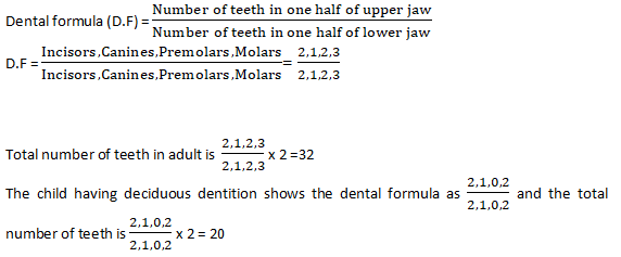
- The number of permanent teeth of mammals is usually indicated in a formula in which the number of
- incisors (i)
- canines (c)
- premolars (pm)
- molars (m) is given strictly in the same order.
J Structure of the Tooth:
- The general structure of all types of teeth is the same.
- Each tooth consists of a crown or the part exposed above the gum and the root or the part embedded in a cup-like socket of the jaw bone.
- The root consists of a single process or fang as in incisors and canines, or of two processes or fangs as in premolars and lower molars, and three in upper molars.
- The bony socket is a slight constriction between the root and the crown. In a vertical section, a tooth shows the following parts:
- Enamel or the “ivory” is the material that covers the crown.
- It is the hardest substance in the body.
- Dentine forms the bulk of the tooth.
- It is harder than bone but not as much as enamel.
- It has minute canals through which run the strands of the cytoplasm of the cells in the pulp cavity.
- Cement is another bone-like structure covering and fixing the root in position.
- Pulp is the soft connective tissue contained in the central space (pulp cavity) of the tooth.
- It consists of blood capillaries, lymph vessels, and nerve fibers which are continuous below those of the body through the opening of the pulp cavity at the base of the root.
The Salivary Glands:
- The saliva is secreted by three pairs of salivary glands:
- parotid glands located just in front of beneath each ear
- submandibular glands lying close to the inner side of the lower jaw on each side
- sublingual glands below the tongue.
- Ducts from each gland transport the secreted saliva into the mouth.
- Small quantities of saliva keep secreting at all times.
- While eating, the salivary flow is considerably increased.
- Sometimes, even the sight, smell, or just a thought of tasty food can cause an increased flow of saliva resulting in “watering of the mouth.
- Saliva is a very slightly acidic (pH 6.8) fluid containing water (about 99%), salts, mucus, and an enzyme salivary amylase (also called ptyalin).
Functions of Saliva:
- Moistens and lubricates the inner lining of the mouth cavity and the surface of the tongue to facilitate the churning of food.
- Acts as a solvent, dissolving some food particles to stimulate the taste buds of the tongue
- Helps food particles to stick together to form bolus so that they can be swallowed in a mass.
- Moistens and lubricates food which again helps in swallowing food.
- Digests starch.
- Its enzyme ptyalin (amylase) converts starch into maltose.
- This explains why if boiled rice is chewed very well it begins to taste sweet.
- Cleans the mouth and tends to destroy germs to prevent tooth decay.
- Dryness in the mouth (due to less water in saliva), gives a feeling of thirst to replenish body water.
- Thus saliva aids in water balance in the body.
Swallowing and Peristalsis:
- The tongue presses upward and back against the roof (palate) of the mouth and this forces the bolus (the ball of chewed food) into the throat or the pharynx.
- The back part of the roof of the mouth cavity (soft palate) closes the opening between the throat and the nasal passage.
- The larynx (“voice box”) which is located at the entrance of the windpipe is pulled upward to bring it close to the back of the tongue when a flap called the epiglottis closes its opening.
- Thus the only passage available to the swallowed food is that of the gullet or esophagus.
- Once the food reaches the esophagus it is conducted behind by a special movement called peristalsis.
- Peristalsis is the wave of constrictions caused by the circular muscles of the gut pushing the food along.
- As the wave passes the circular muscles relax.
- Peristalsis occurs through all regions of the gut.
- The mucus secreted by all the regions of the gut is a slimy fluid that lubricates the food.
Stomach:
- The stomach is an elastic bag located below the diaphragm.
- In an average adult, it can hold 2 to 3 liters of food.
- Its walls are highly muscular and chum the food thoroughly to mix with the gastric juice secreted by the inner lining.
- The opening of the stomach into the intestine is called the pylorus.
- It has a ring of muscles (sphincter) to keep the opening closed like a valve to prevent food from passing from the stomach until it is thoroughly churned up.
- It also prevents regurgitation from the duodenum.
- A similar sphincter termed the cardiac sphincter is present at the front end of the stomach to prevent the backflow of food into the esophagus.
- Gastric juice is secreted by the inner lining of the stomach.
- It is a colorless highly acidic liquid containing water, some salts, hydrochloric acid, and an enzyme called pepsin.
- The acid serves two functions:
- It kills any germs which may have entered along with the food.
- It activates pepsin to act on proteins.
- In fact, the pepsin is first secreted as pepsinogen which is then changed to pepsin by the acid.
- Pepsin digests proteins (about 20% only) into peptides.
- The food stays in the stomach for about 3 hours and it turns a pulp-like form called chyme.
- Now the pylorus opens intermittently to allow the chyme to move to the intestines little by little.
Small Intestine:
- The small intestine is a tube about 7 meters long and about 2.5 mm wide.
- It is coiled and folded in the abdomen.
- Its three sub-regions are as follows:
- Duodenum: Short upper part next to the stomach. (“duodenum” means 12, i.e. twelve finger*-*breadths in length, duo. two deni: ten).
- The common bile duct opens into this.
- Jejunum: Next short region of about 2 meters.
- (“jejunum” means empty because it is nearly always empty after death as found in dissections or in post-mortems.)
- Ileum: About 4 meters. (“ileum” means to twist roll, referring to the twisting movements of this part).
- The inner lining of the ileum is produced into a great number of tiny finger-like projections called villi (singular villus).
- The villi enormously increase the inner surface area of the intestines (nearly 8 times that of the outer body surface) which facilitates the absorption of digested food.
- Between the villi are small holes through which the intestinal juice secreted by its glandular cells is poured into the lumen of the intestine.
- Each villus is covered by a single-cell thick epithelium.
- Inside the villus are contained an artery, a vein, inter-connecting blood capillaries, and a lymph vessel called a lacteal.
- The small intestine serves both for digestion and absorption.
- It receives two digestive juices;
- the bile
- the pancreatic juice in the duodenum and its own walls secrete the intestinal
- Adaptations of the ileum for absorption of digested food:
- It is very long; more surface area is provided.
- A large number of villi further increase the surface area for absorption.
- Single-cell epithelium.
- It is narrow for the slow movement of food allowing absorption.
Bile:
- This is a yellowish green watery fluid produced in the liver, which is transported through the hepatic duct.
- The hepatic duct is joined by the cystic duct to form the common bile duct.
- The bile may flow directly into the duodenum or as it generally happens, gets temporarily stored in the gall bladder.
- The colour of the bile is due to certain pigments (biliverdin and bilirubin) produced by the breakdown of the dead and worn-out red blood cells.
- It contains a lot of sodium bicarbonate which neutralizes the acid content of the food received from the stomach and makes it alkaline to enable the pancreatic and intestinal enzymes to act.
- Bile salts reduce the surface tension of fats and break them into tiny droplets (emulsification) for providing greater surface area for the action of enzymes.
Pancreatic Juice:
- This is produced in a whitish gland, pancreas, located behind the stomach. The pancreatic duct opens into the duodenum by an aperture common to that of the bile duct.
- The pancreatic juice contains three kinds of enzymes —
- Amylopsin: (pancreatic amylase) digests leftover starch into maltose.
- Trypsin It acts on the remaining proteins and polypeptides to produce smaller peptides and amino acids.
- Trypsin is first secreted as inactive trypsinogen which is activated to trypsin by an enzyme, enterokinase (also called enteropeptidase), secreted by the inner lining of the duodenum.
- Steapsin which acts on emulsified fats to split them into fatty acids and glycerol.
- Intestinal juice contains:
- erepsin (peptidases) to convert remaining peptides into amino acids
- maltose to digest maltose into glucose
- lactase to digest lactose into glucose and galactose
- invertase (or sucrase) to split sucrose into glucose and fructose
- some traces of lipase to digest fats into the fatty acids and glycerol.
- Absorption of food. The final products of food are mainly absorbed in the intestine itself.
- The amino acids and the simple sugars (glucose, fructose, galactose) have relatively small-sized molecules which are absorbed through the thin epithelium of the villi and reach their blood capillaries to finally enter the blood circulation first to reach the liver through the hepatic portal vein.
- The fatty acids and glycerol are absorbed into the lymph vessel or the lacteal to enter the lymphatic system which forms a network all over the body to ultimately empty its contents into the blood stream.
- The food passes very slowly through the intestine taking about four hours to enter the last part large intestine.
The Large Intestine:
- The large intestine is about 1.5 metres long.
- It has three parts:
- caecum
- colon
- rectum.
Caecum:
- A small blind pouch situated at the junction of the small and large intestines.
- From its blind end projects a narrow worm- shaped tube called vermiform appendix (when inflamed it causes appendicitis) and today it is a functionless (vestigial) organ.
Colon:
- It is much broader than the ileum and is a little more than a meters long.
- It passes up the abdomen on the right (ascending colon) rosses to the left just below the stomach (transverse colon) and down on the left side (descending colon).
Rectum:
- It is the last pan, about 15 cm long which opens at the anus.
- The anus has circular muscles (sphincters) to keep it closed except when passing bowels.
Functions of Large Intestine:
- The large intestine secretes no enzyme.
- It absorbs much water but very little digested food from the contents which mainly consist of undigested material.
- After much water is absorbed the contents become semi-solid faeces which pass into the rectum and are expelled at intervals.
- The expulsion of the undigested remains of the food from the alimentary canal is called defaecation.
Liver:
- The liver is the largest gland of the body weighing about 1500 gm on an average.
- It is a reddish brown organ located in the upper right side of the abdomen just below the diaphragm.
Functions of Liver:
- Controlling blood sugar levels: It regulates blood sugar by retaining excess glucose received as products of carbohydrate digestion from the intestine and storing it as glycogen, and releasing it again when needed.
- Controlling amino acid levels: Excess amino acids are broken to remove the nitrogen part (deamination) producing urea and sugar.
- Urea is eliminated through excretion and sugar is utilized in metabolism.
- Synthesis of foetal red blood cells: It produces red blood cells in embryo.
- (In adults RBCs are produced in bone marrow).
- It produces fibrinogen and prothrombin which are used in blood clotting.
- It produces heparin (an anticoagulant).
- It regulates blood volume by acting as a temporary store of excess water.
- It destroys dead red blood cells.
- Detoxifies substances including drugs and alcohol.