POM II - Benign Skin Neoplasms - Exam 1
1/41
There's no tags or description
Looks like no tags are added yet.
Name | Mastery | Learn | Test | Matching | Spaced | Call with Kai |
|---|
No study sessions yet.
42 Terms
Benign Nevus
-common mole
-benign skin tumors composed of melanocytic derived cells
-distinctions are based upon the location of melanocytic nests in the epidermis, dermis, or both and age of onset
-junctional nevi are flat
-dermal and compound nevi are elevated
Benign Nevi - Etiology
-typically arise during childhood, adolescence, or very early adulthood, and then senesce in later years
-during pregnancy, existing nevi may darken and become noticeable
-compound nevi are more common in lighter skin phototypes
-not common to get new moles after 50
ABCDE - moles
-A: asymmetrical
-B: borders irregular
-C: colors not uniform
-D: diameter >6 mm
-E: evolving or changing over time
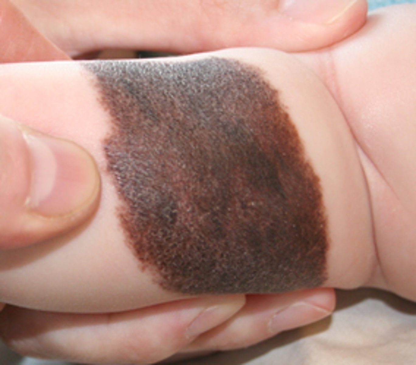
Congenital Nevus
-benign nevus present at birth or within first two weeks of life (1-2% worldwide)
-often flat and tan in color
-may change in color, become papillated, or display hair growth during first few years of life and can vary tremendously in size
Congenital Nevus - Presentation
-been reported to have an increased risk for transformation to melanoma
-risk of melanoma is believed to correlate with CMN size
-most common locations: buttocks, thigh, trunk; can also occur on face, extremities, palms, soles, scalp
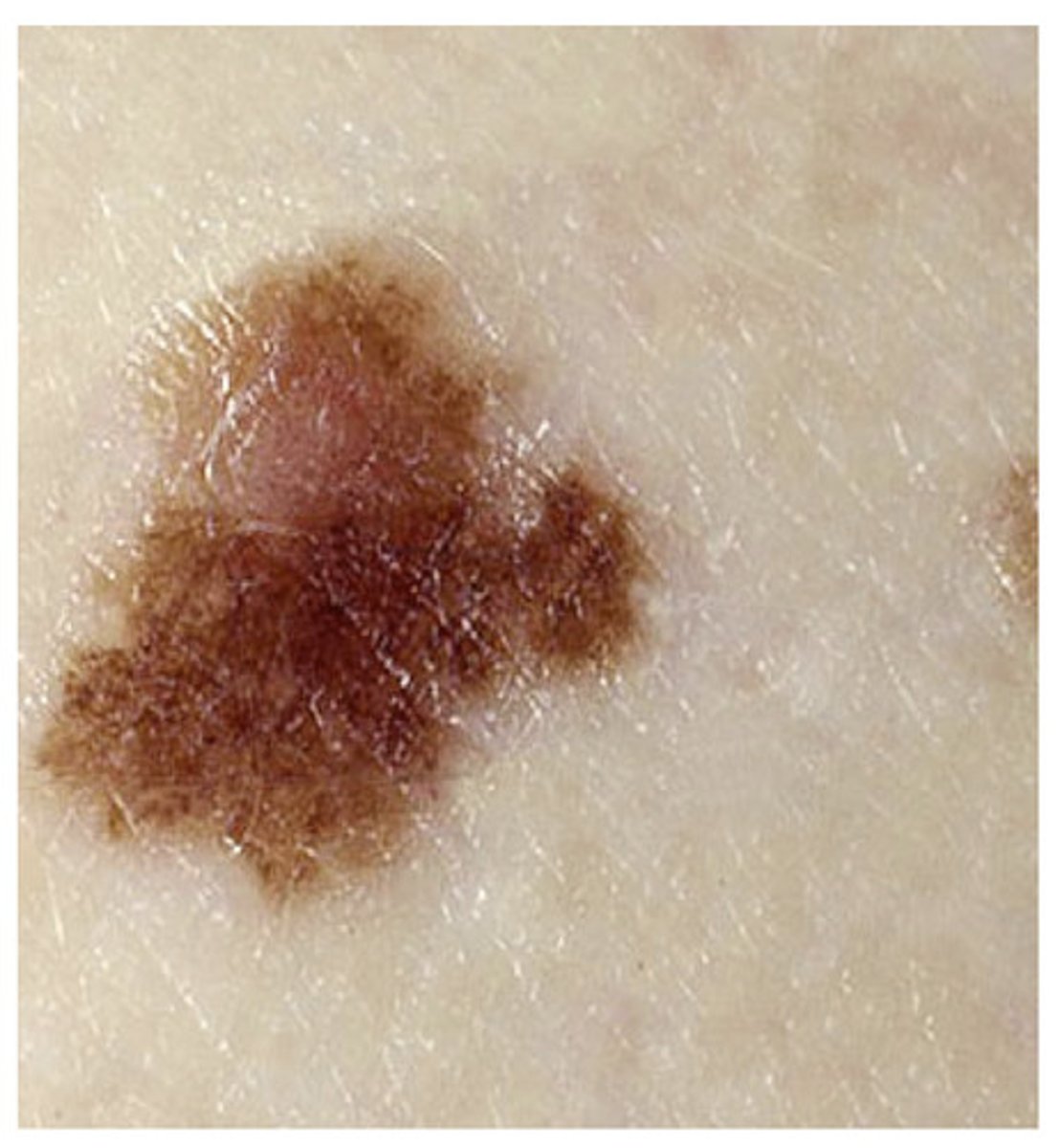
Dysplastic Nevus
-a melanocytic proliferation showing atypia and sharing some features of melanoma
-controversial d/t lack of consensus on how it is defined and what it represents biologically: graded mild, mod, or severe
-believed to correlate with the overall number of melanocytic lesions in an individual
-4-15 fold increased risk of melanoma
Mongolian Spot
-congenital dermal melanocytosis
-common newborn pigmented lesion usually in sacral area
-benign, ill-defined, blue-to-gray patch present at birth or shortly after
-commonly seen in the sacrococcygeal area in infants of Asian or African descent but may be found on any cutaneous surface in infants of all ethnicities
-pigmentation most intense at 1 y.o., reaches peak diameter by 2 y.o., usually fades by adulthood
port wine stain
-a congenital benign capillary malformation
-most common type of vascular malformation
-occurs in about 3 in 1000 people
-swollen blood vessels create a reddish-purple discoloration of skin
-rare cases are a sign of sturge-weber syndrome
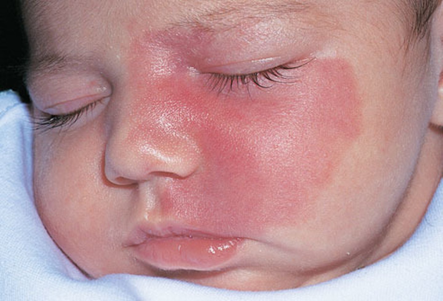
Port Wine Stain - Presentation
-most often on face but can be anywhere
-persists for life
-may become more violaceous and take on a cobblestoned texture with age
-can be cosmetically disturbing to the patient
-lesions may be associated with a number of other findings or conditions
pulsed-dye laser
what is the tx for port wine stain?
Cafe au lait spot
-a well-defined, evenly pigmented brown macule or patch
-light to dark brown depending on pts skin pigment
-onset is usually evident in early childhood as solitary lesion
-a single lesion is present in 10-20% of US population, and 1% of healthy YA have up to 3 spots
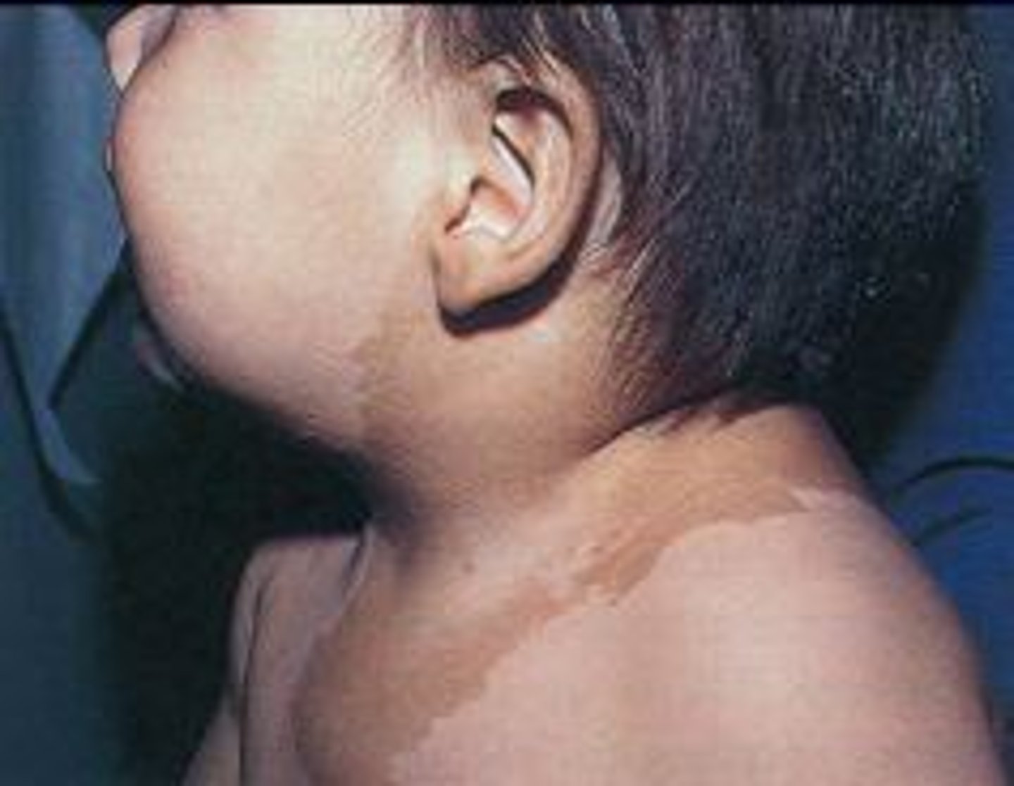
Cafe Au Lait Spot - Presentation
-located anywhere on the body
-typically appear on the trunk or LE and rarely on the face
-increase proportionally in size as a child grows
Neurofibromatosis
-multisystem genetic disorder with hallmark cutaneous findings including cafe au lait macules, neurofibromas, and axillary freckling
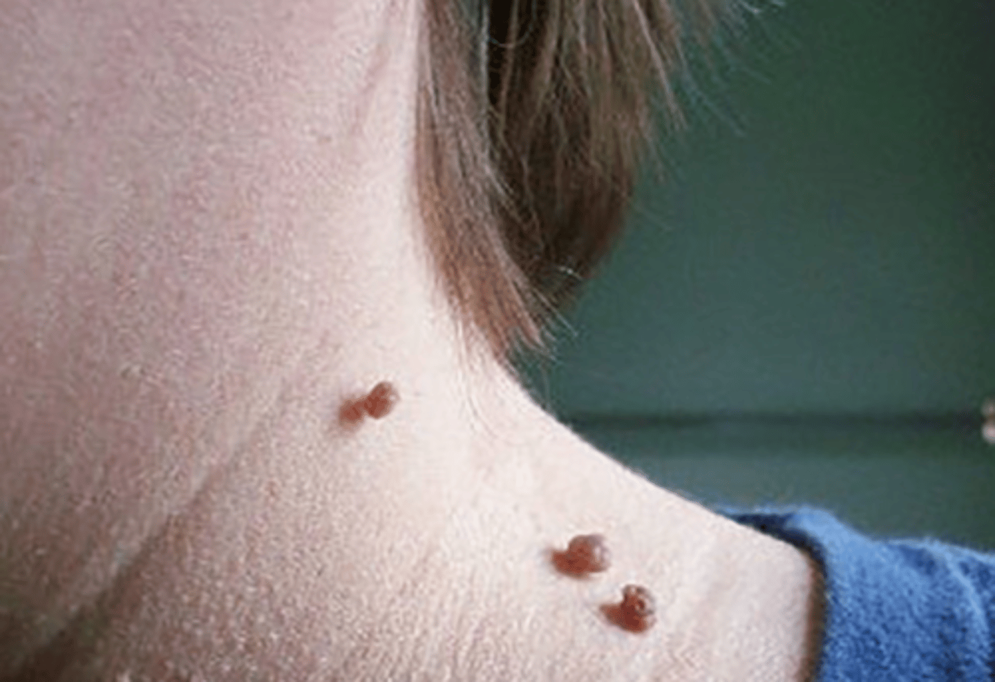
Acrochordon
-skin tags or fibroepithelial polyps
-common, benign, pedunculated growths
-most commonly found in areas of frequent friction such as the eyelids, neck, axillae, and inguinal area
-usually asx but can become irritated by clothing or jewelry
-associated with increasing age, pregnancy, DM, and obesity
Acrochordon - Incidence
-men and women are affected equally
-there is no difference in prevalence among different ethnicities and races
Acrochordon - Tx
LN2 or snip removal
Lipoma
-benign tumors of slow-growing, mature fat cells - most common soft tissue tumor
-present as soft, rubbery, freely mobile subcutaneous masses without overlying skin change
-most often solitary, but can be multiple
-found in areas where there is fat
-can range in size, usually slow growing
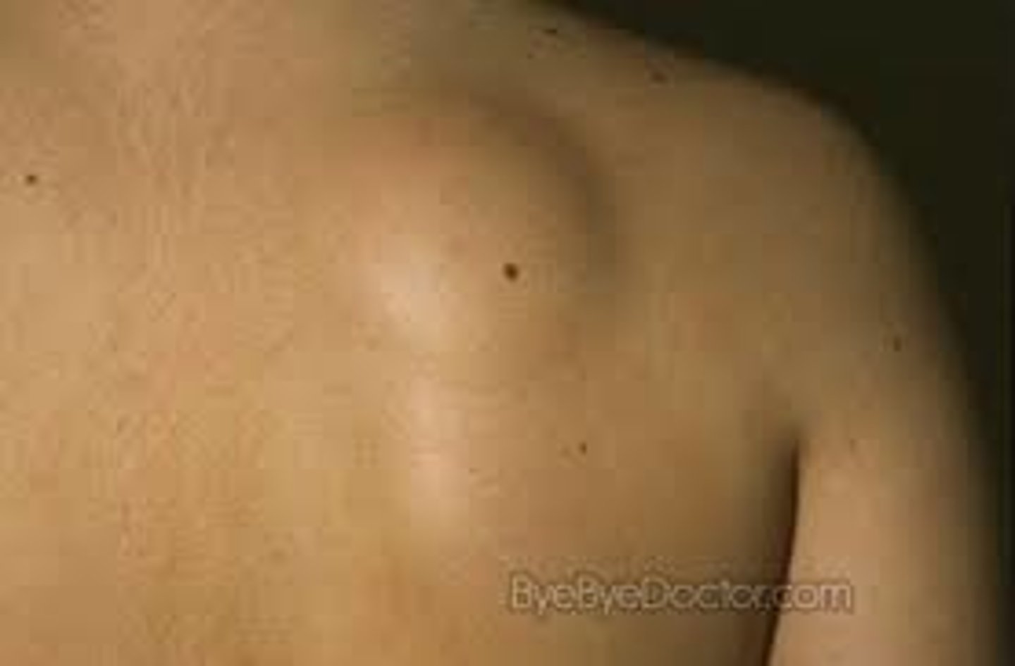
Lipoma - Presentation
-usually asx
-large tumors that compress nerves or limit normal tissue movement can cause lymphedema with discomfort and pain
Lipoma - Tx
-not necessary
-can be surgically excised if bothersome
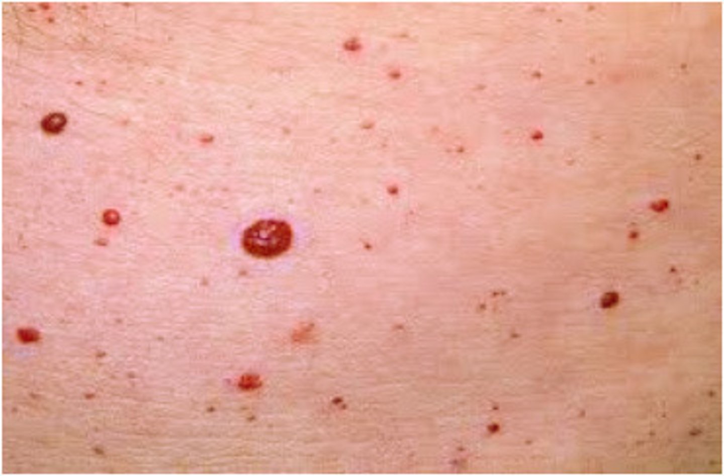
Cherry Angiomas
-most common type of acquired benign vascular proliferation and area composed of thin-walled, dilated capillaries
-often present in early to mid-adulthood and appear as small red or violaceous macules or papules that increase in number and incidence with age
-benign and thus do not require treatment unless irritated or bleeding, but are often of cosmetic concern
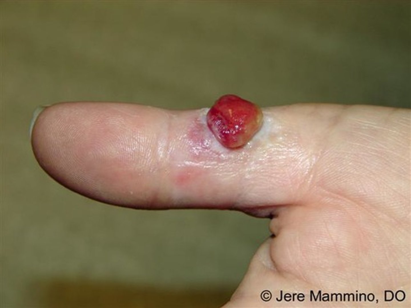
Pyogenic Granuloma
-benign vascular lesion of skin and mucosa, rapidly growing
-a glistening, friable, bright red papule or nodule that bleeds spontaneously or after trauma
-unknown cause
-not infectious or granulomatous
-most often on hand, neck, extremities, trunk, but can also arise on the gingiva or oral mucosa
-can arise in pregnancy
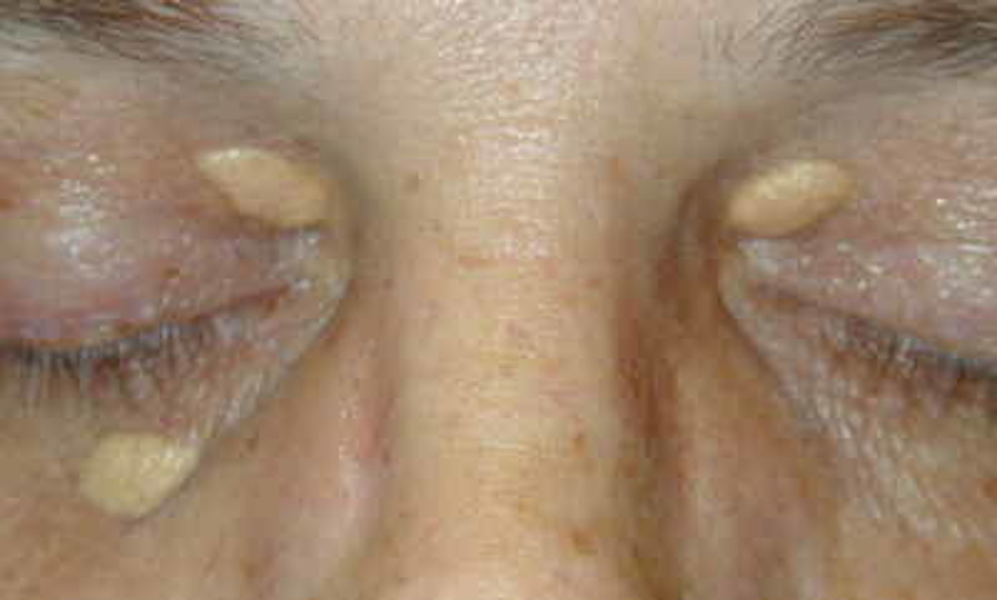
Xanthoma
-a deposit of yellow cholesterol rich material in tendons or other parts of body
-commonly seen on eyelids
-cutaneous manifestations of lipidosis
-associated with hyperlipidemia
Seborrheic keratosis
-most common benign tumor in older individuals
-raised, "stuck-on" appearing papules and plaques with well-defined borders
-unusual before age 30 and increase in amount by age
-etiology unknown
-usually asx but can be itchy or rub on clothing
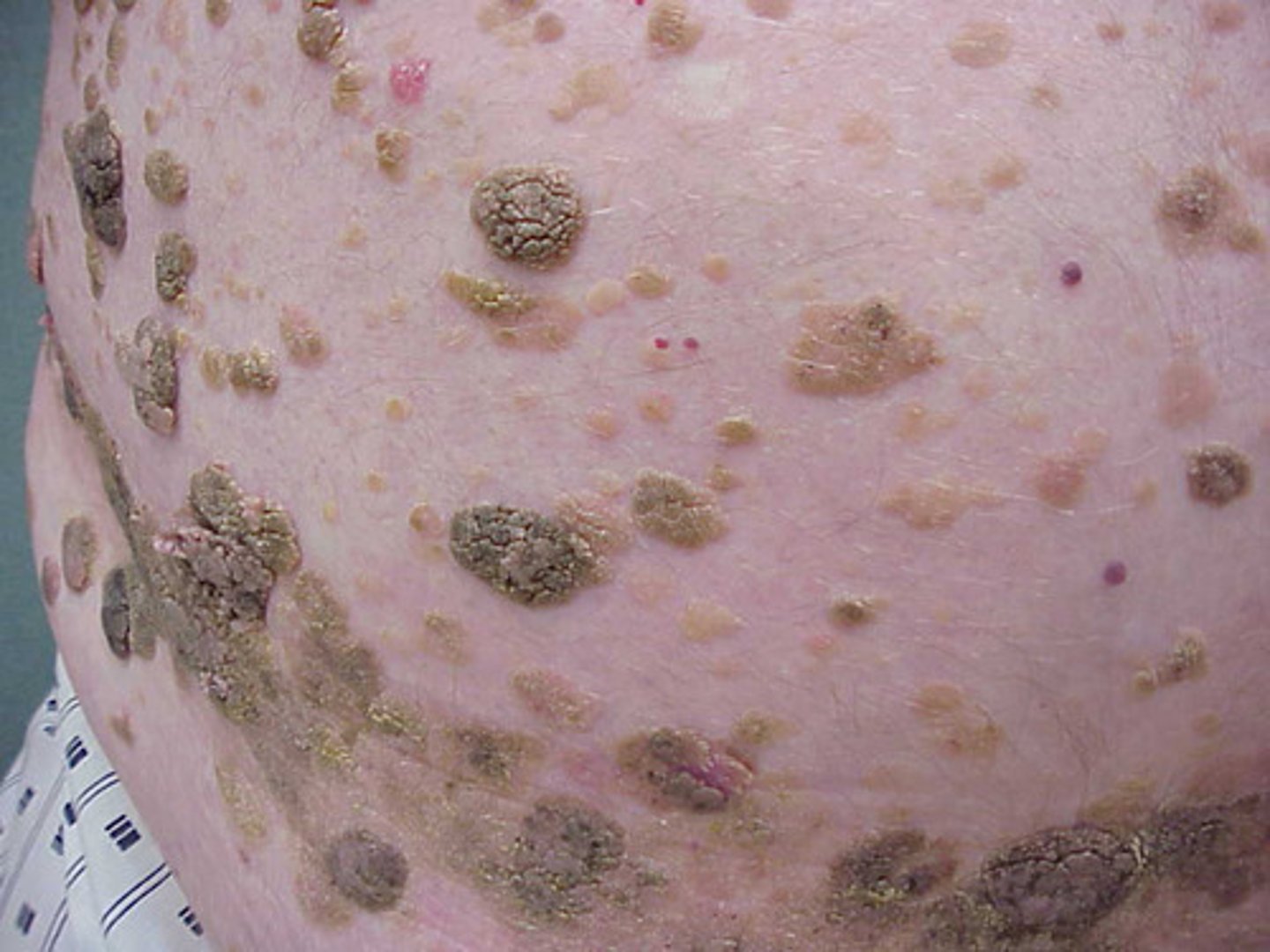
Seborrheic Keratosis - Presentation
-waxy texture
-color can be white, pink, brown, black
-look scary but are benign; will not turn into cancer
-if you are unsure, BX
-can leave them alone or can treat with LN2 if desired
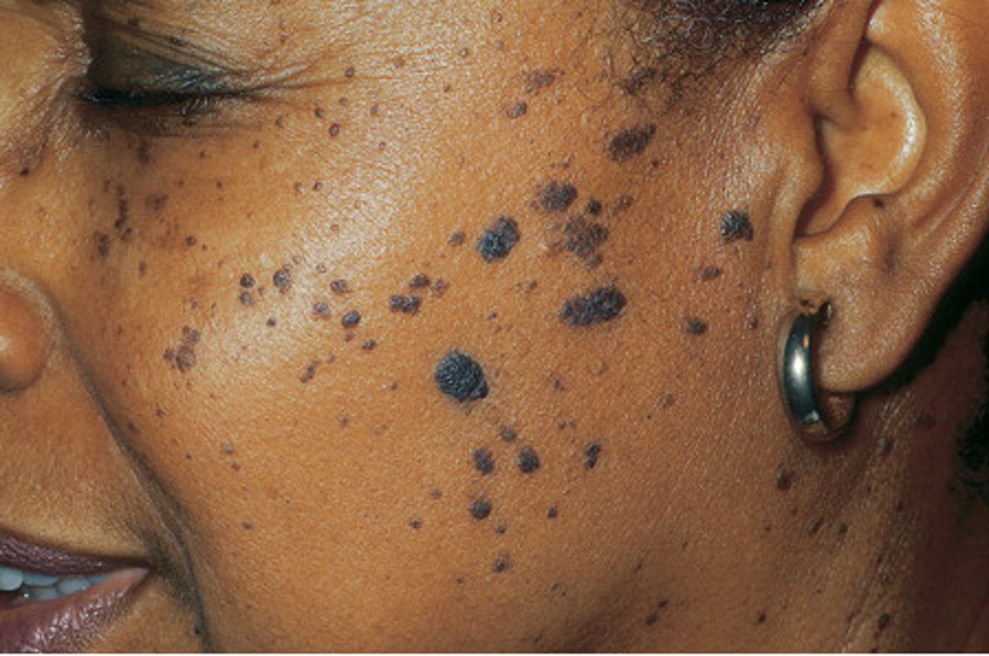
Dermatosis papulosa nigra
common variant of SKs in african americans
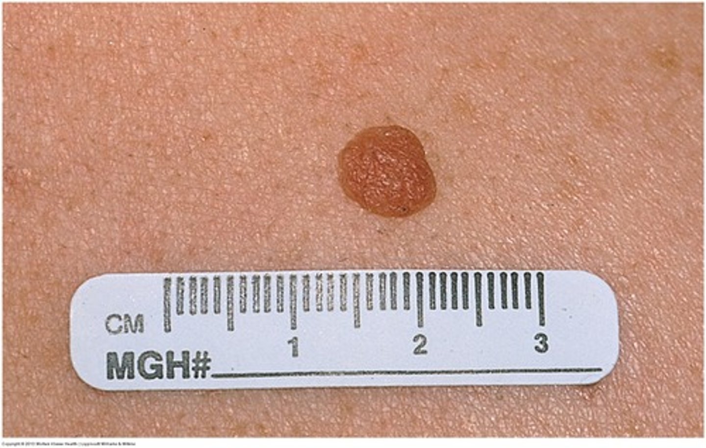
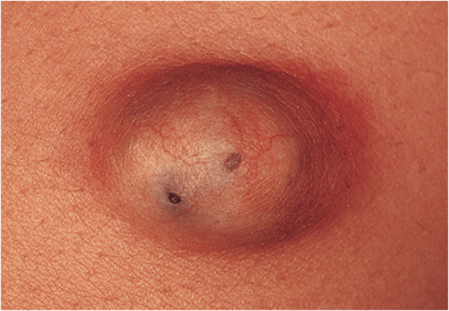
Dermatofibroma
-common, benign skin neoplasms composed of collagen, macrophages, capillaries, and fibroblasts
-etiology is unknown, though some may arise at sites of trauma or insect bites
-most common on the legs of women and usually appear in young adulthood
Dermatofibroma - Presentation
-firm, skin-colored pink or slightly pigmented papules or nodules
-usually asx but may be tender or pruritic
-often persist for life
-tenderness may occasionally be elicited with manipulation of the lesion
-dimple sign
Dimple Sign - Dermatofibroma
-squeezing results in a dimple or puckering inward
-aka Fitzpatrick sign
Dermatofibroma - Tx
-unnecessary
-can be excised if bothersome
-intralesional steroids may help
-cryotherapy less effective
Keratoacanthoma
-rapidly growing, well-differentiated neoplasm of squamous epithelium
-considered by many to be a low-grade SCC
-appear and grow rapidly over a few weeks and spontaneously involute and resolve within 6 months, leaving an atrophic scar
-immune system is thought to play a role in the spontaneous regression of these
Keratoacanthoma - Incidence
-most commonly seen in individuals aged 60 years and older with lighter light skin colors and a history of prolonged sun exposure
-men more common than women
Keratoacanthoma - Etiology
-trauma
-HPV
-genetics
-immunologic status
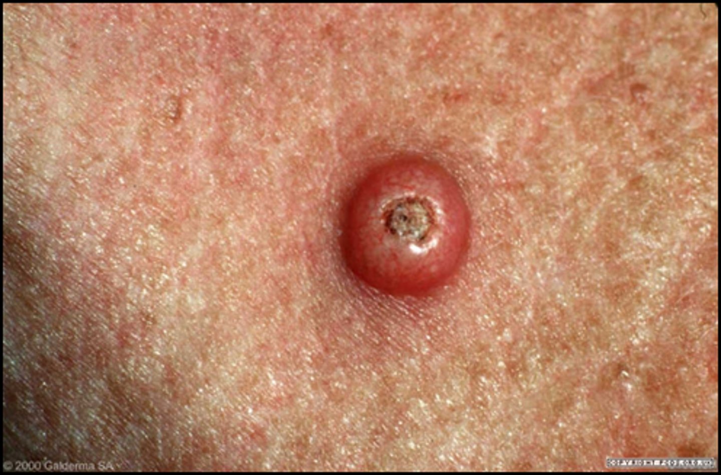
Keratoacanthoma - Presentation
-solitary, crater-shaped nodules measuring a couple centimeters in diameter
-often with a central keratin plug on sun-exposed skin
surgical excision or mohs surgery
what is the tx for keratoacanthoma?
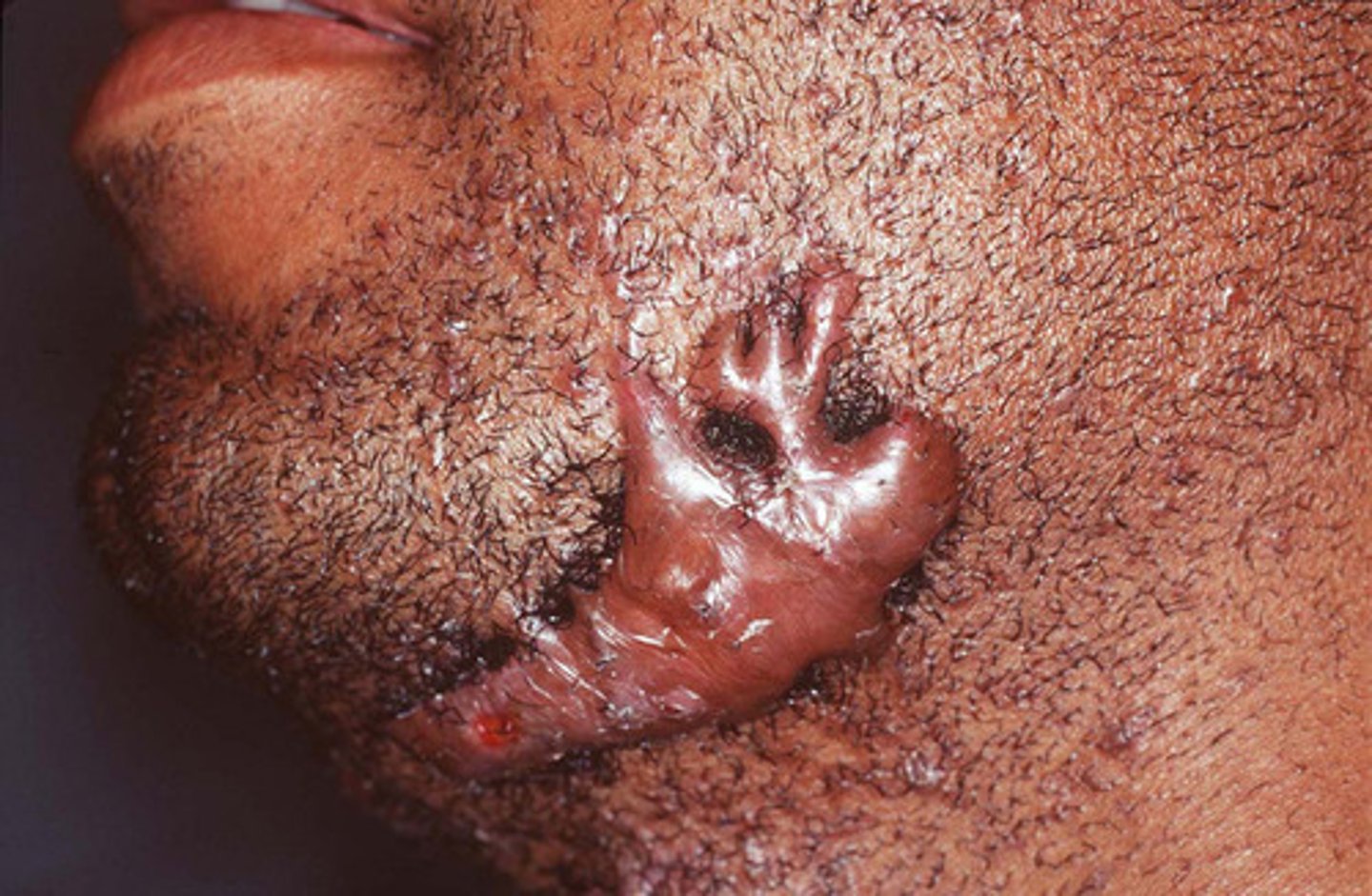
keloid
-hypertrophic scar that extends beyond the area of trauma, injury, or surgical scar
-raised, smooth, firm, hairless, and shiny papules, plaques, or nodules
-found at areas of previously traumatized skin or arising spontaneously on normal skin
Keloid - Presentation
-may be painful, tender, pruritic, and can grow to become very large
-can cause chronic discomfort, be disfiguring, and restrict normal tissue motion
-most often occurring in areas of high skin tension
Keloid - Incidence
-can affect individuals of any race and ethnicity, but most common in african americans
-likely a genetic basis for the tendency to develop
-may occur in acne scars
keloid - tx
-extremely difficult
-prevention is key - surgical wounds should follow skin lines
-try to avoid cutting over joints when there is high mobility or in areas of high tension
-intralesional steroid tx q4 wks
Inclusion Cyst
-dome-shaped, firm, skin-colored nodule that is freely movable on palpation and sometimes has a small, dilated punctum
-filed with thick, cheesy material (keratin and lipid-rich debris) with a foul odor that can be expressed
-can be well-defined or it may have an irregular border and surface d/t prior rupture, scarring, and regrowth
-benign and usually asx, can be painful
Inclusion Cyst - Incidence
-can arise on face, trunk, extremities, in mouth, or in genitals at any age
-more common in men
inclusion Cyst - tx
-not necessary
-can be expressed
-excision (when not inflamed)
-if infected --> incision and drainage +/- packing and definitely abx
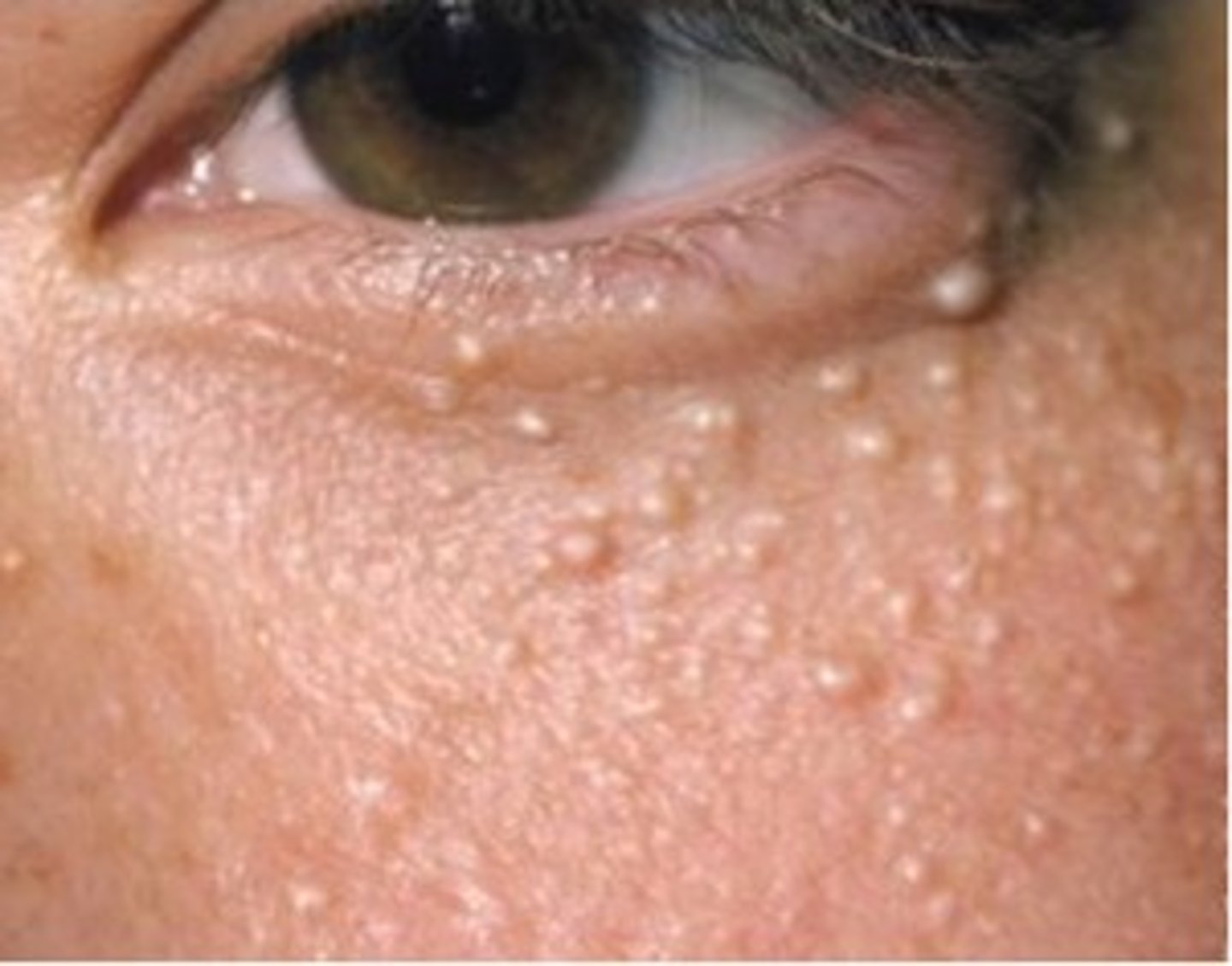
Milia
-very common benign small cysts
-typically seen in infants, but can be seen at any age
-asx, superficial, usually 1-2 mm in diameter
-can easily be punctured with 18 gauge needle and expressed but not necessary