L33 Ureters, bladder and urethra
1/12
Earn XP
Description and Tags
A set of vocabulary flashcards covering key terms and definitions from the HUBS192 lecture material.
Name | Mastery | Learn | Test | Matching | Spaced | Call with Kai |
|---|
No analytics yet
Send a link to your students to track their progress
13 Terms
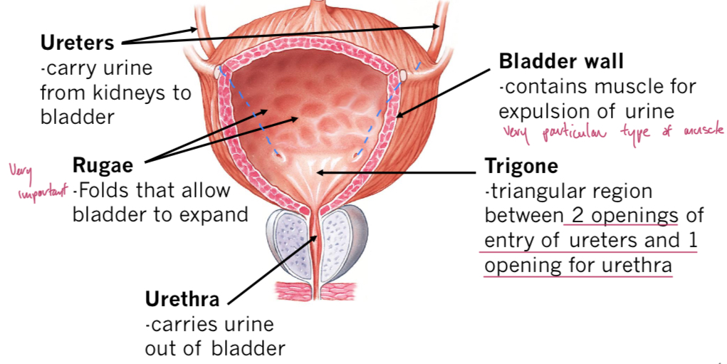
Ureters
Slender tubes that arise from renal pelvis at hilum and carry urine from kidneys to bladder using peristaltic waves. Descend retroperitoneally.
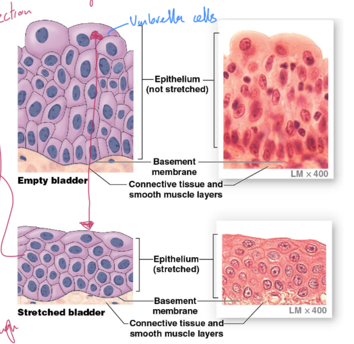
Transitional epithelium
Stratified, rounded cells that flatten when stretched, used for protection.
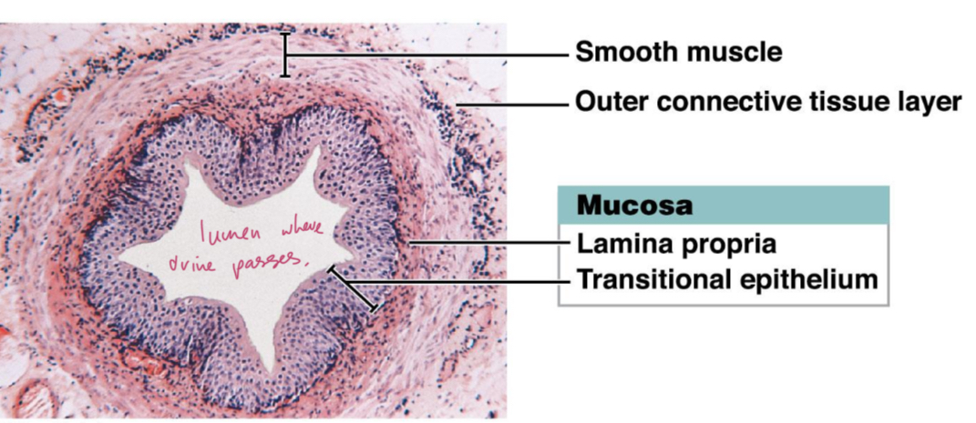
Layers of ureters
Transitional epithelium and lamina propria (mucosa), with folded protein plaques on inner surface. Muscularis made of inner longitudinal, and outer circular smooth muscle (opposite to GI). Adventitia (outer layer of connective tissue)
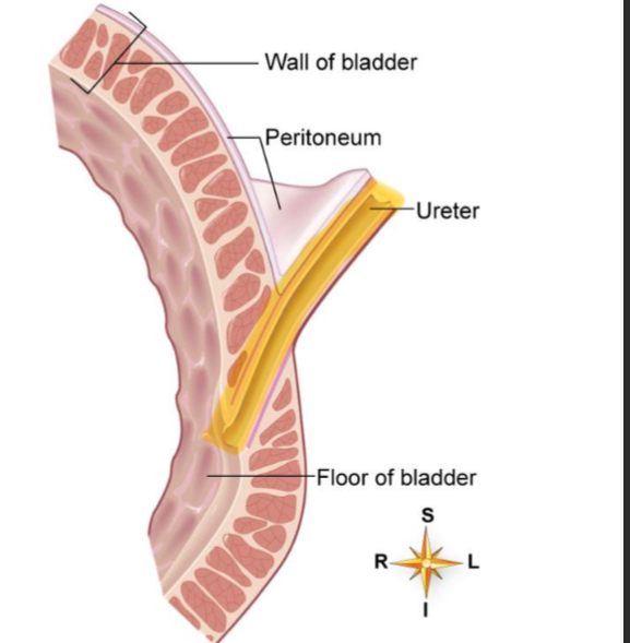
Ureter insertion to bladder
Run obliquely through bladder wall at postolateral corners, which acts as sphincter/valve. Compresses due to increased bladder pressure to prevent backflow.
Bladder
Collapsible muscular sack, stores and expels urine. Walls folded into rugae for expansion, mucosa of transitional epithelium (protection). Detrusor muscle layer.
Detrusor
Thick smooth muscle layer made of longitudinal, oblique and circular fibres. Contracts to expel urine into urethra.
Trigone
Triangular region in the bladder between the openings of the ureters and the urethra.
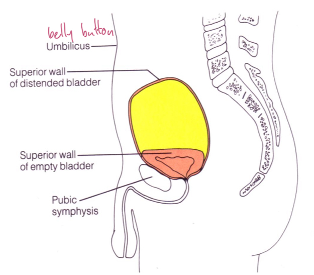
Empty vs full bladder
Empty: pyramidal, lies within pelvis. Filling: spherical, expands superiorly into abdominal cavity, can be palpated above pubic symphysis.
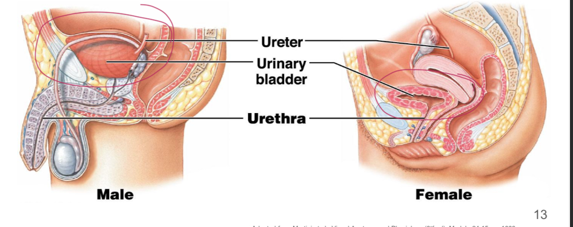
Male vs female location
Male located anterior to rectum, superior to prostate gland (wraps around urethra). Female located anterior to vagina and uterus.
Urethra
Thin walled muscular tube that drains urine from the bladder out of the body. Epithelium is transitional near bladder, columnar (mucous glands for protection), then stratified squamous near external opening.
Internal urethral/urinary sphincter
Involuntary, detrusor muscle, at junction of the bladder and urethra.
External urethral/urinary sphincter
Voluntary, skeletal muscle, located where urethra passes through urogenital diaphragm.
Urination
Bladder fills with urine and expands, APs from stretch receptors to brain. Urgency increases as signals increase, internal sphincters relax. Lastly conscious relaxation of external sphincter expels urine.