Bio II Exam II: Ch 37 "Neurons, Synapses and Signaling"
1/45
There's no tags or description
Looks like no tags are added yet.
Name | Mastery | Learn | Test | Matching | Spaced | Call with Kai |
|---|
No analytics yet
Send a link to your students to track their progress
46 Terms
Neurons
These are nerve cells that provide inputs from sensory systems and outputs to motor or other effector systems, and they make up the information processing network that connects these inputs and outputs. It is a type of cell that exemplifies the close relationship of form and function that often arises in evolution.
electrical; chemical
Neuron cells use two types of signals to communicate throughout the body: _________ signals for long distance, and ________ signals for short distance
Ganglia
Where the processing of signals and information transmitted by the nervous system takes place. This is a simple cluster of neurons that can exist throughout the body
Brain
Where the processing of signals and information transmitted by the nervous system takes place. This is a complex organization of neurons that usually exists in the head
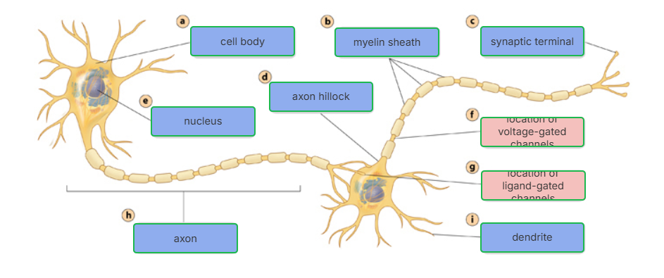
Cell body
Most of a neuron’s organelles are located here
Dendrites
Highly branched extensions that receive signals from other neurons. Most neurons have these.
Axon
A long extension that transmits signals from neurons to other cells
Axon hillock
The cone shaped base of an axon where signals are generated
Synapse
The branched ends of axons that transmit signals to other cells at the junction
neurotransmitters
At most synapses, chemical messengers called this pass information from the transmitting neuron to the receiving cell
glial
The neurons of vertebrates and most invertebrates require this supporting cell. In the mammalian brain, they outnumber neurons 10-50 x
Sensory Input
Integration
Motor Output
The Nervous System processes information in three stages. List them
Sensory
These types of neurons transmit information about external stimuli or internal conditions to processing centers such as the brain, spinal cord, and ganglia
Interneurons
These types of neurons integrate (analyze and interpret) sensory input
Motor
These types of neurons transmit signals to glands or muscles, causing a response
Central Nervous System
This system is composed of the brain and spinal cord and contains mainly interneurons. This system’s primary job is to carry out integration (analyzing and interpreting sensory input)
Peripheral Nervous System
This system is composed of cranial nerves, spinal nerves, and peripheral nerves and contains mainly sensory and motor neurons. This system’s primary job is to carry information into and out of the Central Nervous System
Membrane Potential
The inside of a cell is generally negatively charged compared to the outside of the cell. This difference is a source of potential energy, called this. Changes in this act as signals, transmitting and processing information
Resting Potential
The membrane potential of a neuron not sending signals. K+ and Na+ play an essential role in forming this potential.
Sodium-potassium pump
To maintain resting potential in a neuron cell, these use the energy of ATP to maintain K+ (higher inside the cell) and Na+ (higher outside the cell) gradients across the plasma membrane
Ion channels
The opening of these in the plasma membrane converts the chemical potential energy of the ion gradient to electrical potential energy. They’re selectively permeable, allowing only certain ions to pass through.
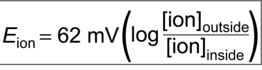
Equilibrium Potential
The membrane voltage for a particular ion at equilibrium. Can be calculated using the Nernst equation. For K+, it is -90mv, though in reality it’s between 60-80 due to a small amount of Na+ diffusion
Gated ion channels
Changes in membrane potential that occur because neurons open or close these in response to stimuli
voltage-gated ion channels
A type of ion channel that opens or closes in response to a shift in the voltage across the plasma membrane of the neuron
Hyperpolarization
This occurs when a gated K+ channel opens and K+ diffuses out, making the inside of the cell more negative (with the Cl- left behind) and increasing the magnitude of the membrane potential
Depolarization
Opening other gated channels (anything but K+) causes this, a reduction in the magnitude of the membrane potential. For example, gated Na+ channels open and Na+ diffuses into the cell.
Graded potential
A change in polarization where the magnitude of the change varies with the strength of the stimulus. It decays with distance from the source
Action Potential
If a depolarization shifts the membrane potential sufficiently, it results in a massive change in membrane voltage called this. It flips on when the threshold is reached, has a constant magnitude, and transmits over long distances.
T
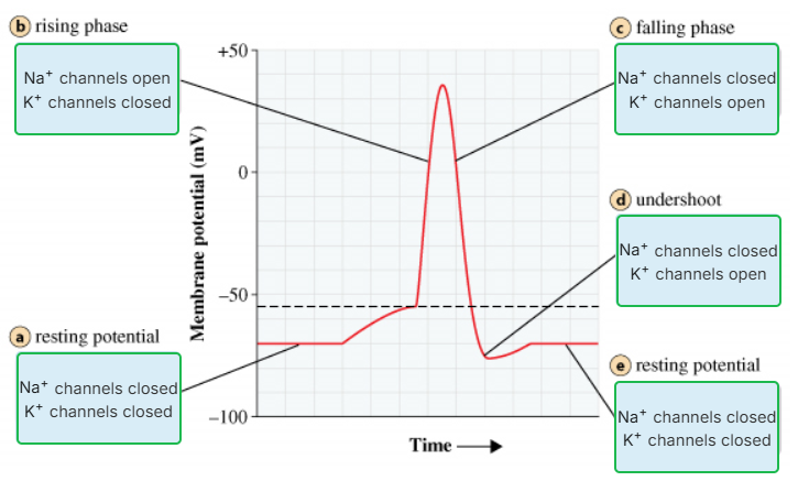
Action Potential Stages:
Resting Potential: All Na+ and K+ channels are closed
Depolarization: Some Na+ channels open, and Na+ flows into the cell. This causes a reduction in the magnitude of the membrane potential (goes from -80mv to 0mv)
Rising Phase: The threshold is crossed, the movement of many types of ions (both inside and outside the cell) alters the distribution of charges near other Na+ channels causing them to open, and the membrane potential increases (0mv to +50mv). More Na+ flows into the cell.
Action Potential: This is reached at the peak of the curve, +50mv
Falling Phase: Voltage gated Na+ cells become inactive, and K+ channels open, allowing K+ to flow out of the cell, leading back to hyperpolarization (-85mv)
Undershoot: The membrane permeable to K+ is higher than at rest, then voltage gated K+ channels close and resting potential is restored (-80mv)
(T/F)
Refractory Period
During this period after an action potential, a second action potential cannot be initiated due to a temporary deactivation of the Na+ channels
depolarizes
At the site where the action potential is usually initiated (usually the axon hillock), an electrical current ________ the neighboring region of the axon membrane. This causes the action potential to travel only towards the synaptic terminals, and inactivated Na+ channels behind this zone prevent the action potential from traveling backwards
diameter
The speed of an action potential increases with the axon’s __________
Mylin Sheath
In vertebrates, axons are insulated by this which enables the fast conduction of action potentials. They’re produced by glia—-oligodendrocytes in the CNS and Schwann cells in the PNS
Nodes of Ranvier
Action potentials are formed only at these depolarized regions, gaps in the myelin sheath where voltage gated Na+ channels are found.
Saltatory Conduction
Action potentials in myelinated axons jump between the nodes of Ranvier in this process.
T
(T/F) In most cases, action potentials are not transmitted from one neuron to another. Instead, information is transmitted at synapses
chemical
Most synapses are ____ synapses, in which a neurotransmitter carries information from the presynaptic neuron to the postsynaptic neuron
Ligand-gated ion channels
Direct synaptic transmissions involve the binding of neurotransmitters to these in the postsynaptic cells. The opening of these generates postsynaptic potential
Excitatory Postsynaptic Potentials
This is one of the two categories under which postsynaptic potential falls. These (EPSP) are depolarizations that bring the membrane potential towards threshold. Results from the movement of Na+ ions into the postsynaptic cell
Inhibatory Postsynaptic Potentials
This is one of the two categories under which postsynaptic potential falls. These (IPSP) are hyperpolarization that move the membrane potential farther from the thresholds. Results from the movement of K+ out of the postsynaptic cell
Summation
A single EPSP is usually too small to trigger an action potential in a postsynaptic neuron. Therefore, individual postsynaptic potentials can combine to produce a larger postsynaptic potential in this process. (Two types, temporal—-two EPSPs produced in rapid succession—-and spacial—-EPSP’s produced nearly simultaneously by different synapses on the same postsynaptic neuron). The combination of the two types can trigger an action potential. IPSP, similarly, can oppose EPSP through this process.
Metabotropic
A type of receptor that neurotransmitters can bind to in some synapses. Movement of ions through a channel depends on one or more metabolic steps, and binding of a neurotransmitter activates a signal transduction pathway in the postsynaptic cell involving a second messenger. Compared to ligand-gated channels, these have a slower onset (due to messenger) but last longer
Acetylcholine
Vital for functions involving muscle stimulation, memory formation, and learning. It is a common neurotransmitter in both invertebrates and vertebrates. In vertebrates, there are two major classes of receptors for these, one ligand gated and one metabotropic
Neuromuscular Junction
In vertebrates, acetylcholine released by motor neurons bind to receptors, ligand-gated ion channels are opened, and EPSP is generated, eventually resulting in muscle contraction. The best known function of the ligand-gated ion channels
Neuron Structure
This is a diagram of what?
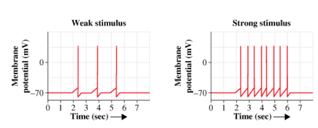
stimulus
The strength of a _________ (for example, whether you feel a slight pain versus an intense pain) determines the number of action potentials sent along an axon. As the graphs show, a strong one of these produces more action potentials spaced more closely together than a weak one of these.
The time between when a first action potential ends and a second action potential can be triggered is determined by the axon's refractory period. A second action potential cannot be triggered until the end of the refractory period.