Looks like no one added any tags here yet for you.
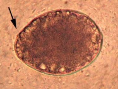
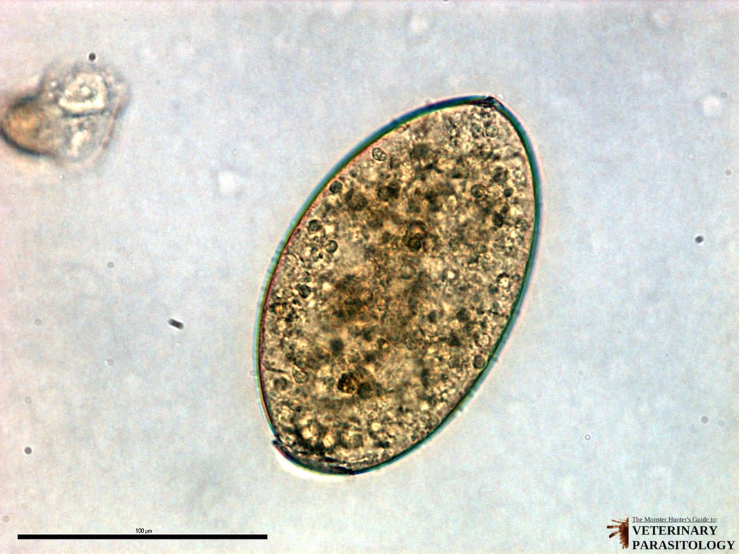
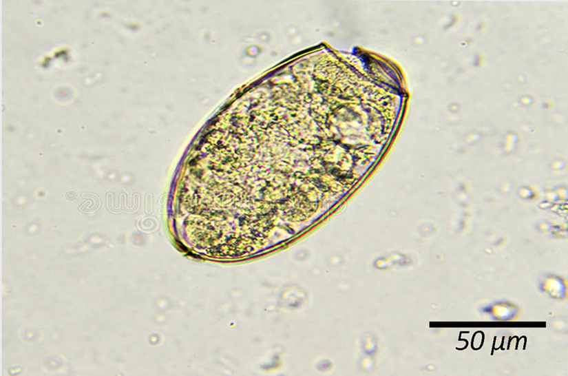
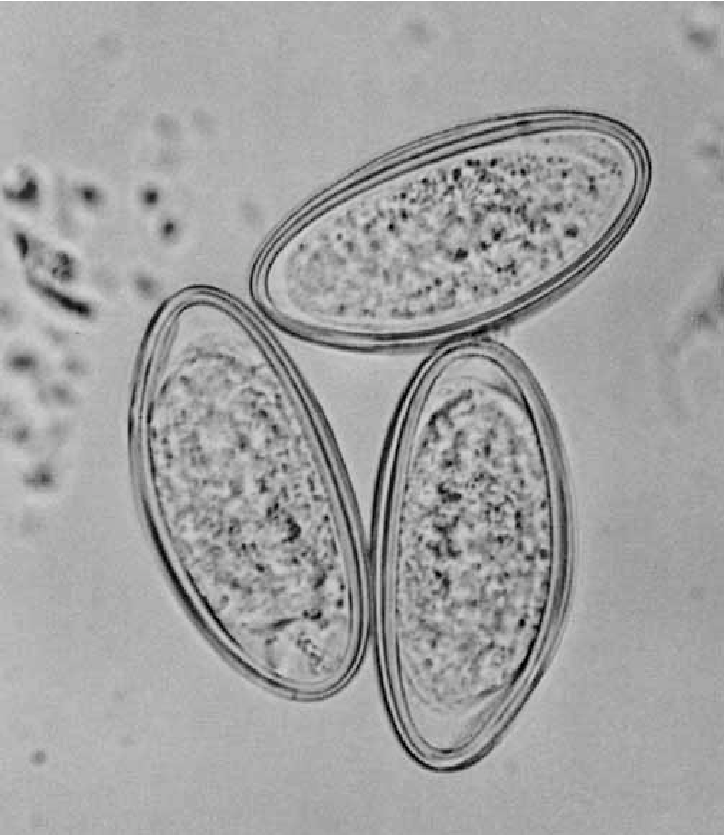
Diphyllobothrium Latum Egg size
60 micrometers (average)
Diphyllobothrium Latum egg characteristics
Broadly oval
Operculated
Abopercular knob
Smooth, yellowish brown
Unembryonated in feces
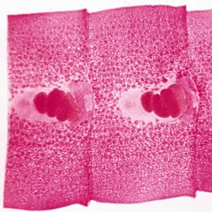
D Latum proglottids
Wider than long
Uterine Structure has rosette configuration
D latum adult
Scolex with two bothria, one dorsal and one ventral
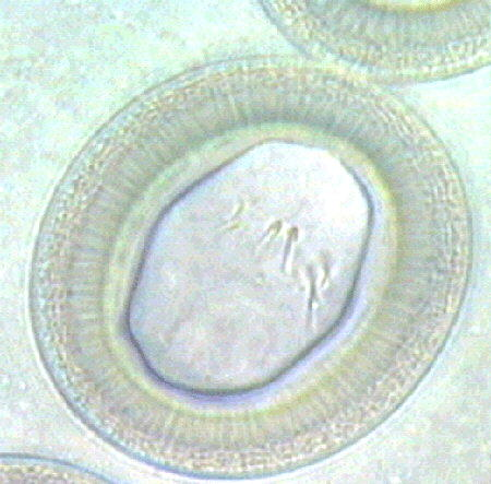
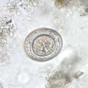
Taenia Species size
~ 35 micrometers
Taenia ssp egg description
Fully embryonated
Infective
Resistant to environmental conditions
Striated shell
Round/oval
Taenia Solium (pork tapeworm)
8-13 uterine branches
Longer than it is wide
Scolex
4 suckers
Hooklets
Taenia saginata size
More than 13 uterine branches
Each segment is longer than it is wide
Scolex
No hooklets
4 suckers
Hymenolopsis nana egg size
~ 38 micrometers
Hymenolopsis nana egg description
Round/oval
Thin shell
Polar thickenings with 4-8 polar filaments between oncosphere and shell
6 hooklets
Hymenolopsis diminuta size
~ 65 micrometers
Hymenolopsis diminuta egg description
round/oval
Oncosphere looks similar to H. Nana
Clear, gelatinous area around oncosphere inside thick shell
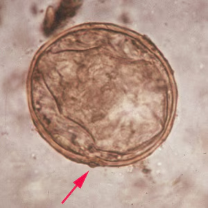
Echinococcus granulosum
Hyatid Tapeworm
Damages bones and organs
Infects human by creating cysts in one of the organs
Clinorchis sinesis size
~ 15 × 31 micrometers
Clinorchis sinensis description
Chinese liver fluke
Infects small ducts of biliary tree
fully embryonated when passed in feces
Ovoid, thick brownish-yellow shell
Distinct opercular shoulders surrounding operculum
comma-shaped appendage at end opposite operculum
Fasciolopsis buski size
~ 75 × 140 micrometers
Can not differentiate from Fasciola hepatica
Fasciolopsis buski description
Large intestinal fluke
Unembryonated when passed in feces
Operculated, ovoid
Yellow-brown
Fasciola hepatica size
~ 75 × 140 micrometers
Can not be differentiated from Fasciolopsis buski
Fasciola hepatica description
sheep liver fluke
unembryonated when passed in fees
Operculated
Brownish-yellow
Ovoid
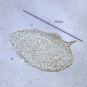
Paragonimus westermani size
~54 × 100 micrometers
Paragonimus westermani description
Lung Fluke
Ovoid, yellow-brown
Unembryonated
Thick-shelled
Operculated with opercular shoulders
Often confused with eggs of D. Latum
Schistosoma Description
Blood Flukes
Cercariae penetrate intact skin in water
Adults reside in circulatory system
Diecious
Eggs not operculated
Schistosoma mansoni size
~ 59 × 147 micrometers
Schistosoma mansoni description
Fully embryonated
No operculum
Light, yellow-brown
Elongate with large lateral spine projecting from the side near one side of the egg
Schistosoma japonicum
~ 50 × 70 micrometers
Schistosoma japonicum description
Fully embryonated
No operculum
Spherical
Minute, lateral spine
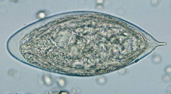
Schistosoma haematobium
~ 55 × 141 micrometers
Schistosoma haematobium description
Urinary schistosomiasis
Fully embryonated
No operculum
Yellow-brown
Large, terminal spine
Enterobius vermicularis
Common name is pinworm
female: 8-13 mm long, with wide pointed tail
male: 2-5 mm long with curved caudal end
Fin at head end
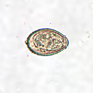
Enterobius vermicularis eggs size
50-60 micrometers x 20-32 micrometers
Enterobius vermicularis eggs description
Foot-ball shaped with one side flattened
Translucent shell of moderate thickness
Fully embryonated larvae
Ascaris lumbricoides - Adult
Intestinal roundworm
Cylindrical
Tapered anterior end
Pass many fertilized and unfertilized eggs (~200,000/day)
Largest roundworm in humans
Females: 20-35 centimeters
Males: 15-30 centimeters
Ascaris lumbricoides - Unfertilized Eggs size
Up to 40 × 90 micrometers
Ascaris lumbricoides - Unfertilized Eggs Description
Oval, elongated
May be largely mammillated or slightly mammillated
Shell is thin and if decorticated
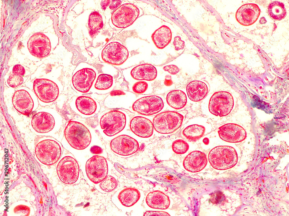
Ascaris lumbricoides Fertilized Eggs size
45-75 micrometers x 30-50 micrometers
Ascaris lumbricoides - Fertilized Eggs description
Broadly oval
Thick, mammilated coat
Bile-stained, golden brown
Sometimes decorticated — loss of mammillation (may resemble hookworm eggs)
Trichuris Trichiura - Adult
Whipworm
Female: 35-50 mm long
Male: 30-45 mm long
Head portion is very small
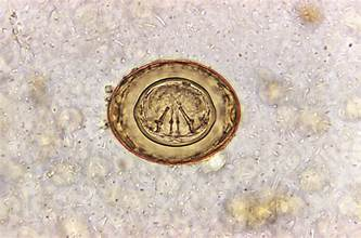
Trichuris trichiura egg size
22-23 × 50-54 micrometers
Trichuris trichiura egg description
Barrel-shaped
Clear, mucoid polar plugs
Hookworm egg size
35-40 micrometers x 50-60 micrometers
Hookworm Egg Morphology Description
Oval with broadly rounded ends
Thin shell
Clear area between the developing embryo and shell
Hookworm Filariform Larvae
300-600 micrometers
Pronounced, pointed tail
Ancylostoma duodenale - Adult
Hooked head
Teeth
Spicules below teeth help it attach
Necator americanus - Adult
Cutting plates
Anterior hook (head is sharply bent from body)
Strongyloides stercoralis - Filariform larvae
Threadworm
Notched tail
Eggs are not seen in stool unless severely immunocompromised
Strongyloides stercoralis - Rhabditiform larvae
Non-infective
Short buccal cavity
Genital primordium
Muscular esophagus
200 micrometers
Wucheria bancrofti Microfilariae size
245-300 micrometers
Wucheria bancrofti microfilariae description
found in blood
sheathed
Body nuclei do not extend to the tip of the tail
Vector: Mosquito
Nocturnal periodicity: 10P-4A
Brugia malayi Microfilariae
Sheathed
Two nuclei distinctly separate from the others in the tail, extend to the tip of the tail
Nocturnal periodicity: 10P - 4A
Vector: Mosquito
Loa Loa Microfilariae
African Eye Worm
Vector: Mango fly or Chrysops spp.
Sheathed
Nuclei are continuous to the tip of the tail
Diurnal periodicity : 10A - 2P
Onchocerca volvulus
Vector: Blackfly or buffalo gnat
Unsheathed
Body nuclei do not extend to tail
Migrate through dermis and connective tissue
Trichinella spiralis
Caused trichinosis
Larvae are coiled in muscle tissue
1 mm in diameter
What are the general characteristics of intestinal protozoa?
Small size
Morphological characteristics overlap among species
Must use permanent stained smear for definitive identification
What are some characteristics of trophozoites?
Motile
Feeds on host
Reproduces
Active Stage
Loose Stool
Diagnostic Stage
Killed by iodine and concentration procedure
Must use trichrome stain
What are some characteristics of cysts?
Non-motile
Dormant Stage
Formed stool
Infectious and Diagnostic Stage
Seen on iodine wm and trichrome
What are the clinical conditions caused by Entamoeba histolytica?
Invasive intestinal amebiasis
Invasive extraintestinal amebiasis
What is morphologically similar to Entamoeba histolytica?
Entamoeba dispar
What is the size of Entamoeba histolytica troph?
10-60 micrometers (15-20 micrometers most common)
Describe the nucleus of the Entamoeba histolytica trophozoite.
Single nucleus
Chromatin: finely granular and evenly distributed around periphery
Karyosome: Small, centrally located
Describe the cytoplasm of the Entamoeba histolytica trophozoite.
Finely granular
May contain bacteria or RBCs
How can you differentiate Entamoeba histolytica troph from Entamoeba dispar under the microscope?
Entamoeba histolytica has bacteria or RBCs while Entamoeba does not.
What is the size of the Entamoeba histolytica/dispar cyst?
10-20 micrometers
Describe the Nucleus of Entamoeba histolytica/dispar cyst
4 nuclei
Peripheral chromatin: fine, evenly distributed
Karyosome: small, discrete, centrally located
Describe the cytoplasm of the Entamoeba histolytica cyst
Chromatoidal bars may be present
Chromotoidal bars have blunt ends
What is the size of the Entamoeba hartmanni troph?
5-12 micrometers
Describe the nucleus of Entamoeba hartmanni troph.
Chromatin: Small granules, evenly distributed
Karyosome: Small, discrete, centric
Single nucleus
Describe the cytoplasm of Entamoeba hartmanni troph.
Finely granular
What is the size of the Entamoeba hartmanni cyst?
5-10 micrometers
Describe the nuclei of Entamoeba hartmanni cyst
1-4 nuclei
Peripheral chromatin: Fine, evenly distributed
Karyosome: Small, discrete, centric
Describe the cytoplasm of Entamoeba hartmanni cyst.
Chromatoidal bars with blunt ends
What is the size of the Entamoeba coli troph?
15-50 micrometers (usually 20-25)
Describe the nucleus of the Entamoeba coli troph
Single nucleus
Chromatin: heavy granules, irregular in size and distribution
Karyosome: Large, may be diffuse, often eccentric
Describe the cytoplasm of Entamoeba coli troph.
Course
Often vacuolated
May contain bacteria or other debris
What is the size of entamoeba coli cyst?
10-35 micrometers
Describe the nucleus of the Entamoeba coli cyst.
8 nuclei
Peripheral chromatin: course, granular
Karyosome: Large, eccentric or central
Describe the cytoplasm in Entamoeba coli cyst.
Chromatoidal bars, usually have pointed ends
What is the size of the Endolimax nana troph?
6-12 micrometer
Describe the nucleus of Endolimax nana troph.
No peripheral chromatin
Karyosome: large, blot-like, centric
Describe the cytoplasm of the Endolimax nana troph.
Granular
May be vacuolates
What is the size of Endolimax nana cyst
5-10 micrometers
Describe the nuclei in Endolimax nana cyst
No peripheral chromatin
Karyosome: large, blot-like, centric
Describe the cytoplasm of the endolimax nana cyst
small granules
rarely have chromatoid bodies
What is the size of Iodamoeba butschlii troph?
8-10 micrometers