12 ATAR Human Bio: The Nervous System
1/68
There's no tags or description
Looks like no tags are added yet.
Name | Mastery | Learn | Test | Matching | Spaced |
|---|
No study sessions yet.
69 Terms
Neurons
- Nerve cells + the basic structural and functional unit of the nervous system
- vary in size + shape
- all neurons consist of a cell body + two types of extensions: dendrite and axon
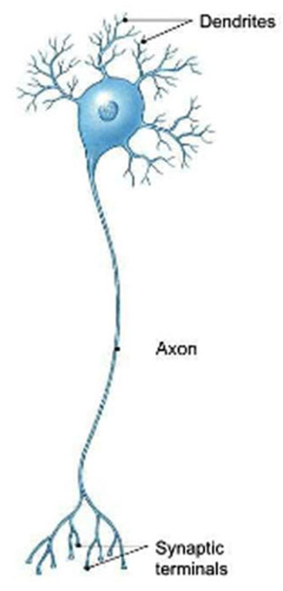
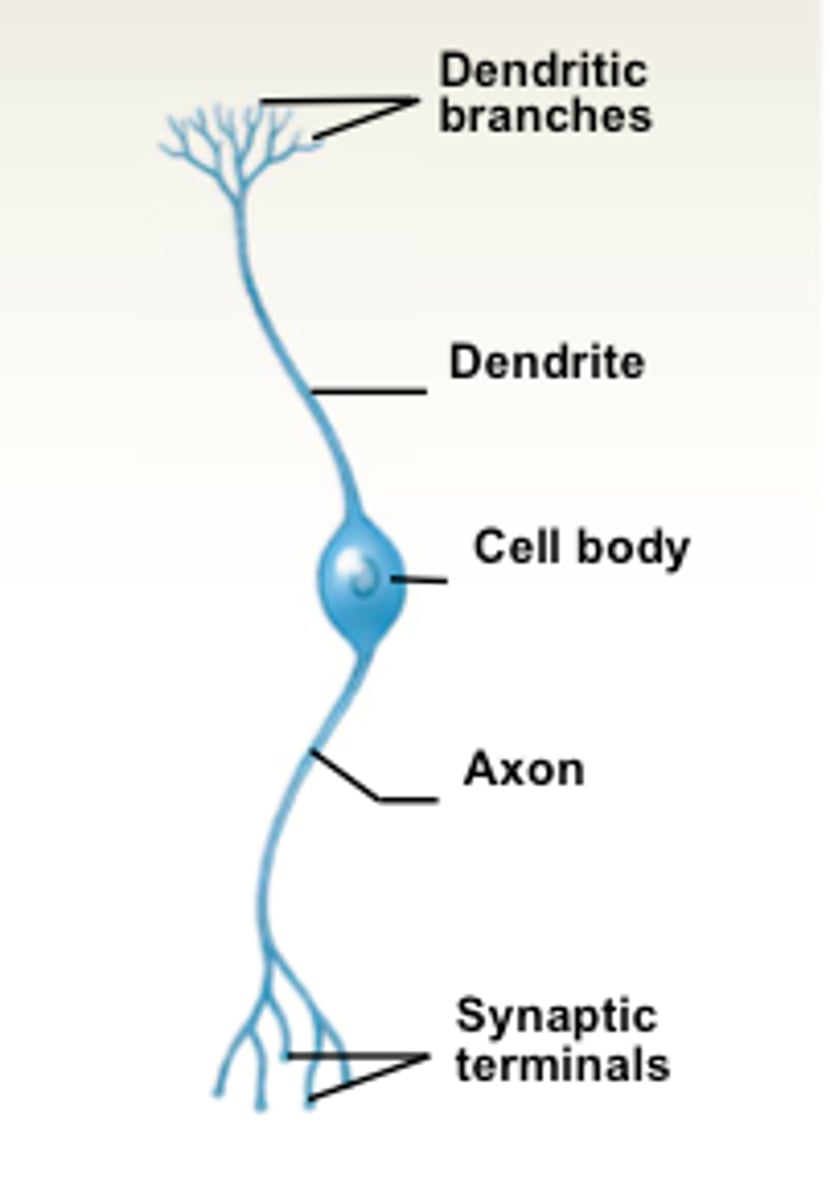
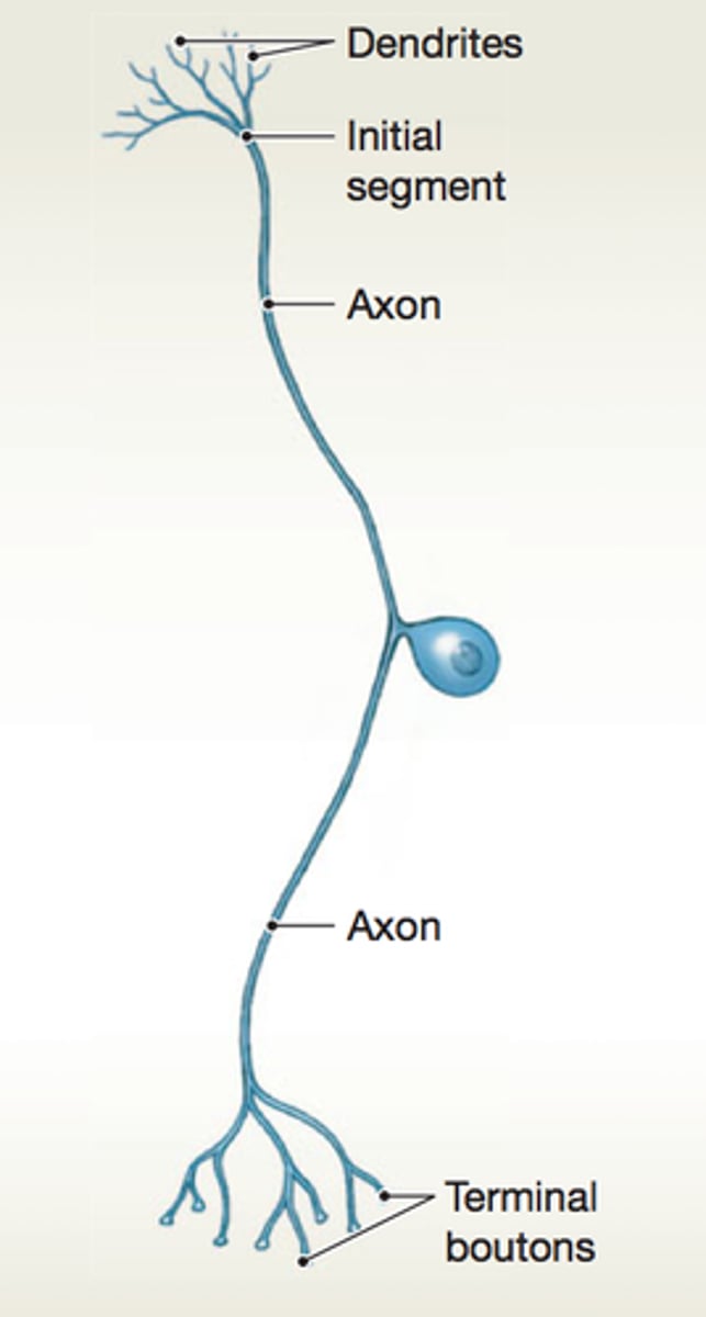
Cell Body, Dendrites + Axon
> Cell body- Contains nucleus, Cytoplasm w other organelles (mitochondria, ER, golgi)
> Dendrite- Short extensions / branches off the cell body (many), Receive messages + carry them TOWARDS cell body.
> Axon- Single (unbranched) long extension from cell body, Vary in length (few mm to 1m in length) , end divides into branches, each ending at an axon terminal= Pass the nerve impulse onto the next neuron, Carries nerve impulses AWAY from cell body
Myelin Sheath
-a layer of lipid (fatty) material covering the axon (not all)
- FUNCTIONS= Insulation, Protection, Speeds up nerve impulses
- Outside of CNS they're formed from SCHWANN CELLS (wrap around axon)
• NEURILEMMA - outermost coil of Schwann cell, helps repair injured fibres (axons)
• Gaps in the myelin sheath=NODES OF RANVIER
Grey vs White matter
GREY MATTER= Grey areas of neurons, Nerve cell bodies and unmyelinated fibres
WHITE MATTER:= White areas (due to lipid myelin), Myelinated fibres

Synapses
• Nerve impulses are passed from neuron to neuron across a synapse.
• At synapse the neurons do not actually join — there is a very small gap between them.
• Messages have to be carried across the synapse.
• A similar synapse exists where an axon meets a skeletal muscle cel= neuromuscular junction.
Neurons vs Nerve fibres vs Nerves
• NEURONS: cells of the nervous system
• NERVE FIBRES: long extension of the cytoplasm of a neuron (it usually refers to an axon and dendrites)
• NERVE: A bundle of nerve fibres held together by CT outside CNS
Types of neurons (functional)
1. Sensory (afferent/receptor)- Carry messages from receptors in sense organs/ skin (the periphery) TOWARDS CNS
2. MOTOR (EFFECTOR/EFFERENT)- Carry messages AWAY from CNS towards muscles and glands (the effectors - put message into effect)
3. INTERNEURONS (Relay / Connector / Association)- Located in CNS, Link between Sensory and Motor neurons
Types of neurons (structural)
multipolar, bipolar, unipolar (not in humans) + pseudo-unipolar
Multipolar neurons
• One axon
• Many dendrites from the cell body
• Motor neurons
• Interneurons
Bipolar neurons
• One axon
• One dendrite from the cell body
• Sensory: eye, ear & nose
Pseudo-unipolar neurons
• One axon that divides in two
• No dendrites connected to the cell body
• Sensory neurons
Potential
- force that pulls unlike charges together can be measured + its strength increases as the charges get closer.
• If a group of +ve and -ve charges are separated they have the potential to come together and release energy.
• The potential/potential difference, between two places can be measured= voltage, measured in volts (V) or millivolts (mV)
Nerve Impulse
an ELECTROCHEMICAL CHANGE that travels along a nerve fibre, brought about by the movement of ions.
Nerves in resting state
> Extracellular fluid (outside cell) - High concentrations of Sodium ions (Na+) and Chlorine ions (Cl- )
> Intracellular fluid (inside cell) - High concentrations of Potassium ions (K+) and various negative ions. And low concentrations of Na+ and Cl-
- A difference between concs. of +ve and -ve ions either side of the cell membrane gives us a POTENTIAL DIFFERENCE, occurs in all body cells
Resting Membrane Potential
- the membrane potential (difference) of unstimulated nerve cells = -70 mV (millivolts= potential of inside of the membrane is 70mV less than that of the outside.
- particularly large in nerve and muscle cells
- due mainly to differences in the distribution of potassium ions (K+) and sodium ions (Na+) on either side of membrane
How does the cell maintain this potential difference?
1) actively moves ions across the membrane- activity described as a sodium-potassium pump= transports sodium ions out of the cell and potassium ions in using carrier proteins (against conc. gradient (3 Na+ for every 2 K+= creates a net reduction in +ve ions inside cell
2) Cell membrane is not equally permeable to all ions - many negatively charged ions trapped inside cell.
- Cell membrane is Highly permeable to K+, Slightly permeable to Na+, Impermeable to the various other large -ve ions= tendency for K+ to diffuse out of cell= inside of the cell more negatively charged (it's losing +ve ions).
Polarised
- combination of: Location of ions, Sodium-Potassium Pump + Permeability of cell membrane means there is a net flow of +ve ions out of the cell as well as a large number of -ve ions inside cell.
- results in the inside being more negative than the outside creating a negative resting membrane potential.
- If this is the case, we say the membrane is POLARISED
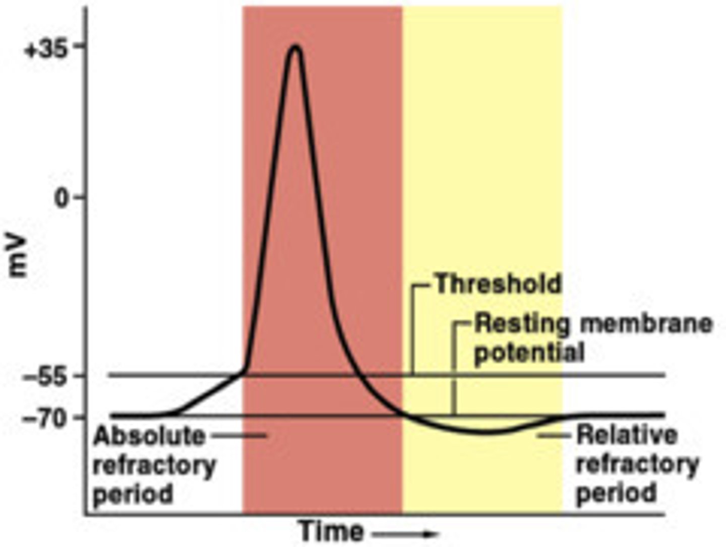
Action Potential (Overview)
- if a sufficiently strong stimulus is applied to a nerve fibre, the membrane becomes more permeable to sodium ions.
- Sodium channels open a little so that Na+ ions diffuse across the membrane and into the cell.
- inward movement is too great to be balanced by an outward movement of K+ ions and membrane becomes depolarised.
- Depolarisation occurs only if level of stimulation exceeds threshold (-55mV)
- If the stimulus is strong enough to cause this change of about 15 mV, then movement of sodium ions proceeds independently of the stimulus (size of response not related to the strength of the stimulus= all-or-none response)
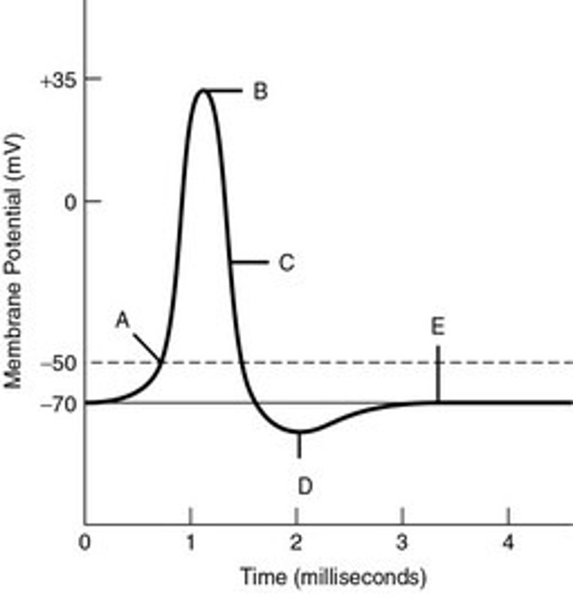
Action Potential- Step 1
• a stimulus (neurotransmitter/receptor) causes sodium to leak into the neuron
• sodium channels open a little so that sodium ions diffuse across the membrane and into the cell (as they move into cell, intracellular fluid becomes more positive /less negative, increasing the potential difference)
Threshold: if enough sodium diffuses in to reach threshold (-55mV), the sodium voltage-gated channels open.
Action Potential- Step 2
DEPOLARISATION- Once cell determines that threshold has been reached, Sodium floods into the neuron as membrane becomes more permeable to Na+ ions
- this causes the original polarity of the membrane to reverse and the inside becomes positive relative to the outside (up to +30mV)
Action Potential- Step 3
REPOLARISATION- At +15mV, the sodium voltage-gated channels close, and potassium voltage-gated channels open, causing K+ to rush out of the cell.
= restores the membrane to negatively charged on the inside
Action Potential- Step 4
HYPERPOLARISATION- At -70mV the potassium voltage-gated channels close, but a little more potassium leaks out than necessary= cell to becomes slightly more negative, dropping lower than resting
How does the cell return to resting membrane potential?
• Running in the background is the sodium potassium pumps (3 Na+ out and 2 K + in), returning the sodium & potassium to their original positions, ready to fire again when needed.
- Almost as quickly, the membrane is restored to its original condition.
- rapid depolarisation and repolarisation of a membrane= ACTION POTENTIAL
- movement of an action potential along a nerve IS the nerve impulse and lasts only 1 millisecond.
Refractory Period
• During an action potential, and for a very brief time afterwards, that part of the nerve fibre cannot be stimulated to respond again= refractory period.
- Period between threshold being reached (-55mV) and resting membrane potential being restored + stabilised (-70mV)
- ensures the impulse travels in only one direction, can't fire on top of each other + cannot be prolonged
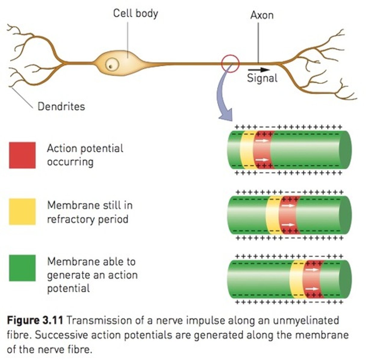
Conduction along unmyelinated fibres (Continuous Conduction)
- depolarisation of one area (channel/gate) of the membrane causes a local current flow between neighbouring areas on the membrane.
- This current flow causes depolarisation immediately adjacent to the site of the original stimulus.
- process repeats itself along the whole length of the membrane so that the action potential moves along the membrane away from the point of stimulation (dominoes)
- nerve impulse is prevented from going backwards by the refractory period
- max speed 2 m/s
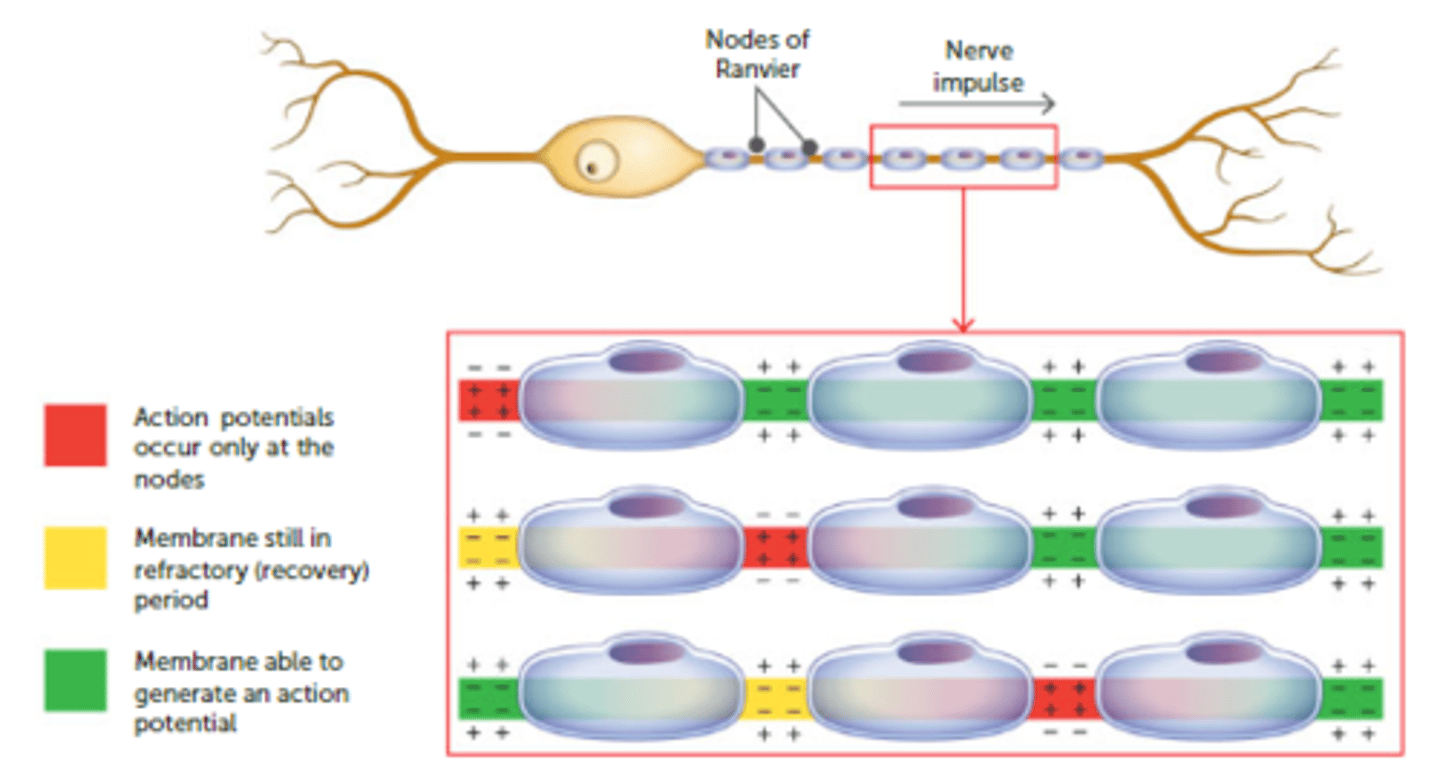
Conduction along myelinated fibres (Saltatory Conduction)
- myelin sheath insulates the fibre from the extracellular fluid so that ions cannot flow between the inside and outside of the membrane, and an action potential cannot form.
- Na+ ions will diffuse along axon, through cytoplasm until they reach the next exposed gate (at the next Node of Ranvier).
- In this case, the action potential jumps from one node of Ranvier to the next because the myelin sheath is absent from the nodes.
- SALTATORY CONDUCTION= 'jumping conduction'= nerve impulse travels much faster along myelinated fibres (less distance to cover)
- max speed 140 m/s
Conduction of nerve impulses (All or None response)
- size of a nerve impulse that travels along a fibre is always the same.
- weak stimulus, provided it exceeds the threshold, produces same action potential as a strong one,= all-or-none response — a stimulus is either strong enough to trigger off an impulse or it is not.
- magnitude of the impulse is always the same.
- 2 things that enable us to determine strength of a stimulus;
1) A strong stimulus causes depolarisation of more nerve fibres than a weak stimulus;
2) A strong stimulus produces more nerve impulses in a given time than a weak stimulus.
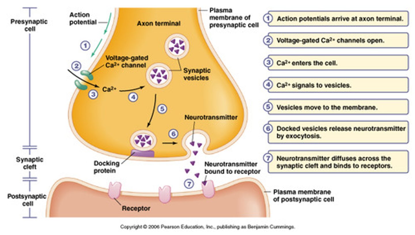
Transmission across a Synapse
- As action potential reaches the axon terminals, depolarisation triggers threshold of calcium voltage-gated channels
- Ca rushes into cell + binds with vesicles (containing neurotransmitters) in the axon terminal.
- causes vesicles to migrate to edge of the cell and attach to the cell membrane.
- Exocytosis occurs, releasing the neurotransmitters into the synaptic cleft.
- neurotransmitters diffuse across the cleft/gap and attach to receptors on the membrane of the next neuron:
• (Usually) initiating new AP/impulse in the next neuron.
• For example: acetylcholine, adrenaline, dopamine and histamine.
• transmission of impulses across a synapse only occurs in one direction — from axon to dendrite or from axon to cell body
Effects of chemicals on the transmission of nerve impulses
- many chemicals, both natural and synthetic influence the transmission of nerve impulses.
- Most work by affecting transmission at the synapse/ neuromuscular junction.
• Stimulants (caffeine, benzedrine) stimulate transmission at the synapse.
• Other drugs (anaesthetics, hypnotics) depress transmission.
• Venom from certain species of snakes and spiders also affects the neuromuscular junction.
3 things happening to neurotransmitter (clearance)
1. DIFFUSE- neurotransmitter diffuses away from synapse
2. ENZYME DEGRADATION- neurotransmitters broken down by enzymes
3. RE-UPTAKE- neurotransmitters reabsorbed (via endocytosis) into the pre-synaptic neuron to be re-used
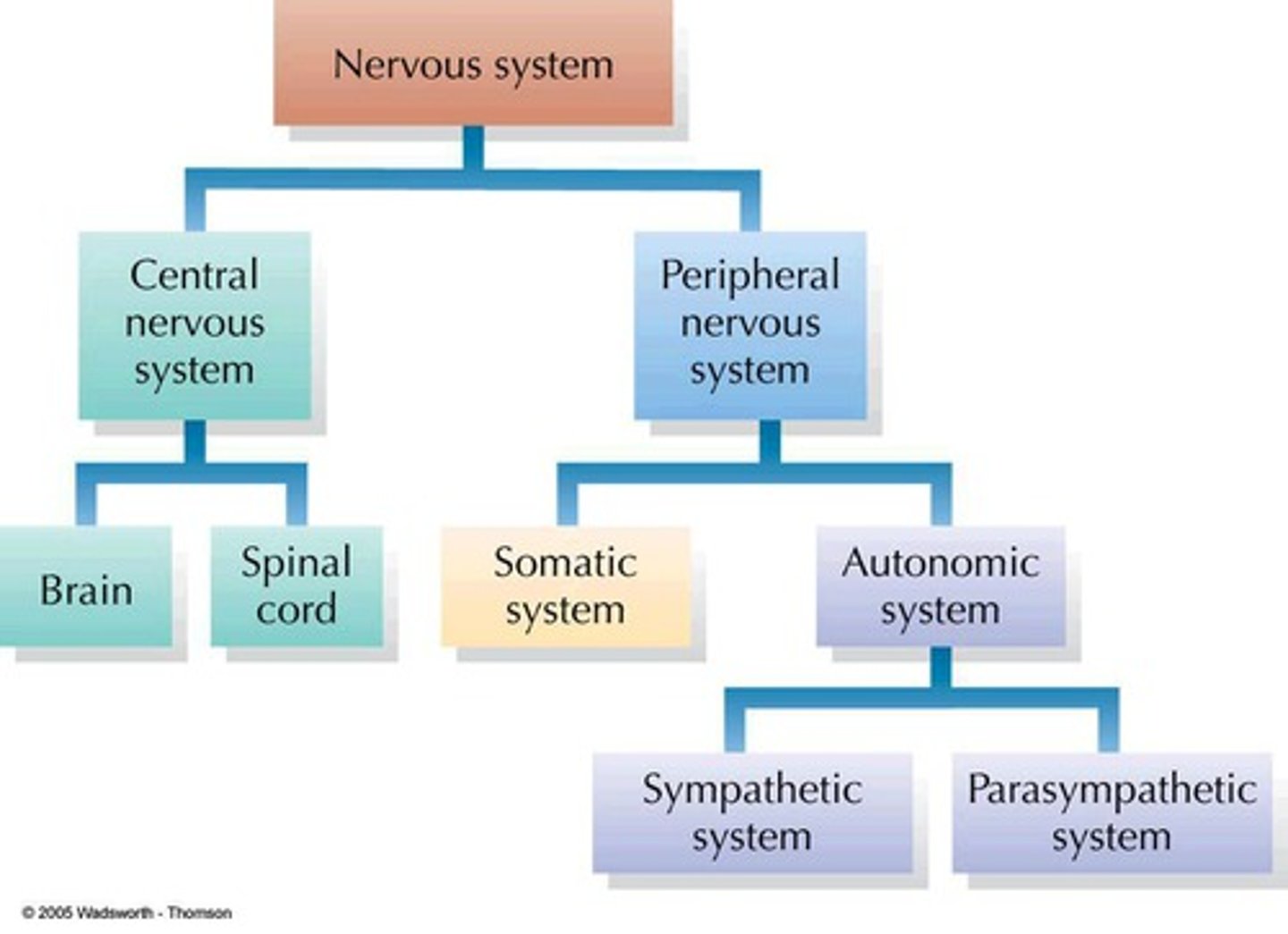
Central Nervous System (CNS)
- The control centre containing the brain (Cerebrum , Corpus Callosum, Cerebellum, Hypothalamus, Medulla Oblongata) and spinal cord
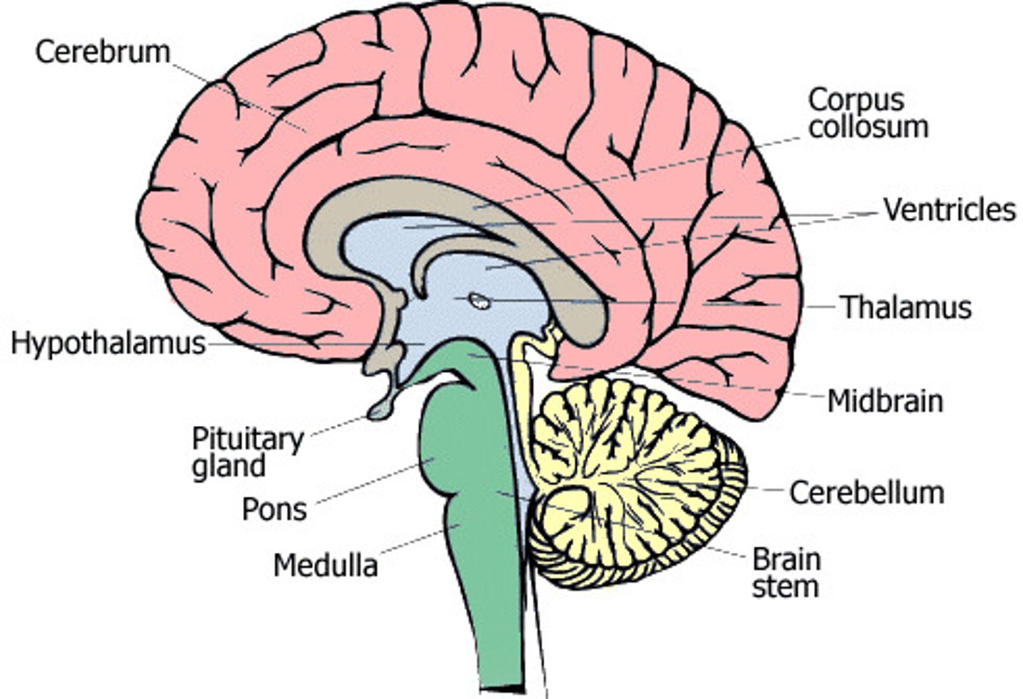
Cerebrum
- the largest part of the brain
- outer layer (2-4mm thick) is made of grey matter= CEREBRAL CORTEX= mental activities - thinking, reasoning, learning, memory, intelligence and sense of responsibility & perception of the senses + initiation and control of voluntary muscle contraction
- has many convolutions= increases the surface area of the brain
- cerebral cortex has shallow folds= sulci , deep folds= fissures, and ridges= gyri.
- deepest fissure= longitudinal fissure, which separates the cerebrum into left and right hemispheres
- 4 main lobes- frontal, parietal, temporal + occipital
Lobes of the brain
FRONTAL - manages thinking, problem solving, emotions, personality, behaviour and control of movement
TEMPORAL- receives auditory information + processes memory and links them to senses
OCCIPITAL- processes visual information
PARIETAL- provides sensory information such as touch, pain, temperature and also controls language
Cerebral Cortex- functional areas
- cerebral cortex receives input from senses and responds accordingly
- can be divided into 3 types of functional areas:
1. SENSORY AREAS - Interpret impulses from receptors, Perception of senses
2. MOTOR AREAS - Control muscular movements, Initiation and control of voluntary muscle
3. ASSOCIATION AREAS - Intellectual and emotional processes, Involved in activities such as thinking, reasoning, learning, memory and intelligence
Cerebrum (white/grey matter)
- Below cerebral cortex is an area of WHITE MATTER made of myelinated nerve fibres
- Underneath the white matter is more grey matter= BASAL GANGLIA
- white matter of the cerebrum contains bundles of myelinated nerve fibres (called TRACTS when in the CNS)
- 3 types of tracts occur in the white matter:
1) Connect various areas in the same hemisphere- regulate and coordinate neural activity in the cortex (Association)
2) Carry impulses between hemispheres (Commissural)
3) Connect cerebral cortex to other parts of brain or spinal cord (Projection)
Corpus Callosum
- a wide band of nerve fibres that lies underneath the cerebrum at the base of the longitudinal fissure
- The nerve fibres cross from one hemisphere to the other and allow the two sides to communicate
Cerebellum
- located under the back of the cerebrum
- folded into a series of parallel ridges; Outer folds= grey matter, inner folds= white matter that branch out like a tree
- Non-conscious control of posture, balance + fine muscle co-ordination
- receives sensory information from the inner ear about posture and balance & Stretch receptors in skeletal muscles about length of muscle
- smooths the contraction of muscles otherwise movements would be jerky and uncontrolled (Impulses do not originate in the cerebellum so wed still move but not smooth)
Hypothalamus
- located in the middle of the brain (cannot be seen from the outside.)
- controls many body activities, mostly homeostasis (maintaining a constant cell environment)
- Other functions= regulation of: ANS, body temp, Food + water intake, Sleeping patterns, Contraction of urinary bladder, Emotional responses (fear, anger, pleasure) + Secretion of hormones (ADH) + coordination of pituitary gland/other endocrine system
Medulla Oblongata (1)
- a continuation of spinal cord, about 3 cm long.
- passage for nerves to pass through going to or from other parts of the brain
- 3 important roles for vital bodily functioning:
1) Cardiac centre - regulates RATE AND FORCE of heartbeat
2) Respiratory centre - control of RATE AND DEPTH of breathing
3) Vasomotor centre - regulates diameter of BVs
- Other roles= swallowing, sneezing, coughing, vomiting.
Medulla Oblongata (2)
- Upper Motor Neurons carry impulses away from PRIMARY MOTOR AREA of cerebral cortex towards appropriate level of spinal cord.
- At this point messages passed to Lower Motor Neurons to carry the impulse to effectors.
- At some point through medulla is where the neurons cross over= motor area of left hemisphere controls contraction of muscles on the right side of the body and vice versa
- All centres in the medulla oblongata are influenced and controlled by higher centres in the brain, particularly the hypothalamus.
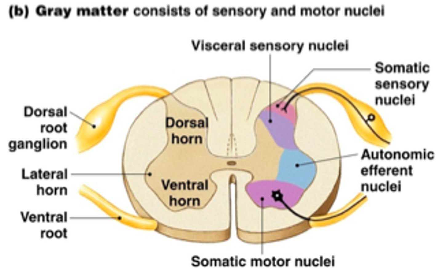
Spinal Cord
- a roughly cylindrical structure, about 44cm long, extends from the foramen magnum (hole in base of cranium) to 2nd lumbar vertebrae
- surrounded by 3 meningeal layers + vertebrae (3rd layer not stuck to vertebrae but made of fat, CT and BVs= padding and allow spinal cord to bend)
- In the middle= CENTRAL CANAL= runs entire length of spinal cord and contains CSF
- contains grey (inside, shape of H/butterfly) and white matter
- FUNCTIONS=
1) Carry sensory impulses towards brain
2) Carry motor impulses away from brain
3) Integrate certain reflexes (fast automatic responses)
Ventral vs Dorsal Root
Ventral= contains axons of motor neurons that have their cell bodies in grey matter of spinal cord
Dorsal= contains axons of sensory neurons that have their cell bodies in a small swelling on root= dorsal root ganglion
Protection of CNS
- brain and spinal cord are very delicate + vital parts of the body and are heavily protected by:
• Bone
• Three membranes called meninges
• A fluid called cerebrospinal fluid (CSF)
1. BONE
- Strong rigid structure that protect from impact/ physical injury
- brain is encased in the CRANIUM
- The spinal cord runs through VERTEBRAL CANAL in vertebrae
2. MENINGES
- 3 layers of CT forming membranes between brain/spinal cord and surrounding bones: protect the brain and spinal cord, help hold them into place + carry BVs that help supply the brain with blood.
1) Outer layer= DURA MATER- tough and fibrous, sticks closely to cranium, inflexibility prevents brain from moving around.
2) Middle layer= ARACHNOID (as it resembles a spider web), loose mesh of fibres.
3) Inner layer= PIA MATER- more delicate, w/ many BVs, sticks close to brain or spinal cord, + creates a seal that protects brain from infection
3. CEREBROSPINAL FLUID (CSF)
- clear watery fluid, containing a few cells and some glucose, protein, urea and salt
- formed from blood, circulates through the CNS, taking nutrients to the neurons and removing wastes and eventually re-entering the capillaries
- fills the space between the middle and inner layers of the meninges + circulates through the cavities in brain and spinal cord
Functions of the CSF
1) Protection - acts as a shock absorber to cushion any blows
2) Support - brain is suspended and floats in CSF in the cranium
3) Transport - it takes nutrients to cells and removes wastes, • regulation of the conc. of H+ and CO2 , which regulates breathing, transport chemical messengers around the CNS
Peripheral Nervous System
- consists of nerve fibres that carry information to and from the central nervous system (CNS).
• These are nerves, and groups of nerve cell bodies= ganglia= lie outside the brain and spinal cord.
• nerve fibres are arranged into nerves that arise from the brain and the spinal cord.
- divided into 2 parts:
1= Afferent (Sensory) division- Carries info to CNS from receptors
2= Efferent (Motor) division- Carries info away from CNS to effectors
Types of nerves (cranial, spinal, phrenic, intercostal, vagus)
Cranial= 12 pairs of nerves arise from the brain.
Spinal= 31 pairs of nerves arise from the spinal cord- all mixed nerves and each is joined to the spinal cord by two roots.
Phrenic= travels to diaphargm
Intercostal= travels to IC muscles (rate + depth of breathing)
Vagus= parasympathetic NS "rest + digest"
Afferent division
- sensory neurons carry info to CNS from sensory receptors
- further divided into 2 divisions:
1) Somatic Sensory Neurons- carry info from receptors in skin, muscles or joints
2) Visceral Sensory Neurons - carry info from receptors in internal organs
Efferent division
- motor neurons carry info from CNS to organs
- further divided into 2 divisions:
1) Somatic Division- carry info to skeletal / voluntary muscles
2) Autonomic Division- carry info to involuntary muscles and glands
Autonomic divided into:
> Sympathetic Nervous system 'fight or flight'- strenuous activity
> Parasympathetic Nervous System 'rest and digest"- rest conditions
Somatic VS Autonomic Nervous systems (SOMATIC)
SOMATIC:
- Effector= Messages to skeletal/voluntary muscles
- General function= Response to external environment
- Control= Conscious (voluntary)
- Efferent pathways (from CNS to effector) = 1 neuron and no synapse, no ganglion
- Nerves to target organ= Effectors only receive 1 nerve fibre
- Effect on target organ= Excitatory effects only
- Neurotransmitter= Acetylcholine
Somatic VS Autonomic Nervous systems (AUTONOMIC)
AUTONOMIC:
- Effector= Messages to heart muscle, involuntary muscles + glands (eg. diaphragm)
- General function= Response to internal environment (homeostasis)
- Control= Not conscious (involuntary)
- Efferent pathways= 2 neurons with a synapse in a ganglion
- Nerves to target organ= Effectors receive 2 sets nerve fibre (parasympathetic + sympathetic)
- Effect on target organ= Excitatory + inhibitory (controls/balances body functions as required)
- Neurotransmitter= Acetylcholine or Noradrenaline
Autonomic Nervous System (ANS)
- Responsible for the control of the body's internal environment= Homeostasis
- Operates without conscious control (involuntary)
- Regulated by medulla oblongata, hypothalamus and cerebral cortex
- carry impulses to heart and involuntary muscles and glands so icontrols: HR, BP, Digestion, Release of energy, Pupil diameter, Air flow to lungs, Defecation and Urination
Effects of the ANS (Structure- Sympathetic effect- Parasympathetic effect) #1
1. Heart- Increases rate + strength of contraction- Decreases rate + strength of contraction
2. Lungs- Dilates bronchioles- Constricts bronchioles
3. Stomach and intestines- Decreases movement- Increases movement
4. Liver- Increases breakdown of glycogen + releases glucose- Increases the uptake of glucose + synthesis of glycogen
5. Iris- Dilates pupil- Constricts pupil
6. Sweat glands- Increases sweat secretion- No effect
Effects of the ANS (Structure- Sympathetic effect- Parasympathetic effect) #2
7. Salivary glands- Decreases secretion of saliva- Increases secretion of saliva
8. Skin BV's- constricts vessels- dilates vessels
9. skeletal muscle BVs- dilates vessels- no effect
10. internal organs BVs- constricts vessels (except in heart and lung)- little effect
11. Bladder- Relaxes muscles of wall- Constricts muscles of wall 12. Adrenal medulla- Stimulates hormone secretion- No effect
Fight or Flight Response
- under normal circumstances we aren't aware of the activities of ANS (both sympathetic + parasympathetic nerves are sending out to internal organs to maintain a stable environment)
- In threatening situations, balance between sympathetic and parasympathetic stimulation is upset and the sympathetic becomes dominant.
- Situations that involve fear, anger, stress, danger or competition provoke what is called a fight-or-flight response or alarm reaction
Responses that result from the activation of the sympathetic division
- ↑ HR + force of contraction= ↑ BP
- Dilation of BVs in necessary organs- skeletal muscle, brain, heart (involved in strenuous activity)
- Contraction of BVs of non-essential organs- kidney, stomach, intestines + skin (pale face) (↓ saliva production= dry mouth)
- ↑ blood glucose levels as liver converts more glycogen to glucose
- ↑ in secretion from sweat glands
- adrenal medulla releases hormone adrenaline + noradrenaline which intensify + prolong above responses
- bronchioles dilate + breathing rate + depth ↑
- pupils dilate, relaxation of bladder
Receptors
- structures in the cell membrane that are capable to detect a change in the body's internal or external environments.
- thermo, osmo, chemo, touch, pain
Thermoreceptors
- detect temperature.
- Skin thermoreceptors detect external temp + send info to hypothalamus and cerebrum. (separate regions of the skin that detect hot and cold).
- Thermoreceptors in hypothalamus detect internal temp
- Through information it collects, the hypothalamus can regulate body temp
Osmoreceptors
- detect osmotic pressure + water balance
- located in the hypothalamus, which can stimulate the posterior pituitary to release ADH to regulate osmotic pressure. (High osmotic pressure = low water conc)
Chemoreceptors
- detect chemicals.
- located in nose and mouth that detect smell and taste.
- also located internally in BVs that detect the composition of body fluids, such as pH, O2 and CO2 levels
- Therefore, messages are sent to the Respiratory and Cardiac centres to regulate heart rate and breathing rate.
Touch receptors
- detect touch.
- mainly located in the skin + base of hair follicle
- Different types + concentrations vary across different locations in body
- eg. some detect light touches (higher concentrations in lips and fingertips) and others detect harder touches/pressure (pressure receptors- deep under skin)
- Touch receptors adapt/desensitize quickly.
Pain receptors
- detect pain + damage to tissues
- Stimulated by poor blood flow, stimulation from excessive heat or chemicals damage to cells, cuts, bumps
- warns us damage is occurring, and receptors will continue to be stimulated for as long as the stimulus is present.
- Located mainly in skin and mucous membranes, also found in most visceral organs (not brain)
Reflexes
- a rapid, automatic response to a change in the external or internal environment.
- one of the ways in which the body achieves homeostasis.
• All reflexes have 4 important properties:
1. triggered by stimulus
2. involuntary.
3. reflex response is rapid — only small number of neurons are involved.
4. stereotyped — occurs in the same way each time it happens
Spinal Reflex
• Some reflexes involve unconscious parts of the brain but most are coordinated by the spinal cord (not carried to brain)
• impulse may be passed to motor neurons at same level in the cord, or may travel a few segments up/down the cord before travelling out through a motor neuron.
• In these cases the reflex is carried out by the spinal cord alone
• pathway a nerve impulse follows in travelling from a receptor to an effector= reflex arc or, in the case of a spinal reflex, a spinal reflex arc.
Reflex Arc
1. receptor is either the ending of a sensory neuron or a specialised cell associated w/ end of a sensory neuron. The receptor reacts to a change in the internal/external environment by initiating a nerve impulse in the sensory neuron.
2. A sensory neuron carries impulses from the receptor TO the CNS.
3. There is at least one synapse. Nerve impulse may be passed directly to a motor neuron or may be 1 or more interneurons, which direct the impulse to the correct motor neuron.
4. A motor neuron carries the nerve impulse to an effector.
5. An effector (muscle/secretory cells/glands) receives the nerve impulse and carries out the appropriate response.
Protective Reflexes
- protect body from injury
- present from birth + determined genetically
- More complex motor patterns appear during a baby's development — including reflexes such as suckling, chewing or following movements with the eyes.
- blinking, sneezing, coughing, constriction of pupil in response to intense light
Learned Reflexes
- ACQUIRED reflexes= Some complex motor patterns are learned
- EG. Muscular adjustments required to maintain balance while riding a bike or catching a ball are all acquired reflexes.
- learned through constant repetition