k2
1/610
There's no tags or description
Looks like no tags are added yet.
Name | Mastery | Learn | Test | Matching | Spaced | Call with Kai |
|---|
No analytics yet
Send a link to your students to track their progress
611 Terms
Most often cancer is detected during
routine exam
malignant neoplasia:
Nursing Diagnosis
◦Ineffective coping
◦Anticipatory grieving
◦Disturbed body image
◦Fatigue
◦Impaired elimination
◦Hopelessness
◦Impaired oral mucous membrane
◦Nausea
◦Impaired nutrition less than body requirements
◦ acute pain
◦Impaired skin integrity
Signs and symptoms of malignant neoplasia:
◦Proliferation of Ca cells
ØPressure
ØObstruction
ØPain ( late sign of Ca )
-Pressure on nerve endings
-Distention of organs/vessels
-Lack of O2 to tissue and organ
-Release of pain mediators
ØPleural effusion and ascites
ØUlceration and necrosis
-As tumor erodes BV and pressure on tissue causes ischemia, tissue damage, bleeding and infection
ØVascular thrombosis, Embolus, Thrombophlebitis
ØTumors tends to produce abnormal coagulation factors
◦Proliferation of Ca cells
ØPressure
ØObstruction
ØPain ( late sign of Ca )
◦Proliferation of Ca cells
malignant neoplasia:
-Pressure on nerve endings
malignant neoplasia:
-Distention of organs/vessels
malignant neoplasia:
-Lack of O2 to tissue and organ
malignant neoplasia:
-Release of pain mediators
malignant neoplasia:
malignant neoplasia:
As tumor erodes BV and pressure on tissue causes
ischemia, tissue damage, bleeding and infection
◦Paraneoplastic Syndrome:
1. Anemia
2. Hypercalcemia
3. Anorexia –
-Ca cells produces chemicals that interfere with rbc production
Anemia
-Iron uptake is greater in the tumor than that deposited in the liver
Anemia
-Blood loss from bleeding
Anemia
Anemia
Ca cells produces chemicals that interfere with
rbc production
- Increases and accelerates bone breakdown and release of Calcium
2. Hypercalcemia
-Final outcome of unrestrained Ca growth
Anorexia
-Ca deprive normal cells of nutrition
Anorexia
-Protein depletion, serum albumin decreases
Anorexia
-Tumors take up Na
Anorexia
-Act in the satiety center causing anorexia
Anorexia
sensation
Anorexia
Goals of Therapy:
1. Curative :
2. Control surgery
3. Palliative Surgery
4. Prophylactic Surgery
- Patients will be disease free & live a normal life expectancy
Curative
Is a “ debulking procedure” that consists of removing part of the tumor
Control surgery
◦Surgery decreases the number of cancer cells & increases the chance that other therapies will be successful
Control surgery
when cure is not possible, the goal of treatment is to make the patient as comfortable as possible and to promote a satisfying and productive life for as long as possible
. Palliative Surgery
Performed to improve quality of life during the survival time
. Palliative Surgery
- performed in clients with an existing premalignant condition or a known family history that strongly predisposes the person to the development of cancer
Prophylactic Surgery
-Removal of non-vital structures that are likely to develop Ca
Prophylactic Surgery
-An attempt is made to remove the tissue organ at risk & thus prevent the development of Ca
Prophylactic Surgery
Therapeutic Modalities for Cancer
◦1. Surgery
◦2. Radiation Therapy
◦3. Chemotherapy
◦4. Immunotherapy
◦5. Bone Marrow Transplantation
= The ideal and most frequently used
1. Surgery
= most successful single therapy if cancer has not yet spread
1. Surgery
= very often performed on an OPD or short stay basis
1. Surgery
Surgery
Diagnostic
Staging
Curative
Reconstructive
Preventive
a.primarily for the purpose of obtaining tissue sample for diagnostic purposes & to determine methods of treatment
Diagnostic
a.= performed to determine the extent of cancer presence & location of metastatic lesions.
Staging
removal of cancer that are blocalized to the area of origin; extent of ressection is determined by the type of tumor
Curative
◦= restoration of the patient’s form, function & appearance of the radical surgery for cancer
Reconstructive
n for patient’s that are in a high risk category, certain surgical procedures that may prevent further development of cancer
Preventive
◦or a permanent paraffin section is prepared to examine the specimen
frozen section
◦is the speed with which the section can be prepared & the dx made because only minutes are required for this test
frozen section
takes about 24 hours however, it provides clearer details than does the frozen section
Permanent paraffin section
CAUSES OF PAIN IN CANCER:
1◦Bone destruction
◦2. Obstruction of an organ
◦3. Compression of peripheral nerves
◦Used to control malignant disease when a tumor cannot be removed surgically
2. Radiation Therapy
◦Destroys the cell’s ability to reproduce by damaging the cellular DNA
2. Radiation Therapy
◦A radiosensitive tumor is one that can be destroyed by a dose of radiation that still allows for cell regeneration in the normal tissue
2. Radiation Therapy
◦is one that can be destroyed by a dose of radiation that still allows for cell regeneration in the normal tissue
radiosensitive tumor
◦to kill or limit the growth of cancer cells. May be internal or external
ionizing radiation
Cells that are rapidly reproducing are vulnerable to the effects of
radiation
Normal healthy cells recover more effectively from the damage caused by
radiation
is a cancer treatment that uses high doses of radiation to kill cancer cells and stop them from spreading.
Radiation
◦used as an x-ray to see inside your body and take pictures, such as x-rays of your teeth or broken bones.
Radiation
use in cancer treatment works in much the same way, except that it is given at higher doses.
Radiation
Radiation therapy is used to:
Treat cancer
Reduce symptoms.
are most vulnerable to radiation during DNA synthesis and mitosis
Cells
Cells are most vulnerable to radiation during
DNA synthesis and mitosis
◦Most sensitive are those body tissue that undergo frequent ______. (BM, Lymphatic, GIT, gonads)
cell division
Tumors that are well oxygenated are more sensitive to
radiation
most sensitive during M and G2 phase
Cells
Cells most sensitive during
M and G2 phase
Radiosensitivity
- ovaries, testes, bone marrow, blood, intestines
◦Highly sensitive
Radiosensitivity
- muscle, brain, spinal cord
◦Low sensitivity
x-rays are used to destroy cancerous cells at the skin surface or deeper
A. Teletherapy (External Beam radiation)
- radiation source is outside the body ( Cobalt)
A. Teletherapy (External Beam radiation)
- radiation source is directed toward the area
A. Teletherapy (External Beam radiation)
- Client is not radioactive during treatment
A. Teletherapy (External Beam radiation)
- Simulation – X-ray or Ct planning session to identify the field which delivers maximum radiation to the tumor and minimal to normal tissue. Involves skin markings
A. Teletherapy (External Beam radiation)
- Administered in fractions of the full dose, 5 days a week for 4-6 weeks
A. Teletherapy (External Beam radiation)
A. Teletherapy (External Beam radiation)
- Administered in fractions of the full dose
5 days a week for 4-6 weeks
Client Education: Teletheraphy
◦* Wash area with water or mild soap & water using the hand rather than a washcloth; rinse the soap thoroughly, & pat dry with a soft towel or cloth
◦*Do NOT remove the radiation markings from the skin
◦* Use no powders, ointments, lotions or creams on the area unless prescribed
◦* Wear soft clothing over the area, avoiding belts, buckles, straps or any clothing that binds or rubs the skin
◦* Avoid sun & heat exposure
◦* Monitor for moist desquamation (weeping of the skin). If moist desquamation occurs, cleanse the area with warm water & pat dry, apply antibiotic ointment or steroid cream as prescribed & expose the site to air
1.The radiation source comes into direct, continuous contact with tumor tissues for a specific time.
B. Brachytherapy ( Implant Therapy) (Internal)( closed therapy) Sealed source Therapy)
2. The radiation source is within the client; for a period of time, the client emits radiation & can pose a hazard
B. Brachytherapy ( Implant Therapy) (Internal)( closed therapy) Sealed source Therapy)
includes an unsealed source or a sealed source of radiation
- Client is radioactive only when implant is in place
- plan cares efficiently to minimize nurses, exposure to implant, use shielding, wear a film badge and maintain safe distance.
B. Brachytherapy ( Implant Therapy) (Internal)( closed therapy) Sealed source Therapy)
◦. Administration is via the oral or IV route or by instillation into body cavities
3. Unsealed radiation source ( Isotope or radiopharmaceutical )
The source is not confined completely to one bodily area, & it enters body fluids & eventually is eliminated via various excreta, which are radioactive & harmful to others;
3. Unsealed radiation source ( Isotope or radiopharmaceutical )
◦most of the source is eliminated from the body within 48 hours, then neither the client nor the excreta are radioactive or harmful.
3. Unsealed radiation source ( Isotope or radiopharmaceutical )
◦C. has a very short half life & because it is not sealed, the body fluids become contaminated
3. Unsealed radiation source ( Isotope or radiopharmaceutical )
is implanted within the tumor target tissues or into a body cavity
Sealed radiation source
◦B. The client emits radiation while the implant is in place, but the excreta are not radioactive.
Sealed radiation source
◦C. this delivers a large amount of radiation to a small area of the body
Sealed radiation source
Removal of sealed radiation sources:
◦A. The client is no longer radioactive
◦B. Inform the client that sexual partners cannot “catch” cancer
◦C. Inform the female client that she may resume sexual intercourse after 7 to 10 days , if the implant was cervical or vaginal
◦D. Provide a povidone –iodine douche if prescribed, if the implant was placed in the cervix
◦E. Administer a Fleet enema if prescribed
◦F. Advise the client who had a cervical or vaginal implant to notify the physician if nausea, vomiting, diarrhea, frequent urination , vaginal or rectal bleeding, hematuria, foul-smelling vaginal discharge, abdominal pain or distention, or a fever occurs.
*Inform all people coming in contact with the patient the specific precautions necessary.
Wear a lead shield to reduce the transmission of radiation
◦* A nurse should never care for more than one client with a radiation implant at one time
◦* Do not allow a pregnant nurse to care for the client
◦* Do not allow children under the age of 16 or a pregnant woman to visit the client
◦* List on the chart:brachtherapy
1◦Type of radiation
◦2. Time inserted & where
◦3. Anticipated removal time
◦4. Specific precaution for the type of radiation
◦Private room & bath
◦Plan care so that minimal time is spent in the room
◦When prolonged care is required, wear a lead shield or apron
Do not touch a dislodged radiation source with
bare hands
◦If the radiation source dislodges, use ______ to place the source in the lead container kept in the client’s room, & call the radiation therapist & the physician
long handled forceps
A Dislodged Radiation Source
Body fluids of clients treated with systemic radioactive iodine are
radioactive
Radiation Dosage
dose is defined as the dose that will eradicate 95% of the tumor yet preserve normal tissue
lethal tumor dose
◦= radiation source is absorbed into the circulation & travels throughout the body
Systemic Radiation Therapy
◦Systematically administered _____ ( radioisotope) may cause radioactive body secretions.
radionuclitides
Radiation Safety
◦Distance -
◦Time -
◦Shielding -
◦Standards -
◦Monitoring device -
the greater the distance the lesser the
◦exposure ( 6 feet)
- the less time spent close to radiation the less
exposure (max of 30 min per shift)
use lead aprons and gloves
Shielding
kept as low as reasonably achievable
Standards
film badge (measure the whole exposure of the nurse)
Monitoring device
Adverse Effects of Radiation Therapy
Skin : Itching, redness, burning, sloughing
B. GI Disturbances
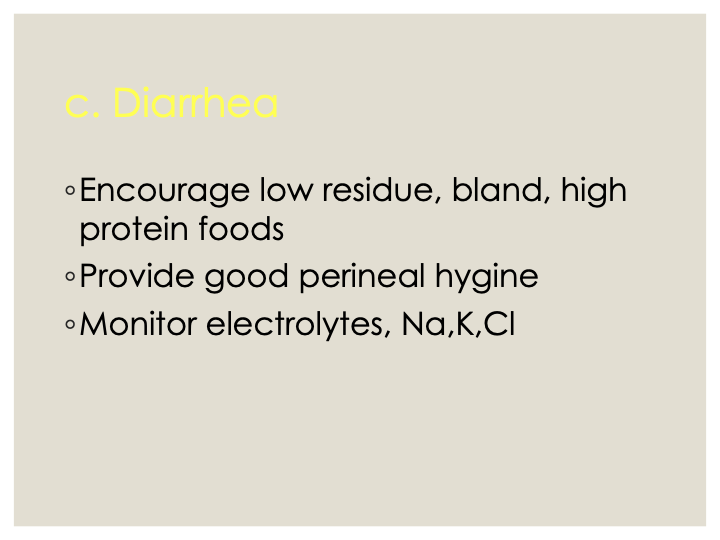
c. Diarrhea
d. Anemia. Leukopenia, thrombocytopenia
1.Keep skin free of foreign substance
2.Avoid use of medicated solutions
3.Avoid pressure, trauma, infection
4.Avoid exposure to heat, cold or sunlight
Skin: Itching, redness, burning, sloughing
1.Provide small, attractive feedings
2.Avoid extremes of temperatures
3.Administer antiemetics before meals
B. GI Disturbances
A. Anorexia, Nausea & Vomiting