fundamentals of neuroscience unit 1
1/230
There's no tags or description
Looks like no tags are added yet.
Name | Mastery | Learn | Test | Matching | Spaced | Call with Kai |
|---|
No analytics yet
Send a link to your students to track their progress
231 Terms
bell and magendie discovered
dorsal and ventral roots carry information in opposite directions
franz J gall
phrenology: bumps on the surface of skull reflect brain surface and related personality traits
Paul Broca discovered
broca aphasia/expressive aphasia
broca / expressive aphasia
acquired damage to the frontal regions of brain such as broca area/frontal lobe of dominant hemisphere
discrete region of human cerebrum for speech
Charles Darwin and evolution of the nervous system
nervous systems of diff species may share common mechanisms
common ancestors
damage to the broca area means
struggle with speech
in the 19th century the neuron was discovered and this allowed us to study
cell theory
cells
nerve cells
levels of analysis in modern neuroscience
molecular, cellular, systems, behavioral, cognitive
Alzheimers disease
a progressive degenerative disease of the brain characterized by dementia and always fatal
autism
impairments in communication and social interactions and restricted and repetitive behaviors
cerebral palsy
a motor disorder caused by damage to the cerebrum before during or soon after birth
neuropathologist vs neurosurgeon
neuropathologist trained to recognize the tissues that result from disease
neuroethologist
studies the neural basis of species-specific animal behaviors in natural settings
Glia cells
insulate, support and nourish neurons
neurons do what
process information
sense environmental changes
communicate changes to other neurons
command body response
Santiago ramón y Cajal and Camilo Golgi
sanitgo = neuron doctrine
golgi = reticular doctrine
obstacles to study cellular neuroscience
small cell size .01 and .05 mm
soft tissue
colorless
histology workflow
the gain in the brain is mainly in the stain
fixation
embedding
sectioning
staining
microscope
nissl staining / cresyl violet
franz Nissl
class of basic dyes stain the nuclei of all cells
clump of material surrounding the neuronal nuclei: nissl bodies
facilitates the study of cytoarchitecture in the cns
golgi staining/ silver stain
invented by Italian histologist Camillo golgi
silver chromate solution
thicker section
small fraction of neurons became darkly colored
reveals the entire neuronal cell
great for studying morphology
golgi stain revealed two parts of neurons
cell body, soma or perikaryon
neurites: axons, dendrites
axons
usually one axon
Uniform in diameter
any branches extend at right angle
longer distance
carry outputs
dendrites
usually multiple
short
antennae of neuron to receive signals
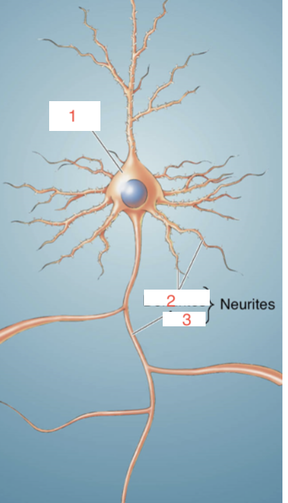
1 - soma
2- dendrites
3- axon
cajals neuron doctrine
use of golgi stain
neural circuitry in fine details
neurons communicate by contact not continuity
brain adheres to cell theory
golgis reticulum doctrine
use of golgi stain
neurites of different cells are fused together to form a continuous reticulum or network
brain is an exception of the cell theory
neurites of different neurons are not continuous with one another and this was found by
the electron microscope in the 1950s owing to the increased resolving power
the soma/cell body/ perikaryon
spherical central part
cytosol
watery fluid inside the cell
organelles: membrane enclosed structures in the neuron include
nucleus
rough endoplasmic reticulum
smooth endoplasmic reticulum
golgi apparatus
mitochondria
the cytoplasm
contents within a cell membrane excluding the nucleus
the nucleus of a neuron
contained with a perforated double nuclear membrane aka nuclear envelope
5-10 um
inside chromosome
dna is highly packed 2 meter linear length
the reading of the DNA is known as
gene expression
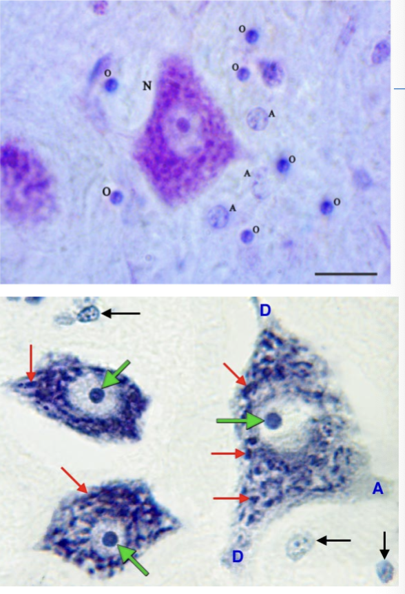
nissl staining
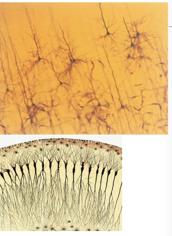
golgi staining
different cell types have the same dna but
different gene expression
in neurons translation into protein happens where
in the cytoplasm
ribosomes in neurons
convert the information in genes by synthesizing proteins
mrna transcripts bind to the ribosomes and the ribosomes translate the information in the mRNA to assemble a protein molecule
rough ER
abounds in neurons
nissl bodies are staining of rough ER
there are free floating ribosomes
free ribosome proteins are destined to reside within _____ of the neuron
cytosol
rough ER proteins inserted into the membrane of a cell or an organelle and then they are
synthesized on the rough ER
why is there a high number of rough ER in neurons
brain function heavily counts on membrane protein
smooth endoplasmic reticulum
stacks of membranous organelles without ribosomes
some is continuous with rough ER to process proteins
others may regulate the internal concentrations of substances such as calcium in muscle cells
Golgi apparatus
described by golgi
membrane enclosed disks farthest from the nucleus
sites for post translation chemical processing
sorting proteins for delivery to different cell regions
mitochondria in neurons
very abundant in the soma
about 1um long
inner membrane folds: cristae
inner space: matrix
site of cellular respiration
generates atp, which is cells energy source
the cytoskeleton of the neuron
shape is not static
internal scaffolding of neuronal membrane
the three structures of the cytoskeleton of a neuron
microtubules
neurofilaments
microfilaments
microtubules in the cytoskeleton
20 nm in diameter run longitudinally down neurites
a thick walled hollow pipe that is composed of strands
each strand consists of tubulin
polymerization and depolymerization can regulate neuronal shape
MAP (microtubule associated protein) is implicated in
alzheimers
neurofilaments
consists of multiple subunits that are wound into rope like structure
each strand consists of long protein molecules making neurofilaments mechanically strong
microfilaments in neurons
5 nm in diameter
numerous in the neurites
two strings of polymers of actin
constantly assemble and disassemble
run longitudinally down the neurite and closely associated with the membrane
the axon: only found in neurons
axon hillock: beginning
proper: middle
terminal: end
axon collateral: branch
differences between axon and soma
ER does not extend into axon, needs protein from soma
protein composition is unique
axon terminal:
bouton
where the axon contacts with other neurons
the synapse
when a neuron makes a synaptic contact with another cell it is called
innervation
axon terminal is called
presynaptic
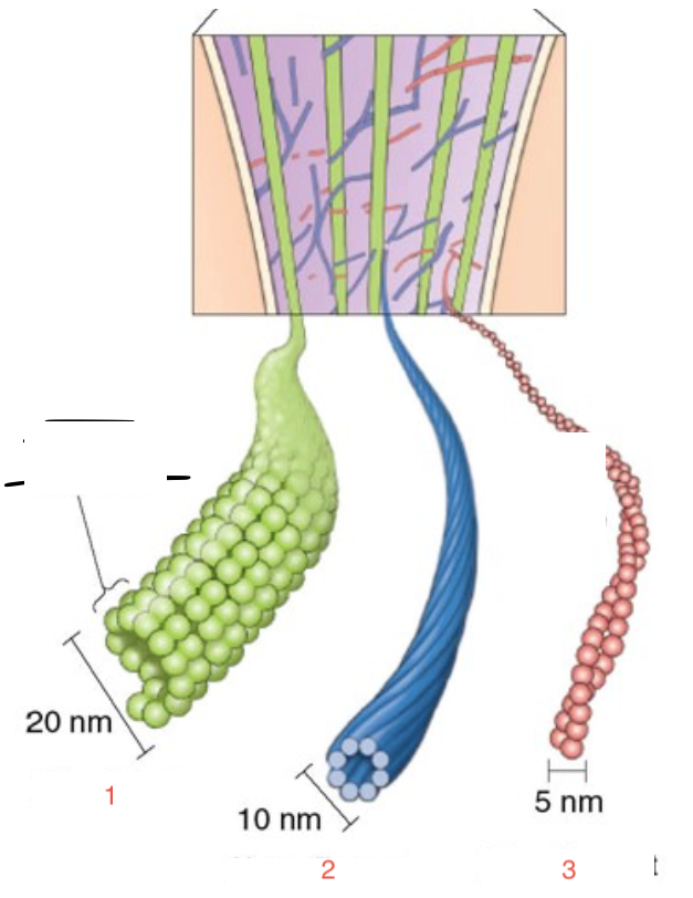
1= microtubule
2= neurofilament
3=microfilament
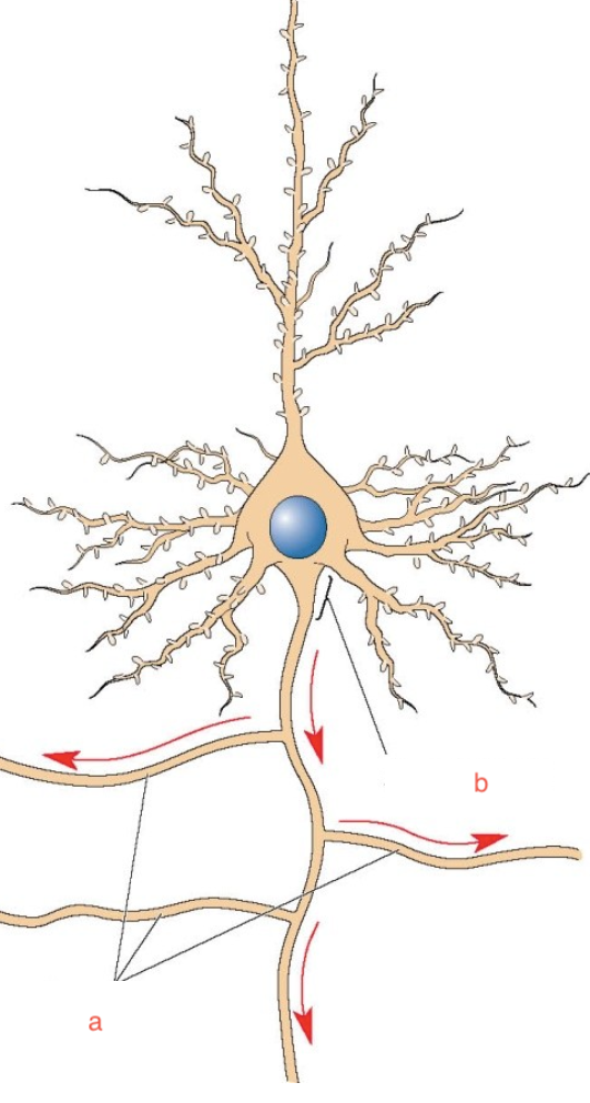
a= axon collaterals
b=axon hillock
postsynaptic is where
dendrite
differences between the cytoplasm of axon terminal and axon
no microtubules in terminal
presence of synaptic vesicles in the terminal
abundance of membrane proteins in the terminal
large number of mitochondria inthe axon terminal
the synaptic transmission
electrical → chemical → electrical
disfunction of synaptic transmission leads to
mental disorders and is the target of toxin or drug development
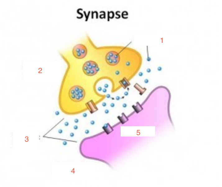
1 = neurotransmitters
2 = presynaptic terminal
3 = synaptic cleft
4= postsynaptic terminal
5= receptors
axoplasmic transport
no ribosome proteins ship down the axon
anterograde (soma to terminal)
wallerian degeneration 1-10mm per day
transport is fast with radioactive amino acid injection, 1000 mm per day
walk down microtubules thru kinesin and atp
retrograde transport
the enzyme horseradish peroxidase is selectively taken up by axon terminals and then transported retrogradely to the soma
some viruses exploit retrograde transport to infect neurons
regulated by dynein protein
dendrites
antennae of neurons
dendritic tree: all dendrites from a single neuron
synapse - receptors
dendritic spines
dendritic spines
post synaptic
isolate various chemical reactions
dynamic and sensitive to synaptic activity
unusual changes in disorders
classifications of neurons based on number of neurites
single neuritis = unipolar
two or more neurites = bipolar or multipolar
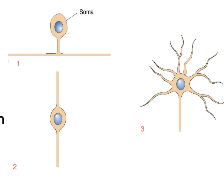
1= unipolar
2= bipolar
3= multipolar
classifying neurons based on dendritic and somatic morphology
stellate cells (star shaped)
pyramidal cells
spiny or aspiny
classification can overlap
neurons connected within the pns
primary sensory neurons, motor neurons, interneurons
golgi type 1 (classifying neurons based on axonal length)
projection neuron; from one part of brain to another
golgi type II neuron
local circuit neuron, short axon, only in vicinity
functions of glia
support neuronal functions, nutrition, insulation
astrocytes
most numerous glia in the brain
fill spaces between neurons
influence neurite growth
regulate chemical contents of extracellular space
express neurotransmitter receptors
myelinating glia types
Oligodendrocyte (CNS)
Schwann cells (PNS)
function: insulating axons
Oligodendrocytes and nodes of Ranvier
Nodes= region where axonal membrane is exposed
Oligodendrocytes= myelinated sheath protecting axon
microglia as phagocytes
can migrate out from blood and take away debris
ependymal cells
line up the ventricles
the reflex from stepping on a thumb tack
signals travel up sensory nerve fibers
spinal cord some send axons to brain where pain is felt and others synapse on motor neurons which send signals to muscles to retract
the motor commands trigger muscle contraction and withdrawal
challenges in neurons conducting information through electric impulse
cytosol is far less conductive
axon is bathed in salty ECF: leaking
axon alone is not well insulated
action potential refers to
potential refers to separation of electrical charges across the membrane
action potential and distance
they are signals of fixed size and duration, they do not diminish over distance
the frequency of action potentials of individual neurons gives us
information that is encoded
there is excitable membrane for action potential in
both neurons and muscle cells
resting potential is described as
when the neuron is not generating impulses
in neurons the inside surface of the membrane is _______ charged
negatively
the electrical charge across the membrane is called the
resting membrane potential
action potential
brief reversal of resting potential and for an instant (example 1/1000 of a second)
factors of resting potential
salty fluids on either side of the membrane, cytosol, ECF
the membrane
the proteins that span the membrane,: ion channels, pumps
water as it relates to ICF and ECF
polar solvent, uneven distribution of electrical charge
allows for solvent of other charged or polar molecules
ions in the intracellular and extracellular fluid
NA+, K+, CA2+, Cl-
the membrane of a neuron
phospholipid bilayer
proteins that span the phospholipid bilayer include
enzymes, cytoskeletal elements, receptors
control resting and action potentials
ion channel proteins components
polar r groups hydrophilic exposed to water on either sides of the membrane
nonpolar R groups: hydrophobic inside the membrane
ion channel selectivity
ion channel gates
enzymes use energy from atp breakdown and these pumps include
neuronal signaling
sodium/potassium pump
calcium pump
ion channel driving forces
diffusion and electricity
movement of ions with diffusion
dissolved ions distribute evenly
diffusion is a net movement of ions from regions of high con to low con