POCUS Fundamentals Test
1/91
There's no tags or description
Looks like no tags are added yet.
Name | Mastery | Learn | Test | Matching | Spaced |
|---|
No study sessions yet.
92 Terms
Freq Range-Infrasound
0-25 Hz
Freq Range-Audible
20Hz to 20 kHz
Freq Range - Ultrasound
20-100 kHz
How the image is created
Ultrasound images are created by electrical energy being converted into sound waves via the transducer. The transducer sends sound waves into the body, the sound waves bounce off or penetrate through various structures/organs within the body and return to the transducer to be converted back into electrical energy and to create an image on the screen. The machine knows where to place the reflected sound waves due to the time it took for the sound wave to return to the transducer. This determines the depth, the type of reflector, solid or fluid filled, determines the color of the reflector (echogenicity).
Most commonly used probe for the abdomen
Curved Array
Curved Array - used on abdominal, thoracic and pelvic patients due to frequency of probe. The frequency is typically 2-5 MHz, best for deep imaging.
Gain
Receiver gain (overall gain) allows the operator to increase (make brighter) or decrease (make darker) the echoes that are received back from the body. The operator can either darken or brighten the echogenicity of the entire image. If the gain is set too high, artifactual echoes or 'noise will be displayed throughout the image. Adjusted by: Gain button- rolling knob/toggle or single button on compact machines
Time Gain Compensation (TGC)
Uniform brightness-
TGC allows the operator to increase or decrease the brightness of the echoes that are received back from the body at different depths. The operator can either darken or brighten the echogenicity of the image at specific locations. The operator must adjust the gain to compensate for the time the sound wave takes to travel back to the machine. The depth of the structure being imaged is determined by the amount of time it takes for the transmitted sound wave to return to the transducer. The sound wave weakens with the increased depth of the image.
*Reminder, this is due to attenuation. The weakening of the original ultrasound signal as it leaves the transducer and courses through the body.
Color Doppler
Color doppler is a motion doppler that detects movement either towards or away from the transducer.
It is used to determine presence or absence of blood flow and is also beneficial in differentiating an anechoic structure from a vessel. Color doppler is adjusted by the sonographer.
Doppler shift is defined as the change in frequency of sound wave due to a reflector moving towards or away from the transducer. When the transducer emits soundwaves at a certain frequency and they are reflected back at the same frequency, this tells us the reflector is not in motion.
However, if the reflecting source is in motion either toward or away from the emitting source the frequency of the sound waves received will be higher (positive Doppler shift) or lower (negative Doppler
shift) than the frequency at which they were emitted, this is called the doppler shift and is how the ultrasound unit determines direction of flow.
Color doppler has many helpful uses in the field. It can help you confirm the presence or absence of blood flow, differentiate vessels from non-vascular structures and determine the presence and direction of flow.
Top color = towards the transducer. Bottom color = away from the transducer
The same principles of overall gain are used with color Doppler gain. The brightness of the red or blut color can be increased by increasing the doppler gain.
Freeze
Freeze
The freeze button allows the operator to pause the image, typically before measuring an object or printing an image
M-mode (motion mode)
M-mode scanning is used predominantly for documenting the fetal heart: in a first trimester ultrasound, proving fetal viability, but can also be helpful in lung examination.
The fetal heart rate is measured by placing a caliper at the peak of each wave (sometimes 2 waves).
Various features of B mode and M mode
Various features of B mode and M mode
B mode or brightness modulation, is the result of an ultrasound wave being sent into the body, bouncing off a reflector and returning back to the ultrasound system. The image is displayed in brightness, based on the amplitude of the reflected echo. M mode or motion mode, is the result of a series of B mode scans rapidly repeated to represent motion of the image over time.
A device that converts one form of energy into another to create an ultrasound image.
Transducer/probe
_________describes a structure that is completely black, lacking any echos at all
Anechoic
________ describes an organ or object that reflects echoes with a bright intensity
Hyperechoic
The M in M-mode scanning stands for
Motion
A common transducer used to scan the abdomen would be a(n)
Curved array
_______ allows the operator to amplify or boost ALL ECHO SIGNALS and make the entire image brighter
Receiver gain
The image ________ is typically displayed in centimeters on the side of the image
Depth
Color Doppler is used for all of the following except:
a. Confirm the presence or absence of blood flow
b. Differentiate vessels from non-vascular structures
C. Determine the presence and direction of flow
-d. IMAGE BONES MORE ACCURATELY
Doppler shift is defined as the change in frequency of sound wave due to a reflector moving towards or away from the transducer.
True
At what frequency is considered ultrasound?
1-20 MHz
The loss of sound energy due to converting into thermal energy is known as
Absorption
When imaging through a weak structure, the image beyond the structure will be very bright due to the increased attenuation of the structure. This artifact is called
Enhancement
No confirmation of harmful bioeffects from exposure to diagnostic ultrasound have ever been reported
True
All of the following are ultrasound artifact assumptions except
a. Sound travels in a straight line
b. Sound travels directly to a reflector and back
C. Sound travels in soft tissue at exactly 1,540m/s
d. THE IMAGE PLANE IS VERY THICK
The artifact is caused by the sound beam hitting a strong reflector and bouncing back and forth between the strong reflector and the transducer
Reverberation
This artifact is caused by the sound beam being blocked by a rib.
Shadowing
The greatest risk with ultrasound is using a probe with cracked housing
True
The leading edge of the probe is typically pointed towards the patient's _______ side or toward the patient's_____
Right
Head
The most frequently used position to scan the abdomen is _________
Supine
When scanning a patient lying supine in the area of the pelvis in a transverse scan plane, the leading edge of the probe is pointed towards the _______
Operator; patient's right side
The beam is ideally reflected when the transducer is ______ to the structure being imaged
Perpendicular
The following motion(s) can be helpful when obtaining ultrasound images
a. Fanning
b. Angling
C. Rotating
d. All of the above
D. All of the above
Ultrasound images containing patient information should remain confidential at all times and only be presented to other healthcare professionals on a 'need to know' basis.
True
All of the following are considered image annotation EXCEPT:
a probe position
b. patient gender
c. area/object imaged
d. position on area/object
b. patient gender
Ultrasound images can be nearly impossible or impossible to obtain due to which of the following factors?
a. inability to move patient
b. body habitus
c. bowel gas
d. all of the above
D. ALL OF THE ABOVE
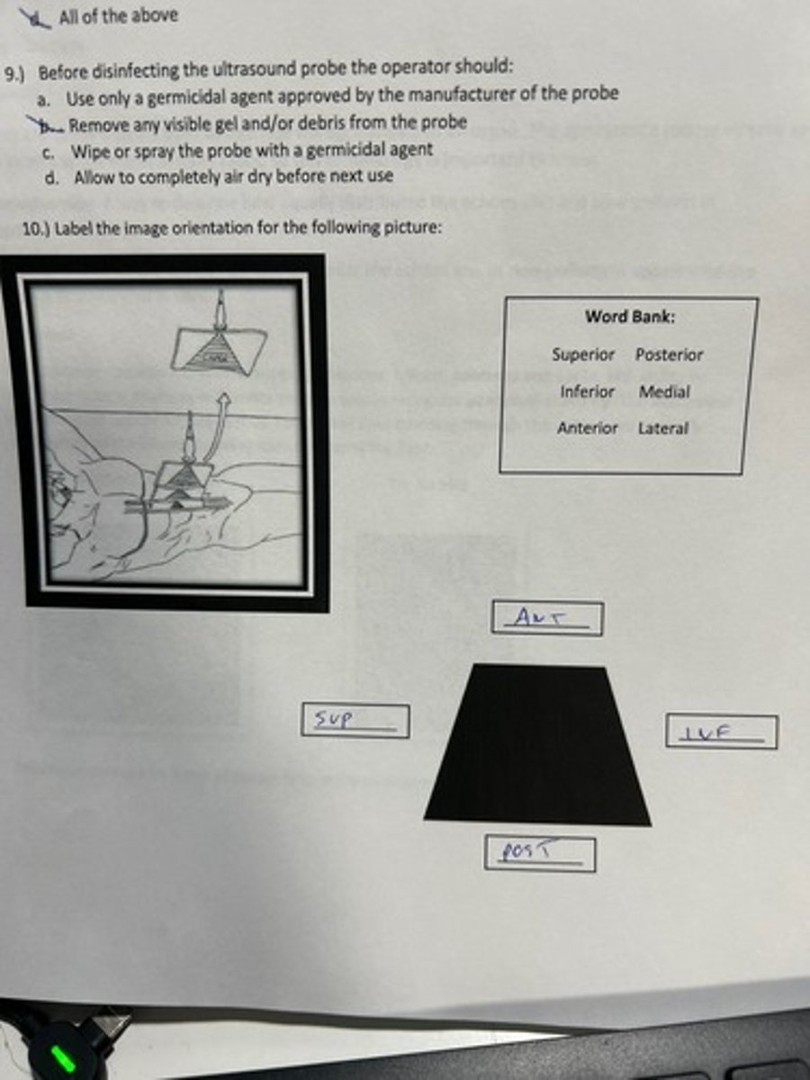
Before disinfecting the ultrasound probe the operator should:
a. use only a germicidal agent approved by the manufacturer of the probe
b. remove any visible gel and?or debris from the probe
c. wipe or spray the probe with a germicidal agent
d. allow to completely air dry before next use.
B. REMOVE ANY VISIBLE GEL AND/OR DEBRIS FROM THE PROBE
10.) Label the image orientation for the following picture:
1.) The abdomen contains all of the following organs except:
a.) Liver
b.) Kidneys
c.) Gallbladder
D.) Uterus
D. Uterus
2.) This is a way to describe how equally distributed the echoes are, and/or how uniform appearance then organ is as a whole or in part.
a.) Homogeneous
b.) Hyperechoic
c.) Hypoechoic
d.) Heterogeneous
A. Homogeneous
The interface between the sag right kidney and the sag liver is called
Morison's pouch
What region of the abdomen is the spleen located int?
LUQ
The space posterior to the uterus is called:
Pouch of Douglas
1. The uterus lies in the:
Pelvic cavity
Small amounts of fluid seen posterior to the uterus during ovulation is normal
true
The female pelvis contains all of the following except
a bladder
b. uterus
c. prostate
d. ovaries
c. prostate
Which organs are included in the thorax?
a. liver
b. heart
c. lungs
d. both b and c
d. both b and c
The vascular system is composed of all the following except
a. heart
b. ligaments
c. veins
d. arteries
b. ligaments
2. Which hyperechoic muscular structure separates the thoracic cavity from the abdominal cavity?
a. Mediastinum
b. Pleural cavity
c. Diaphragm
d. Pericardial cavity
Diaphragm
3.) The diaphragm is seen ______ to the liver and spleen
a. Inferior
b. Superior
C. Lateral
d. Medial
Superior
The accumulation of serous fluid in the peritoneal cavity is called
Acites
The space above the bladder can be described as
supravesical space
The space behind the uterus where a small amount of fluid can collect in ovulating females and still be considered normal is called
pouch of Douglas
The femoral vein is a branch of the exernal iliac vein
true
__________are compressible, __________ are not compressible
Veins
Arteries
The ______ pulses with the beat of the heart, whereas the _ is responsive to breathing movements
Aorta
IVC
A non-copressible common femoral vein is indicateive of
deep vein thrombosis
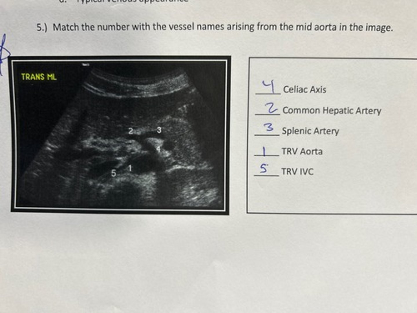
5.) Match the number with the vessel names arising from the mid aorta in the image.
Which vessel is circled in the image?
SMA
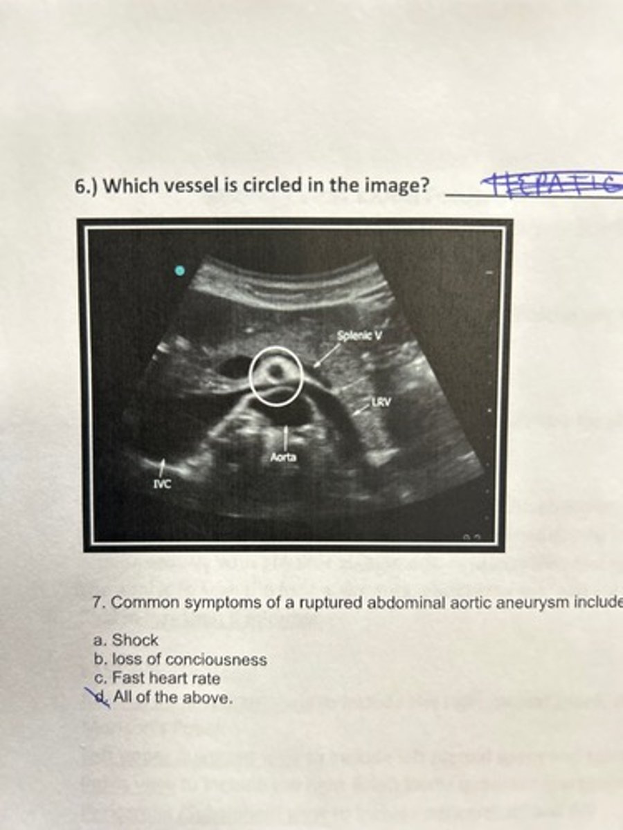
The Focused Assessment by Sonography in Trauma exam is also know as
FAST exam
The FAST exam includes evaluating which of hte following organs?
a. liver
b. splenorenal recess
c. paracolic gutters
d. all of the above
d. all of the above
The right and left paracolic gutters are located____ to the ascending and descending colon
lateral
The ideal time frame to perform a FAST exam is
5 min or less
The potential space between the liver and kidney where fluid can collect is known as:
Morrison's pouch
Ultrasound exam of the heart, the IVC, Morison's Pouch, Aorta and Pulmonary view is the protocol for which emergent exam?
RUSH
The RUSH exam is indicted when the patient is experiencing
Undifferentiated non-traumatic hypotension
HIMAP stands for
Heart
IVC
Morrison's Pouch
Aorta
Pulmonary view
The RUSH exam is used to help the first responder identify the reason for shock when there is not an obvious cause after examination of the patient
true
Oxygen poor blood enters the ____ atrium through the ____
right
vena cava
Which view of the hear can be imaged by placing the probe just below the left nipple?
apical 4 chamber
Typically, the wall of the left ventricle is_____ than right
thicker
When fluid is seen around the heart within the pericardial sac, this abnormality is called
Pericardial effusion
An enlarged right ventricle with an enlarged IVC is suggestive of ______
Pulmonary embolism
All of the following signs are associated with an abnormal lung except
a. absent lung sliding
b. Barcode sign
c. comet tail artifacts
d. greater than 3 B-lines per frame
c. Comet tail artifacts
_____ can be useful in detectin lung sliding because it picks up motion on ultrasound
M-mode
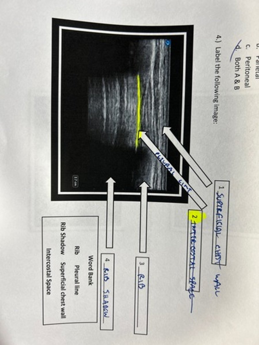
On a lung ultrasound the two layers of peura we observe are called
a. visceral
b. parietal
c. peritoneal
d. both a & b
d. both a & b
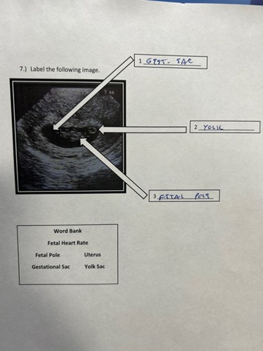
Label the following image
An exam used to determine the patient's fluid volume status
FSA- fluid status assessment
A collapsed IVC and normal/dry ling sounds are signs that your patient can benefit from fluids
IVC
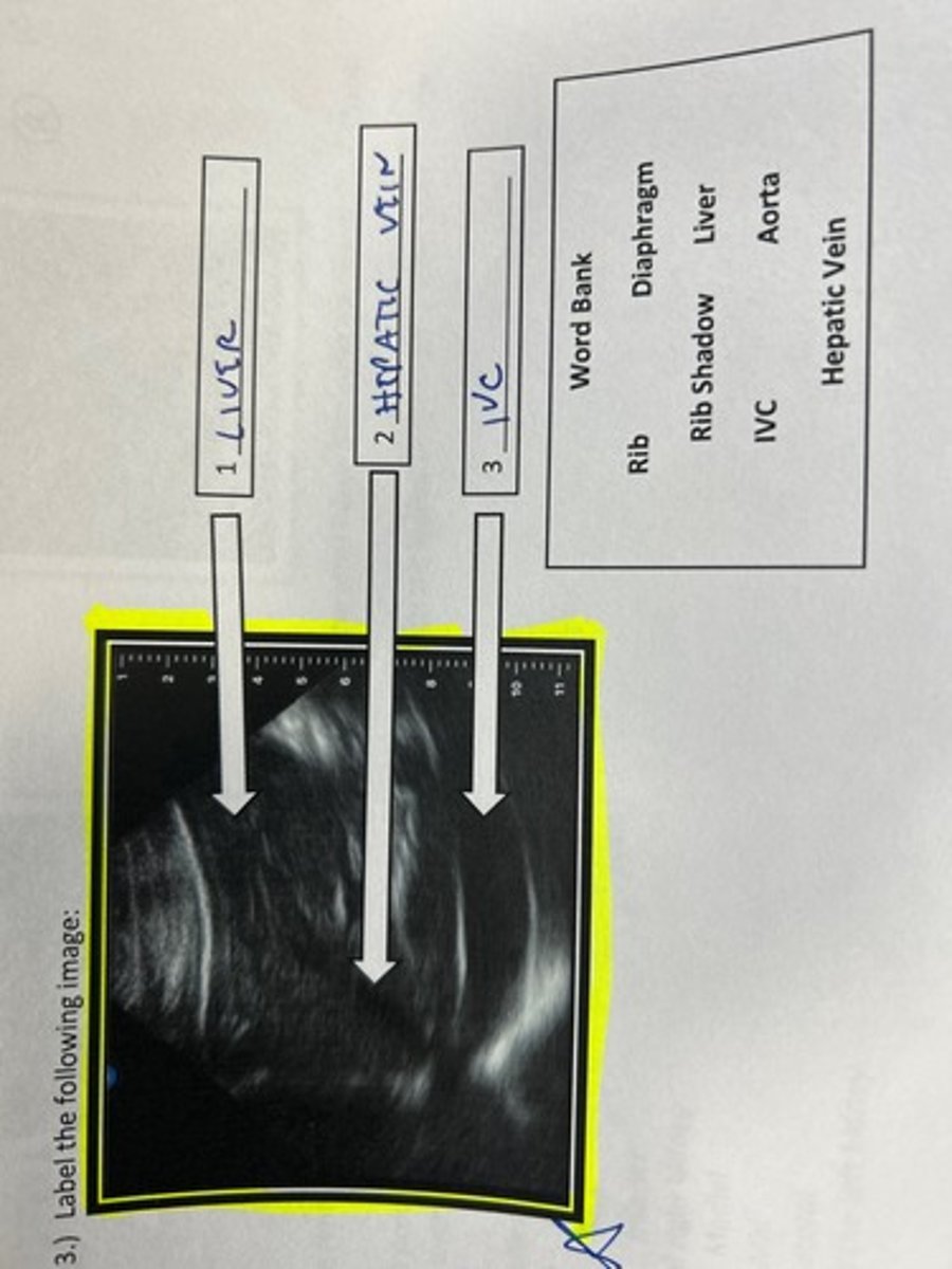
Label the following image
The cortex of the normal kidney is ____ to the liver
hypoechoic
Renal stones will appear as bright structures in the kidneys that can cause _____
shadowing
The renal pelvis is _______ to the cortex in a normal kidney
Hyperechoic
The renal arter, vein and ureter all enter the kidney at the renal _____-
Hilum
In the case of an obstructing renal stone, urine can back up into the kidney and cause hydronephrosis
True
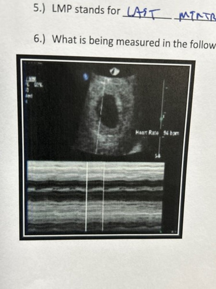
Thsi mode of imaging is best to evaluate the fetal heart rate
M mode
All of the following are indicateive of an intrauterine pregnancy EXCEPT
a. gestation sac seen within the uterus
b. yolk sac seen within the gestational sac
c. fetal heart rate captured within the gestational sac
d. free fluid with debris seen lateral to the uterus
d. free fluid with debris seen lateral to the uterus
subchorionic hemorrhage is a frequent cause of vaginal bleeding in the first trimester
true
LMP stand for
last menstrual period
The fetal heartrate cannot be visualized before the 5th week when scanning transabdominally
true
What is being measured in the following image
Fetal HT
Label the following image