EXAM 3 Chapter 9 The Muscular System 1: Skeletal Muscle Tissue and Muscle Organization単語カード | Quizlet
1/126
There's no tags or description
Looks like no tags are added yet.
Name | Mastery | Learn | Test | Matching | Spaced | Call with Kai |
|---|
No study sessions yet.
127 Terms
skeletal muscle
attaches to skeleton
has single, long & cylindrical striated cells
the only multinucleate type of muscle
controlled voluntarily
cardiac muscle
occurs in the heart wall
has branching chains of striated cells
one nucleus per cell; some binucleate
intercalated discs contains several types of cell jun.
cardiac cells are electrially coupled by gap junctions
controlled involuntarily
smooth muscle
occurs in walls of hollow organs
single, fusiform nonstriated cells
uninucleate (one cell)
controlled involuntarily
1. excitability
2. contractility
3. extensibility
4. elasticity
What are the 4 specialized properties the three types of muscles share?
excitability
ability of muscles to respond to nerve signals or stimuli
causes electrical impulses to travel along the muscle cells' plasma membrane
contractility
ability to generate a strong pulling force while muscle cells shorten (contract)
elasticity
ability of a muscle, after being stretched (or contraction), to recoil passively to its original (or resting) length
extensibility
the ability to continue to contract over a range of resting lengths
1. movement
2. maintain posture and body position
3. supports soft tissue
4. regulates entrance and exit of materials
5. heat generation
6. joint stabilization
What are the functions of skeletal muscles?
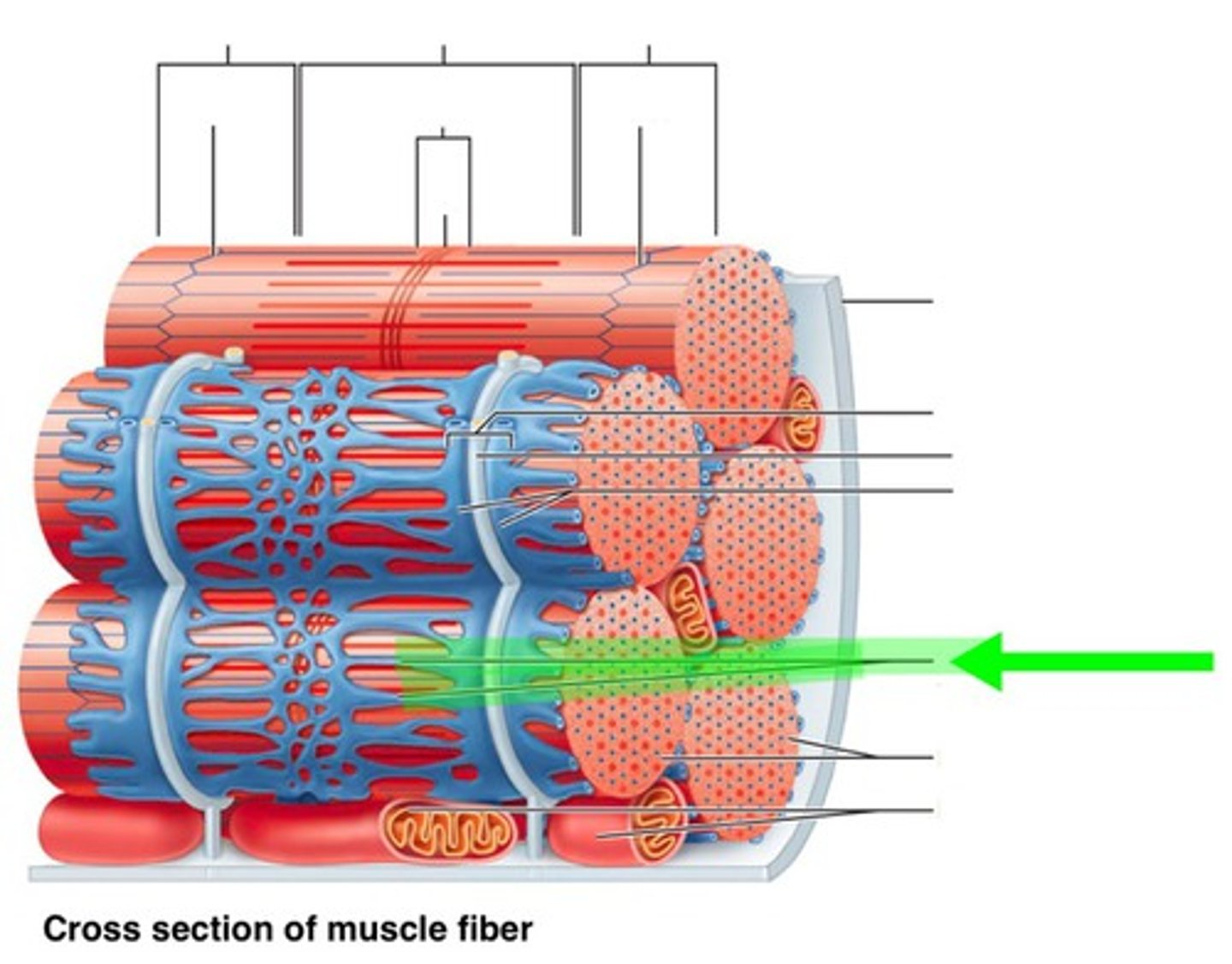
epimysium
What surrounds skeletal muscles?
bundles of muscle fascicles
What is skeletal muscle comprised of?
bundles of muscle fibers
What are muscle fascicles comprised of?
perimysium
What surrounds muscle fascicles?
muscle fiber (myofiber or muscle cell)
a highly elongated cell (in skeletal or smooth but NOT in cardiac muscle) holds together many myofibrils
endomysium
What surrounds muscle fibers/myofiber/muscle cells?
sarcolemma
plasma membrane of a muscle fiber (muscle cell)
comprised of abundant myofibrils
sarcoplasm
cytoplasm of muscle fiber (muscle cell(
contains numerous myofibrils
myofibril
a cylindrical structure, as long as entire muscle fiber (muscle cell)
consists of sarcomeres
surrounded by sarcoplasmic reticulum
myofibrils because they can shorten
What structure is responsible for contraction of the skeletal muscle fiber and why?
because a myofibril is attached to the sarcolemma at each end of the cell
Why does myofibrils contraction lead to shortening of the entire cell?
sarcoplasmic reticulum (SR)
an internal membrane complex similar to the smooth endoplasmic reticulum of other cells
STORES calcium ions
sarcoplasmic reticulum (SR)
What structure in skeletal muscle is closely associated with the transverse (T) tubules?
sarcoplasmic reticulum (SR)
What structure in skeletal muscle plays an important role in controlling the contraction of individual myofibrils via the release of calcium ions?
- STORES calcium ions
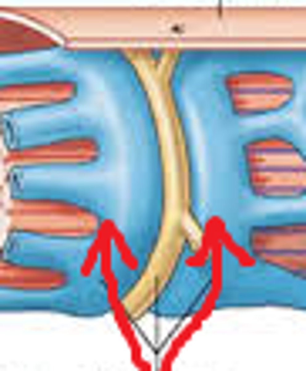
transverse (T) tubules
deep invaginations of the sarcolemma (into the sarcoplasm) which allow electrical impulses (that stimulate the membrane to contract) to quickly travel to the interior of the cell
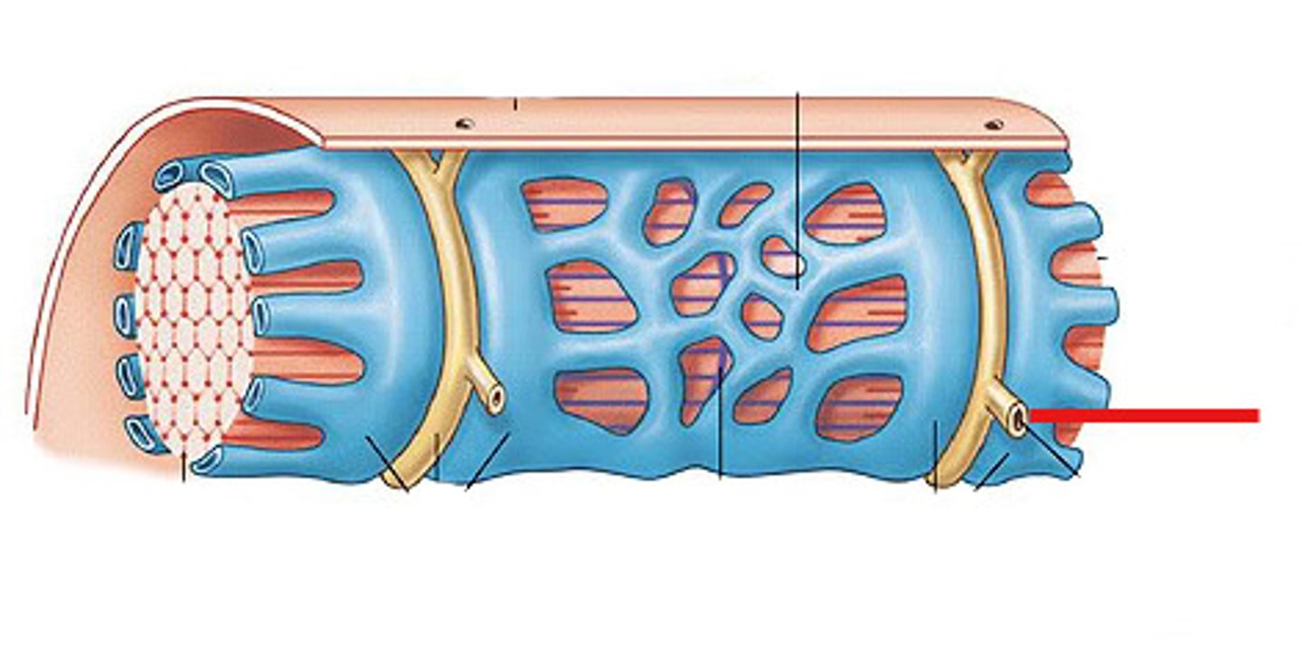
terminal cisternae
expanded chambers on either side of a transverse tubule where the tubule of the SR has enlarged and fused
triad
the combination of a pair of terminal cisternae plus a transverse tubule
sarcomere
myofibrils consists of sarcomeres, which are repeating units of myofilaments; the smallest functional unit of muscle fiber
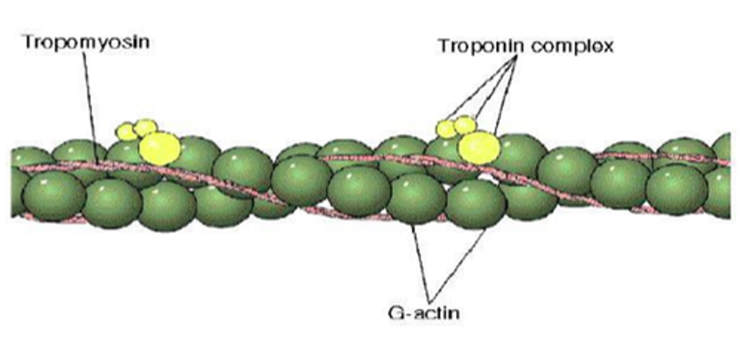
myofilaments
What is the smallest functional until of muscle fiber?
myofilaments
the sarcoplasm of muscle contains protein filaments, which generate contractile force
- fills up most of the sarcoplasm of each muscle fiber
- organized in the repeating units called sarcomeres
myofilaments
What determines the striation pattern in skeletal muscle fibers?
actin and myosin
What are the 2 primary types of myofilaments in muscle cells?
actin filaments
protein filaments found in THIN filaments
myosin filaments
protein filaments found in THINK filaments
parallel to the long axis of the cell
sarcomeres arranged side to side (dark and light band appearance)
How are myofibrils arranged in a muscle cell/fiber?
M line
a group of proteins, which link the thick filaments that lie in the center of the sarcomere
Z lines
open meshwork of interconnecting proteins called ACTININS, which occur where thin filaments from adjacent sarcomeres join
actin filaments (thin) attach to the Z lines and extend towards the M line
Where do thin filaments (actin) attach and extend towards?
Zone of Overlap
area where the thin filaments pass between the thick filaments
thin and thick overlap
A band (anisotropic band)
the area containing THICK filaments, including the:
- M line
- H line
- Zone of overlap
appears as a dark band
H band
area contains THICK filaments ONLY
I band (isotropic band)
the area containing THIN filaments only
appears as a light band
Z lines move closer together
I and H bands shorten
What happens to the Z lines during contraction?
What happens to the I and H bands during contraction as well?
Tropomyosin
protein molecules that form a long chain, which overs the active sites, preventing actin-myosin interaction
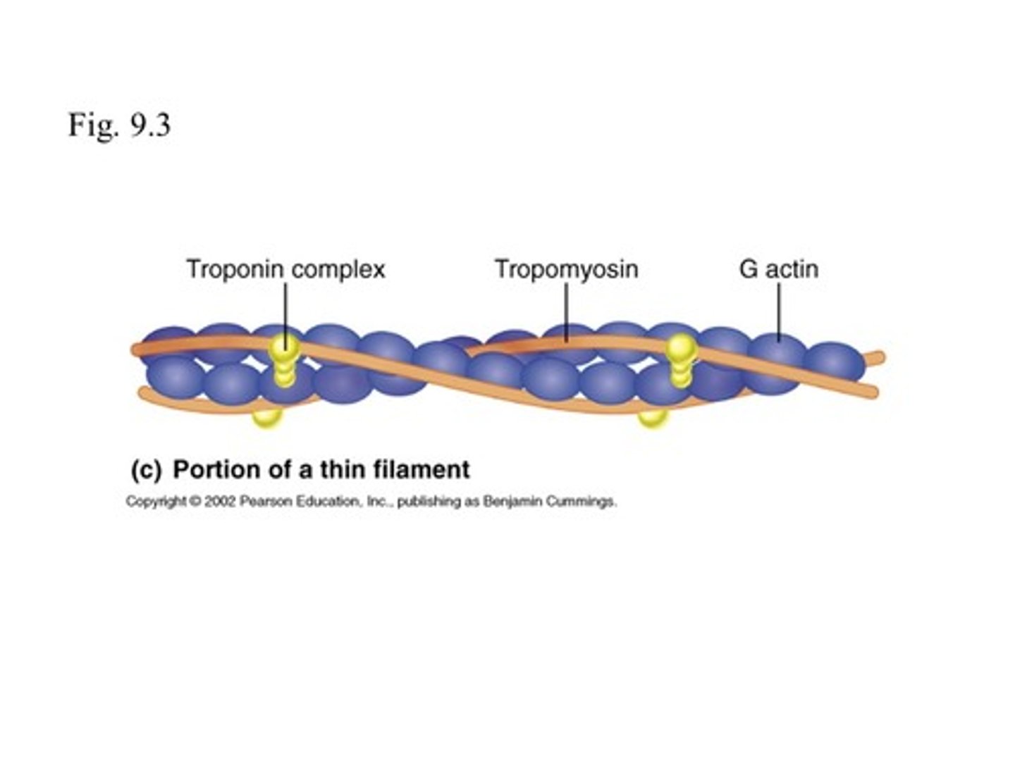
Troponin
protein molecules that hold the tropomyosin strand in place
- changes position to move the tropomyosin molecules, exposing the active site, prior to a muscle contraction
- act as the regulator molecule of a muscle contraction
Cross-bridges because they connect the thick and thin filaments during a muscle contraction
What are myosin heads also known as and why?
spindle-shaped cylinders
triangles
sheets
What are the various shapes of skeletal muscle?
whole muscle (organ)
muscle fascicles
muscle fibers (cells)
myofibrils
sarcomere
myofilament (actin and myosin)
What are the levels of organization in skeletal muscle? (gross to microscopic)
epimysium
dense irregular connective tissue sheath wrapped around a whole muscle
perimysium
fibrous connective tissue sheath wrapped around a fascicle
endomysium
thin reticular fiber connective tissue sheath wrapped around each muscle fiber
origin
immovable (or less movable) attachment from which a muscle extends
insertion
more movable attachment of a muscle
tendons, aponeuroses, or direct (fleshy) attachment
Skeletal muscles attach to bones through what?
muscle contraction
exerting a pull, or tension, and shortening the muscle fibers in length
muscle fibers are stretched to near-optimal length before stimulation of contraction occurs
Explain near-optimal length in muscles.
Calcium ions
The presence of what is needed to TRIGGER for a contraction?
ATP
The presence of what is REQUIRED for the contraction to occur?
Sliding Filament Theory
theory or mechanism that explains the physical changes that occur between the thin and thick filaments during muscle contraction
H band and I band get smaller
Zone of overlap gets larger
Z lines move closer
A band remains constant throughout contaction
What happens in the Sliding Filament Theory of muscle contraction?
bind to active sides on thin filaments causing sliding to occur
What do the myosin heads of thick filaments do during the Sliding Filament Theory
Cross-bridge binding
the myosin heads pivots toward the M line, pulling the thin filaments toward the center of the sarcomere
detaches and returns to its original position
What happens to the cross bridge after it pivots towards the M line?
binds, pivot, detach, and return
What is the cycle in which the myosin heads follow in the Sliding Filament Theory?
Z lines move towards the M line when the think filaments pull on the thin filaments
How does the sarcomere shorten?
1. an impulse from the sarcolemma signals the sarcoplasmic reticulum to release Ca2+
2. Ca2+ initiates the sliding of the myofilaments
(muscle contraction)
What happens when a nerve cell stimulates a muscle fiber?
neuromuscular synapse
a specific synapse between a motor neuron and a muscle cell
synaptic terminal
the expanded tip of the motor neuron's axon, at the neuromuscular synapse
synaptic vesciles
small secretory vesicles (filled with acetylcholine) in the cytoplasm of the synaptic terminal
neurotransmitter
a chemical released by a neuron to communicate with another cell
Acetylcholine
a neurotransmitter that signals the muscle cell to contract; released at axon terminal
synaptic cleft
a narrow space that separates the synaptic terminal from the motor end plate of the skeletal muscle fiber
Acetylcholinesterase
an enzyme that breaks down ACh molecules and is released by the basal lamina of the cell into the synaptic cleft
1. electrical impulse arrive at synaptic terminal
2. ACh is released and binds to receptor sites on motor end plate
3. action potential (AP) is generated
4. AP travels all over the surface of the sarcolemma and into each T tubule
5. APs continue to be generated until AChE removes the bound ACh.
(ACh is immediately broken down by AChE after it signals a single contraction)
What are the steps during Neural Stimulation of a Muscle?
motor unit
consists of one motor neuron and all the skeletal muscle fibers it innervates (controls)
- contains different numbers of muscle fibers distributed widely within a muscle
simultaneously
All muscle fibers in a motor unit contract _________.
level of control of the movement
The size of a motor unit indicates what?
muscle twitch
a single, momentary contraction, which is a response to a single stimulus
contain different numbers of muscle fibers distributed widely within a muscle
all or non principle
a characteristic in which each muscle fiber either contacts completely or does not contract at all
how many motor units are activated
The amount of force, exerted by the muscle as a whole, depends on what?
recruitment
the smooth but steady increase in muscular tension produced by increasing the number of motor units that is activated
muscle tone
resting tension in a skeletal muscle
muscle spindles
specialized muscle cells that are monitored by sensory nerves, which control the muscle tone in the surrounding muscle tissue
muscle hypertrophy
enlargement of skeletal muscles that undergo excessive repeated stimulation that produces near-maximal tension
muscle atrophy
reduction in skeletal muscle size, tone, and power as a result of inadequate stimulation to maintain resting muscle tone in the affected area
slow oxidative fibers (red fibers / type I fibers)
thin fibers that are red because of their abundant myoglobin (oxygen-binding pigment in sarcoplasm)
obtain energy from AEROBIC metabolic reactions
abundant mitochondria and rich capillary supply
contract slowly; extremely resistant to fatigue as long as enough oxygen is present
deliver prolonged contractions
best for maintaining postures
intermediate (fast oxidative) fibers / type II fibers
contract quickly
oxygen dependent and have a high myoglobin content
abundant mitochondria and rich capillary supply
fatigue resistant but less so than Type I
best for long-term production of fairly strong contractions, such as required in locomotion of lower limbs
fast glycolytic fibers (white fibers/ type IIx fibers)
pale fibers because they contain little myoglobin
twice the diameter of Type I fibers
contain more myofilaments
generate much more power
depend on ANAEROBIC pathways to make ATP
few mitochondria and capillaries present
contract rapidly; fatigue quickly
best for short bursts of power (lifting heavy; upper limbs)
fascicle
bundle of muscle fibers
parallel muscle
the long axes of the fascicles are parallel to the long axis of the muscles, and the muscle extends from origin to insertion
has a central body, or belly
ex: biceps brachii muscle
convergent muscle
the origin is broad, and the fascicles converge toward the tendon of insertion, its common attachment site
fibers may pull on a tendon, tendinous sheet, or a slender band of collagen fibers known as a raphe
ex: pectoralis major muscle
circular muscle (sphincter)
the fascicles are arranged in concentric rings around an opening or recess
when muscle contracts, the diameter of the opening decreases (close-tighten)
ex: orbicularis oris muscle
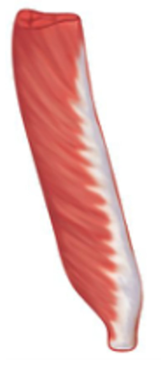
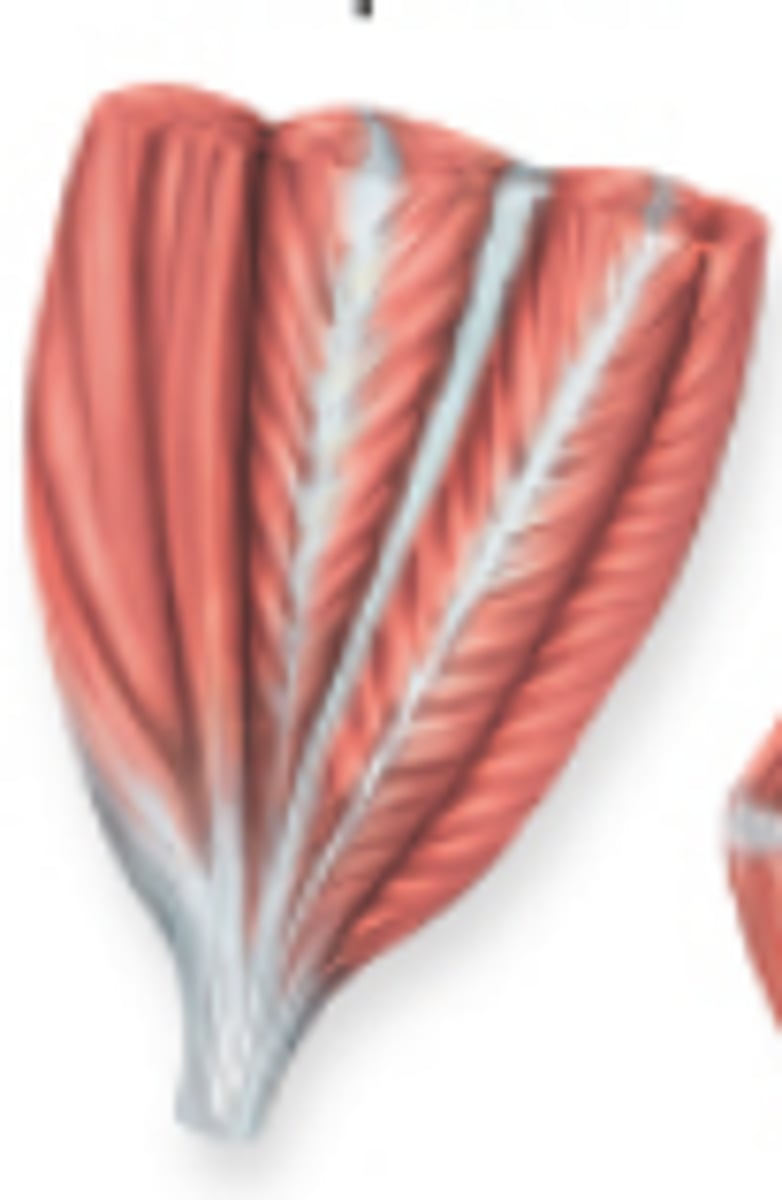
pennate muscle (penna, feather)
the fascicles are short and attach at an oblique angle to a tendon that runs through the body (the whole length) of the muscle
unipennate muscle
the fascicles insert into one side of the tendon
ex: extensor digitorum longus muscle of the anterior leg
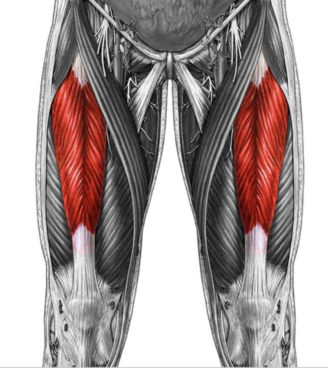
bipennate muscle
the fascicles insert into both sides of the tendon
ex: rectus femoris muscle of the thigh
multipennate muscle
the fascicles insert into a tendon that branches within the muscle
looks like many feathers situated side by side, whose quills are all inserted into one tendon
ex: deltoid muscle
agonist (prime mover)
a muscle whose contraction bears the main responsibility for a PARTICULAR MOVEMENT
ex: biceps brachii for forearm flexion at the elbow
antagonist
group(s) of muscles whose actions oppose that of the corresponding agonist
ex: tricepts brachii, during forearm flexion, are stretched and maybe slightly relaxed, and stablizes the flexion movement; BUT THEY ALSO ACT AS AGONISTS for extension of the forearm
synergist
aid the agonists, either by adding a little extra force to the same movement or by reducing undesirable extra movements that the agonist may produce; they stabilize joints, as fixators
externus or superficialis
muscles visible at the body surface
internus or profundus
muscles lying beneath the body surface