2nd and 3rd trimester
1/109
There's no tags or description
Looks like no tags are added yet.
Name | Mastery | Learn | Test | Matching | Spaced | Call with Kai |
|---|
No analytics yet
Send a link to your students to track their progress
110 Terms
in femur length do not measure
femoral head or distal femoral condyles
in abdominal circumference you should NOT SEE
kidneys or lungs
AC should show
round abdomen w flat ribs on each side
stomach and J view of umbilical/portal vein
placenta previa
leading edge of placenta within 2cm
BPD calipers
outer to inner
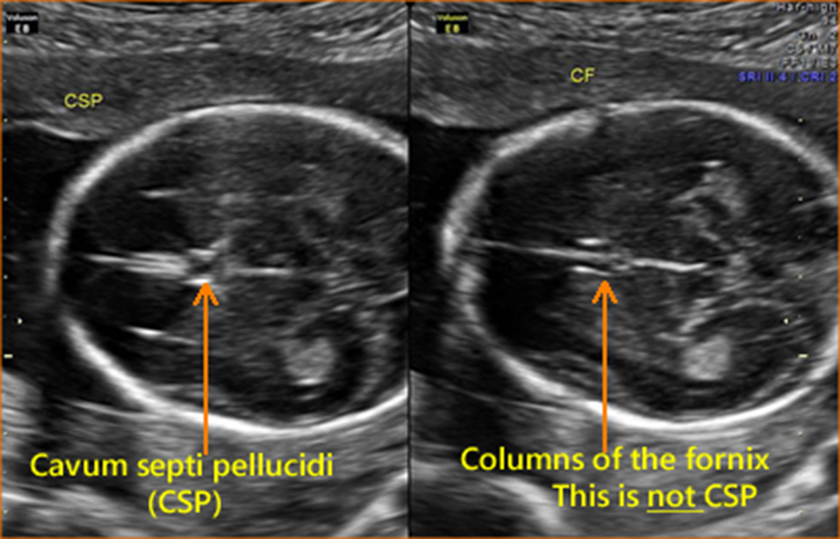
CSP sits _____ to thalamus
anterior
cerebellum and cisterna magna
cerebral peduncles behind thalamus
when measuring nuchal fold what do you have to have in image
CSP
nuchal fold where to measure
outer skull to outer skin
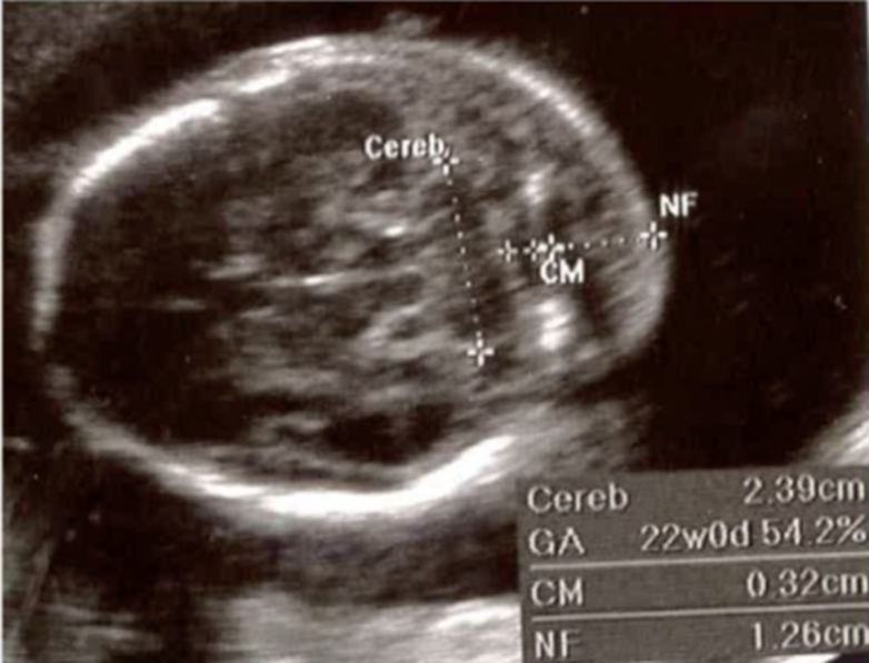
thickened nuchal fold
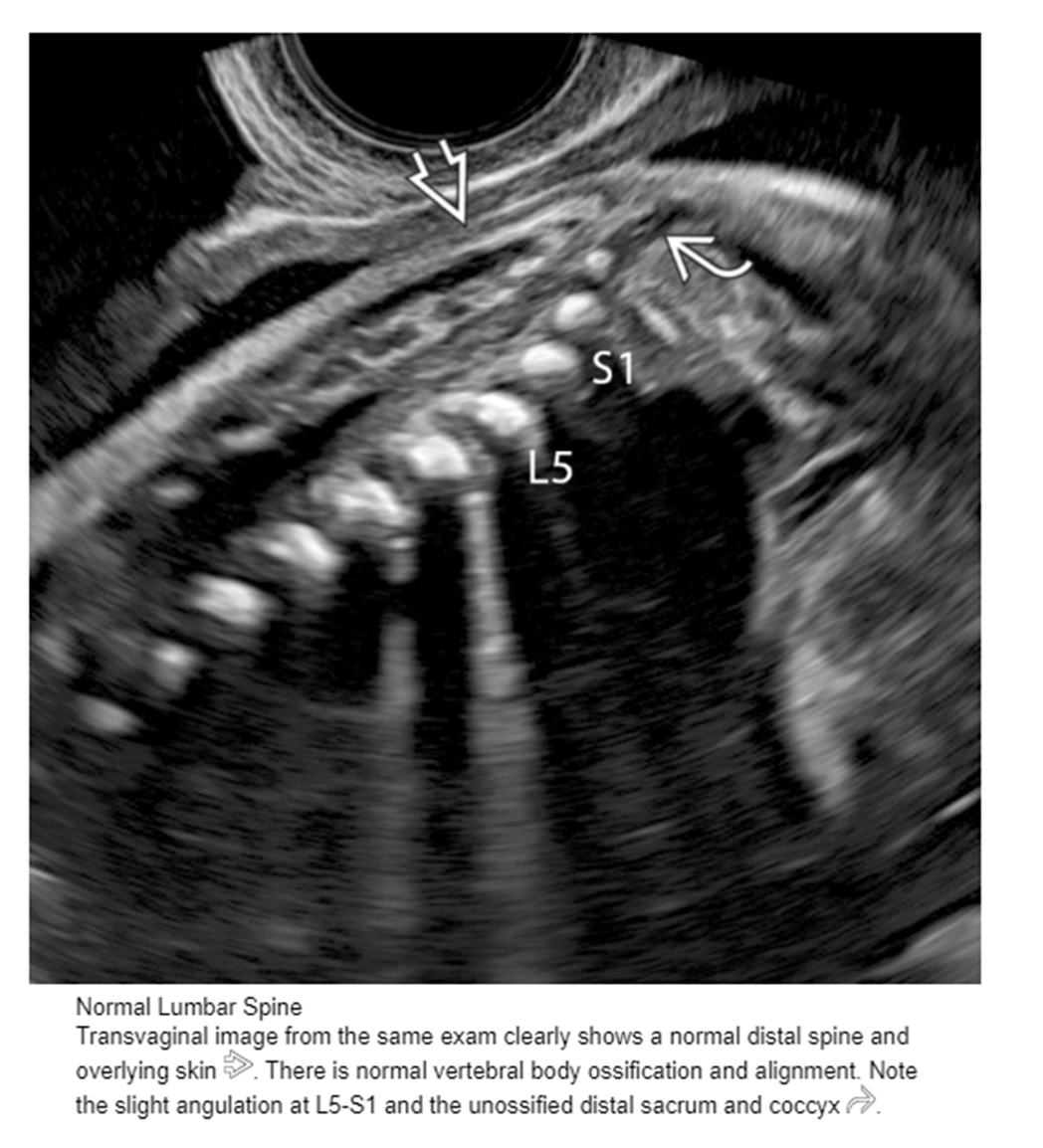
vertebrae numbers
7 cervical
12 thoracic
5 lumbar
5 sacral
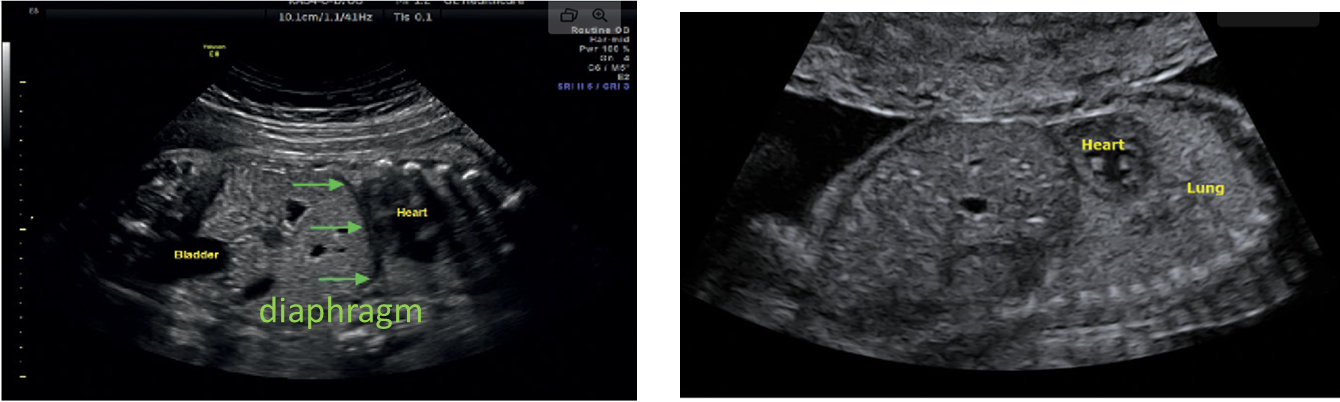
stomach and heart on ____ side
left
in sagittal _______ should be imaged to ensure you are imaging the cervical spine
occipital bone
in transverse, the ______ should be visualized to ensure you are imaging cervical spine
fetal clavicles
thoracic spine, _____ or _____should be seen
heart or stomach
lumbar spine, ____ should be seen
kidneys
sacral spine, _____ is landmark
iliac crest
BPD is ____ to _____ caliper placement
outer to inner
in HC do not include ____ ____
skin line
BPD and HC do not include
cerebellum or orbits
what landmarks in HC and BPD
thalamus, cavum septum pellucidum (CSP), portion of falx
3V cord be sure to show
split around bladder and not just 2 vessesls
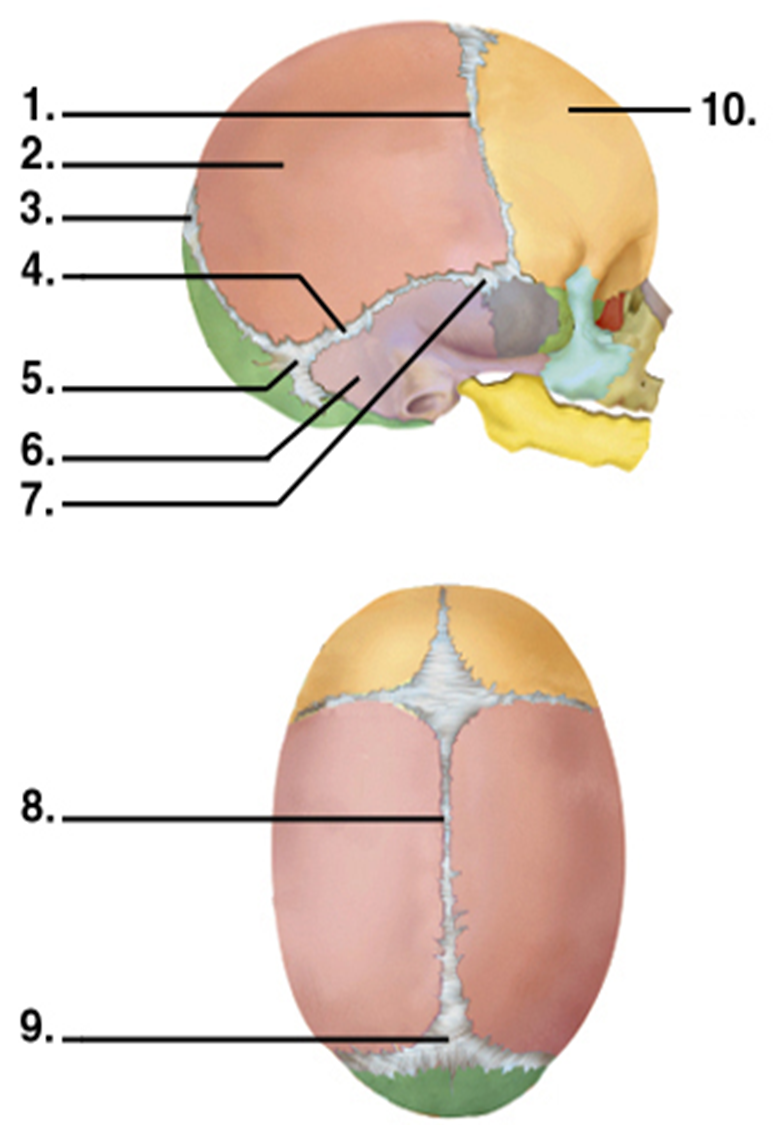
1.Coronal Suture
2.Parietal Bone
3.Posterior Fontanel
4.Squamous Suture
5.Mastoid Fontanel
6.Temporal Bone
7.Sphenoid fontanel
8.Sagittal Suture
9.Posterior Fontanel
10.Frontal Bone
cerebral aqueduct aka
aqueduct of sylvius
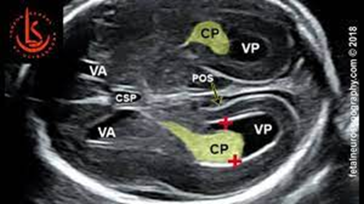
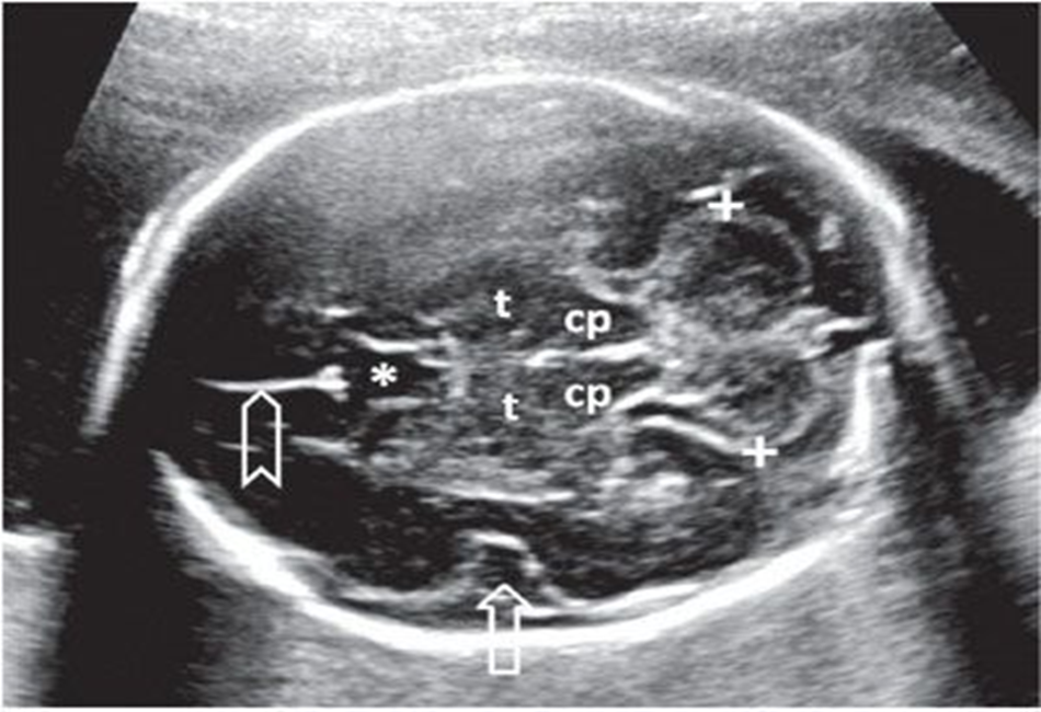
lateral ventricles should not exceed
1-1.1 cm
calipers of choroid plexus and lateral ventricles
inner to inner
measure _____ portion of lateral ventricle through the most _____ portion of choroid plexus
posterior, posterior
back of brain / posterior fossa includes
cerebellum, cisterna magna, vermis, 4th vent, cerebral peduncles, brainstem, and tentorium
cisterna magna should be less than
1cm
if you see the distal femoral epiphysis, the baby is at least
33 weeks
bimetry not typically done until
13-14 weeks
cannot reliably use femur length after ??? weeks
14 weeks
AC typically not done until ??? - ??? weeks
13-14
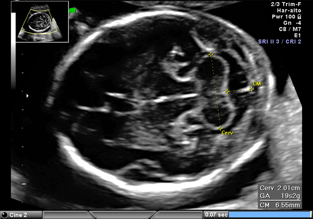
cervical length measurement typically done ??? - ??? weeks
16-24
AFI typically done around ??? weeks +
28
what separates the cerebrum from the cerebellum and brainstem
tentorium
cerebellum has a ___:___ ratio in mm to gestational age from ___-___ weeks
1:1 ratio
16-24 weeks
nuchal fold measurement routinely performed ___ - ___ weeks
15-19.6
nuchal fold measurement should be less than ___ mm
6 mm
nuchal fold calipers
outer skull to outer skin
what is important to include in spine images
skin line
echotexture of lungs
symmetrically homogeneous
increases as gestation progresses
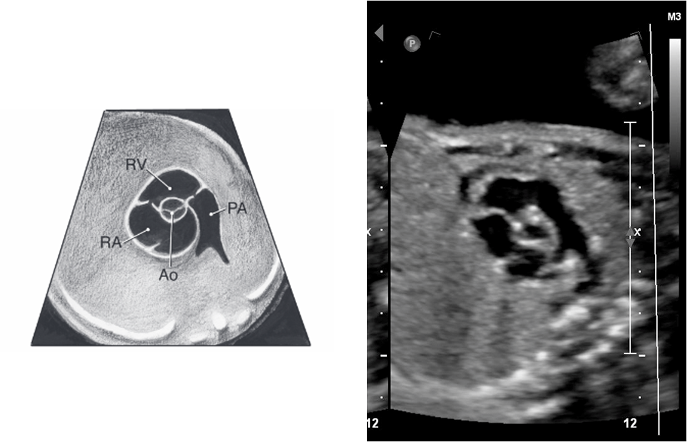
pulmonary artery comes out of ???
right ventricle
pulmonary artery quickly passes _____ and ____ of ascending aorta
anterior and left
right pulmonary artery passes ____ to the aorta
posterior
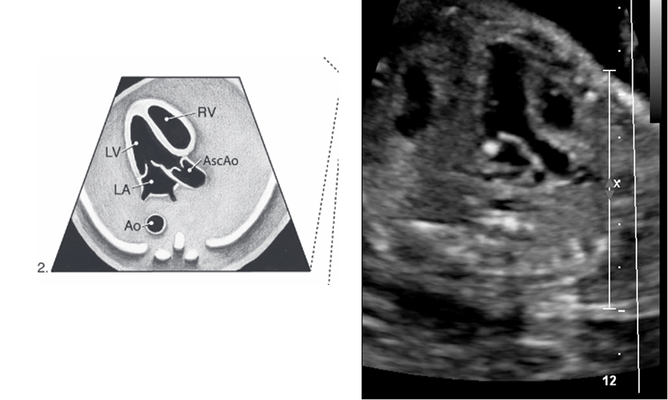
normally what is closest to anterior abdominal wall
right ventricle
what is closest to spine
left atrium
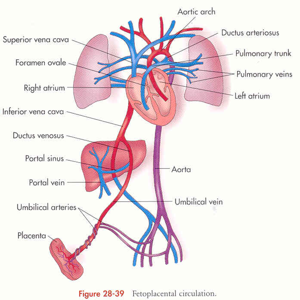
blood flow through foramen ovale
right to left
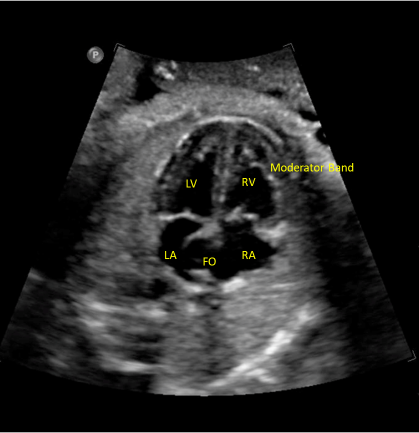
where is moderator band located
right ventricle
tricuspid valve on ____ side
right
mitral valve on ____ side
left
what is foramen ovale
fetal shunt allowing connection between atrium
4 chamber heart should sit at a ___ degree angle and take up about ____ of the chest
45 degree
1/3
___ of the herat located in left chest with apex pointing ____
2/3
left
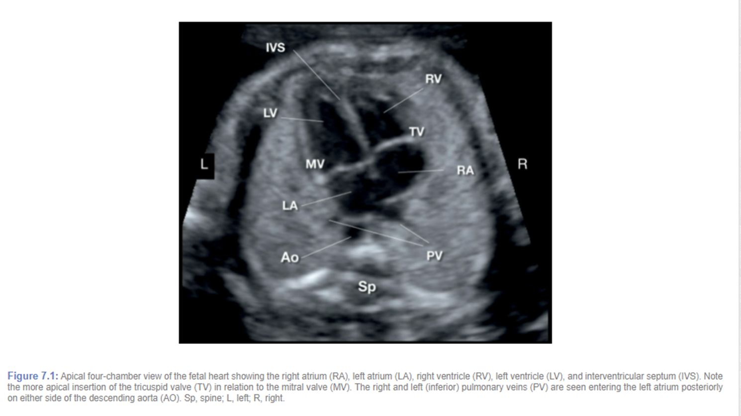
pulmonary veins drain into
left atrium
there are 4 but we usually see 2
tricuspid valve is slightly displaced _____ vs mitral valve
apically
best way to evaluate interventricular septum (IVS) and why
sub-costal bc angle is closer to 90 degrees
checklist for 4 chamber heart
situs
heart position
cardiac size
squeeze ok?
chamber ID and symmetry
septum appearance
AV valve offset
foramen ovale flap
area behind heart
rate and rhytm
what does pulmonary artery split into
right and left pulmonary artery and ductus arteriosus
right pulmonary artery goes _____ aorta
behind
MPA always ____ or ____ ____ than aorta
equal or slightly bigger
RVOT
LVOT
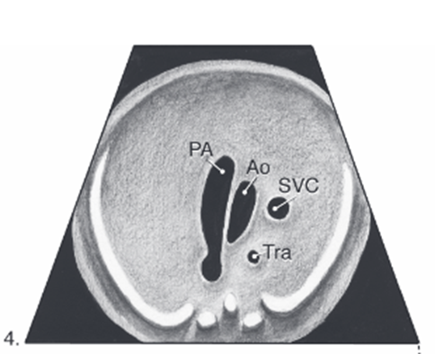
3 vessel view (3VV)
pulmonary artery and aorta junction
ductus arteriosus
Where pulmonary artery and aorta join (ductus arteriosus) in the 3VT view is sometimes also referred to as the
transverse arches
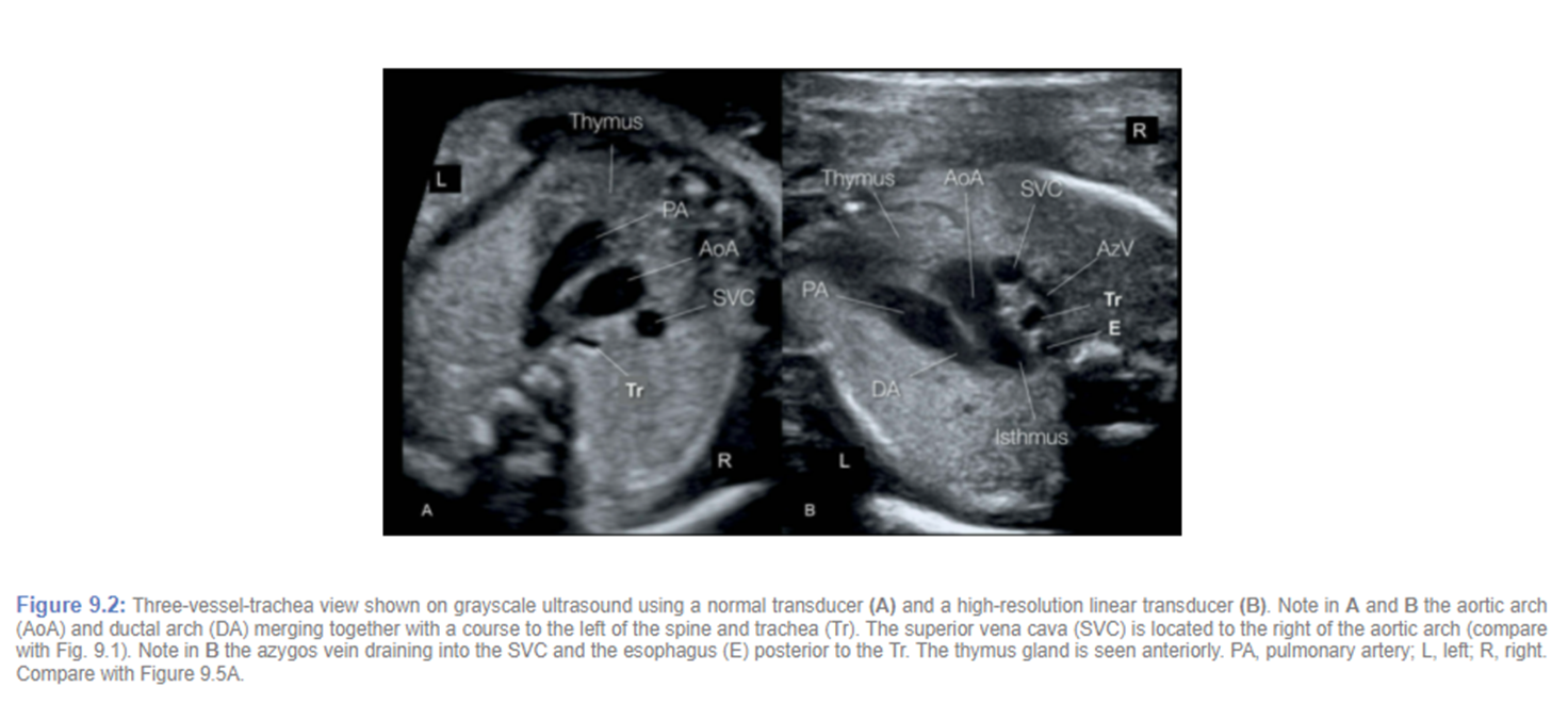
transverse arches
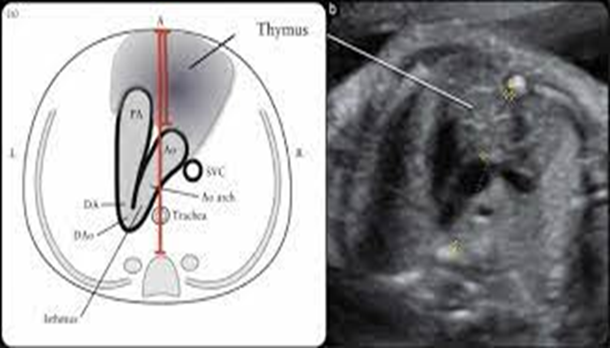
3vt (thymus)
pulmonary artery vs aorta color flow
same color and flow towards spine
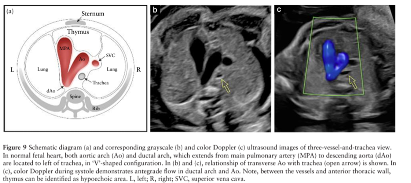
3VV with trachea
in-flow tracts
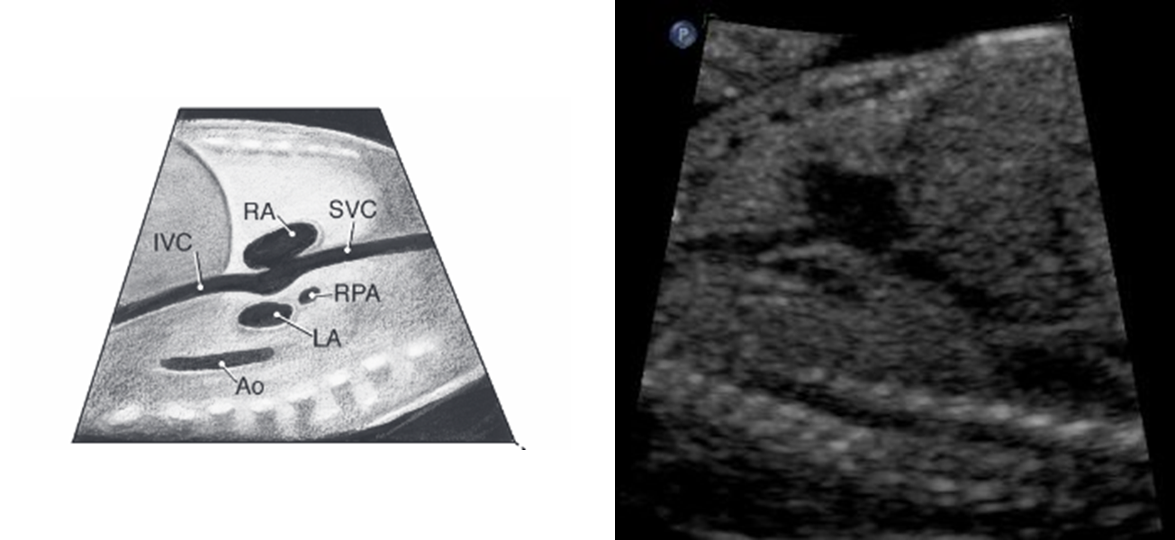
SVC/IVC
normal HR in 2nd and 3rd trimesters
120-160 bpm
what can be observed with XDCR pressure
bradycardia
persistent bradycardia below ___-___ required timely evaluation
100-110
persistent tachycardia above ___ requires timely evaluation
180
mild tachycardia ___-____ can occur with fetal movement
160-180
what is it called when organs in the chest and abdomen may be in the wrong position
heterotaxy
heart and stomach on ____ side
left
inability to see fetal stomach raises the concern of
tracheoesophageal fistula or esophageal atresia
tracheoesophageal fistula and esophageal atresia are typically accompanied by ???
polyhydramnios due to inability to swallow correctly
GB typically seen as early as ___-___ weeks
16-20
bowel is ____ echogenic than the liver and ____ echogenic than bone
more, less
what is the material occasionally seen in the stomach
vernix (skin cells or blood if placental bleeding)
the material collecting in the fetal intestines is termed
meconium
kidney echogenicity
1st trimester nearly isoechoic but then decrease until they become hypoechoic in comparison to the liver
what makes up 90% of amniotic fluid by 20 weeks
fetal urine
MPV of less than __ cm or an AFI less than _ is typically categorized as oligohydramnios (too little fluid)
2 cm
5