Neurons and Glia
1/18
There's no tags or description
Looks like no tags are added yet.
Name | Mastery | Learn | Test | Matching | Spaced | Call with Kai |
|---|
No study sessions yet.
19 Terms
What 2 major cell types make up the human brain?
Neurons — specialised for electrical/chemical signalling.
Glial cells — support, regulate and protect neurons; include astrocytes, microglia, oligodendrocytes/Schwann cells, and ependymal cells.
What were the competing historical theories explaining how neurons are connected?
Reticular theory (Golgi): neurons fuse into a continuous network.
Neuron doctrine (Cajal): neurons are individual cells that communicate via contact, not continuity.
The electron microscope eventually proved Cajal right by showing synaptic gaps.
Electron microscopy gave nanometre-scale resolution → could directly visualise synapses and membranes.
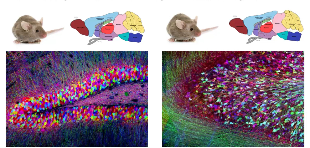
What is the “Brainbow” technique and what does it allow?
Combines fluorescence microscopy with genetic manipulation (e.g., Cre-Lox) to make individual neurons express different coloured fluorescent proteins.
Produces multicolour labelling → visualises neuron morphology, connectivity, and glial interactions in exceptional detail.
Brainbow uses stochastic recombination so each cell expresses unique fluorophore combinations.
Why do we study microanatomical features of the brain?
To understand connectivity, synaptic organisation, disease pathology (e.g., synapse loss, demyelination), and cell-type specific functions.
These microscopic features drive global brain function and dysfunction.
Neurons
Information processing cells within the nervous system, highly specialised for the conduction and transmission of electrical and chemical signals
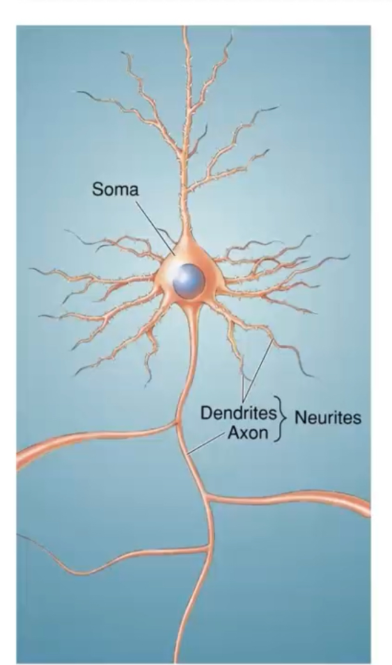
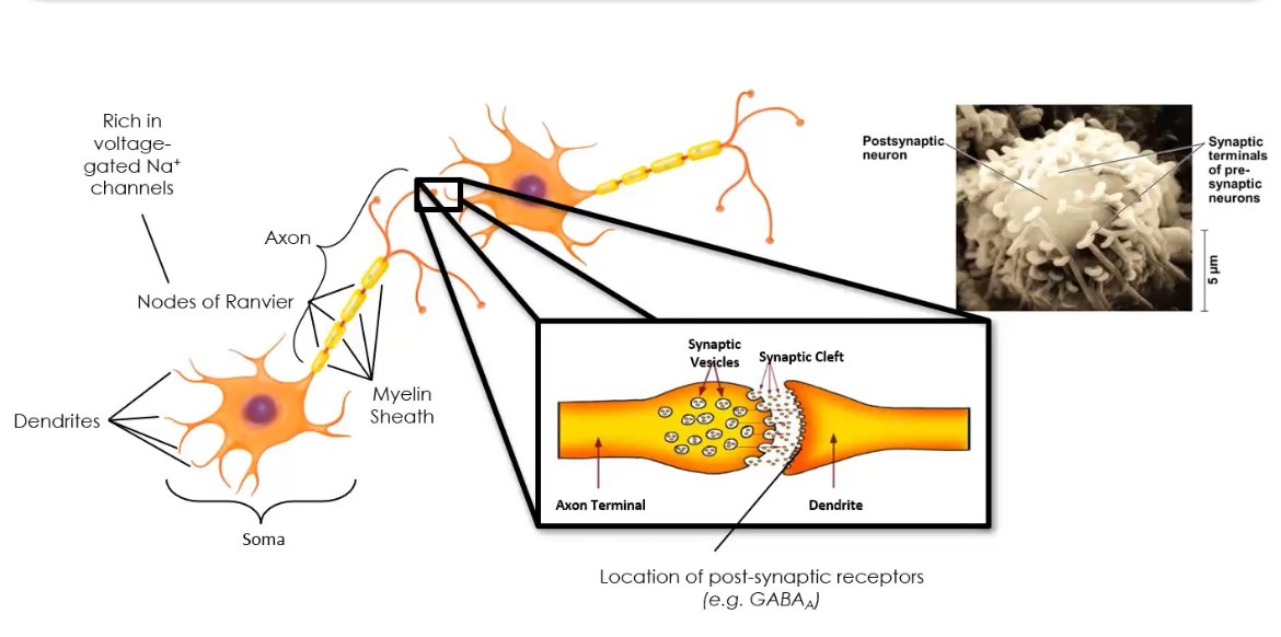
What are the three main structural components of a prototypical neuron?
1. Cell body (soma)
2. Dendrites — receive inputs
3. Axon — conducts action potentials
What is the primary function of neurons?
To process and transmit information using electrical impulses (action potentials) and chemical signalling (neurotransmission).
Neurons are polarised cells, with information flowing from dendrites → soma → axon.
What organelles are found in the neuronal soma?
Nucleus
Rough ER (Nissl substance)
Smooth ER
Golgi apparatus
Mitochondria
Neurons are extremely energy-demanding → dense mitochondrial content.

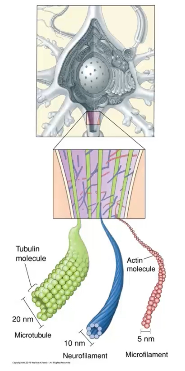
Neuronal Cytoskeleton
What three structures make up the neuronal cytoskeleton?
Microtubules — transport cargo (kinesin/dynein), maintain axon structure
Neurofilaments — provide tensile strength
Microfilaments (actin) — support growth cones, synaptic plasticity
Microtubules = dynamic; neurofilaments = stable, abundant in large axons.
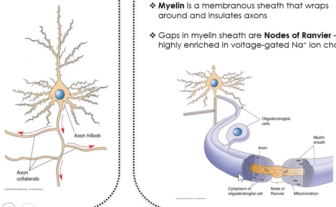
What are the key features of axons?
Neuronal signals are action potentials that are initiated at the axon initial segment, then propagate along the axon, and are speeded up by myelin, which forces the signal to jump between Nodes of Ranvier.
Step-by-step explanation of the pathway
Step 1: Signal arrives at dendrites and soma
• Dendrites receive chemical signals from other neurons.
• These cause graded potentials in the dendrites and soma.
• These signals move towards the axon hillock.
Step 2: Axon hillock / initial segment (decision point)
• The axon hillock tapers from the soma into the axon.
• This region has the highest density of voltage-gated Na⁺ channels.
• If the summed input reaches threshold → an action potential is generated.
• This is why arrows in the diagram point away from the soma.
Step 3: Action potential travels along the axon proper
• Once generated, the action potential self-propagates along the axon.
• It only moves in one direction (away from the soma) due to Na⁺ channel inactivation behind it.
Step 4: Axon collaterals (branching)
• The axon can branch into axon collaterals.
• Each collateral carries the same action potential to different targets.
• This allows one neuron to signal multiple postsynaptic cells simultaneously.
Step 5: Myelination by glial cells
• In the CNS, oligodendrocytes wrap around axons to form myelin.
• Myelin electrically insulates the axon and prevents ion leakage.
• This means depolarisation cannot occur under the myelin.
Step 6: Nodes of Ranvier and saltatory conduction
• Nodes of Ranvier are gaps between myelin segments.
• They are packed with voltage-gated Na⁺ channels.
• The action potential is regenerated only at the nodes, so it appears to “jump” node to node.
• This is saltatory conduction, which is faster and more energy-efficient.
Step 7: Axon terminals (end point)
• The action potential reaches the axon terminal.
• Voltage-gated Ca²⁺ channels open.
• Neurotransmitter is released into the synapse.
What do dendrites do?
Receive synaptic inputs via post-synaptic receptors.
Form a dendritic tree—critical for integrating thousands of synaptic signals.
Dendritic spines change in learning & memory.
Decreased spines linked with AD
What is neurotransmission?
The process by which neurons communicate using chemical messengers released at synapses.
Requires:
Presynaptic neurotransmitter synthesis & storage
Vesicle fusion
Receptor activation postsynaptically
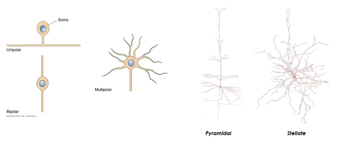
How can neurons be classified by structure?
Number of projections:
Unipolar
Bipolar
Multipolar
Structural class often aligns with function—e.g., bipolar neurons in retina.)
Based on dendrite/axon arrangement and morphology.
Neurons can be classified by connections or axon length.
By connections: sensory neurons carry information from the periphery to the CNS, interneurons process information within the CNS, and motor neurons transmit signals from the CNS to muscles or glands.
By axon length (Golgi classification): Golgi type I neurons have long axons for long-distance signalling, while Golgi type II neurons have short axons and function in local circuits.
How can neurons be classified by gene expression?
Based on genes coding for neurotransmitter synthesis enzymes, vesicle transporters, ion channels, etc.
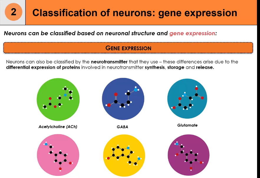
What are the major neurotransmitter phenotypes?
GABA (inhibitory)
Glutamate (excitatory)
Dopamine
Serotonin
Noradrenaline
Acetylcholine
Determined by enzyme expression—e.g., GAD for GABA, ChAT for ACh.)
Enzymes are needed to synthesise neurotransmitters
Neurotransmitters are not stored ready-made.
Neurons must manufacture them from precursors.
Specific enzymes catalyse these reactions.
Examples:
Choline acetyltransferase → makes acetylcholine
Glutamate decarboxylase → makes GABA
Without these enzymes → no neurotransmitter → no synaptic signalling.
Enzymes ensure cell-type specificity
Only neurons expressing the correct enzymes can make a given neurotransmitter.
This is how neurons become cholinergic, GABAergic, glutamatergic, etc.
Gene expression of enzymes defines neuronal identity.
Enzymes are needed to terminate the signal
Neurotransmitters must be removed quickly after release.
Some are broken down by enzymes in the synapse.
Example:
Acetylcholinesterase breaks down acetylcholine.
This prevents:
Continuous receptor activation
Overstimulation of the postsynaptic neuron
Enzymes allow tight regulation
Enzyme activity controls:
How much neurotransmitter is available
How long the signal lasts
This allows fine control of neural circuits.
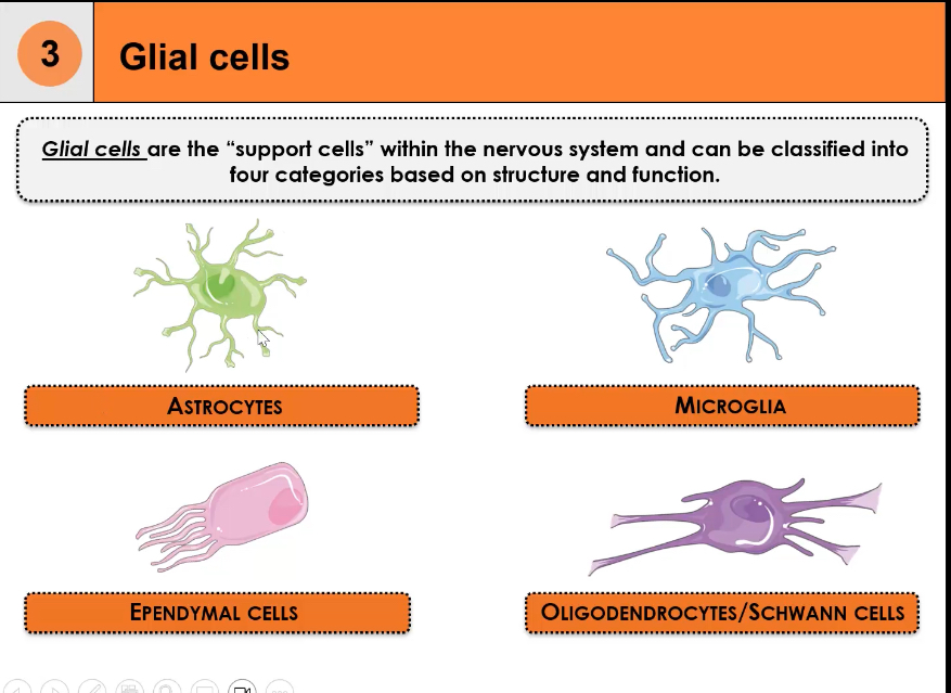
What are glial cells?
Glial cells are non-neuronal support cells in the nervous system.
They outnumber neurons.
They maintain the environment neurons need to function.
Four major classes:
astrocytes
microglia
oligodendrocytes/Schwann cells
ependymal cells.
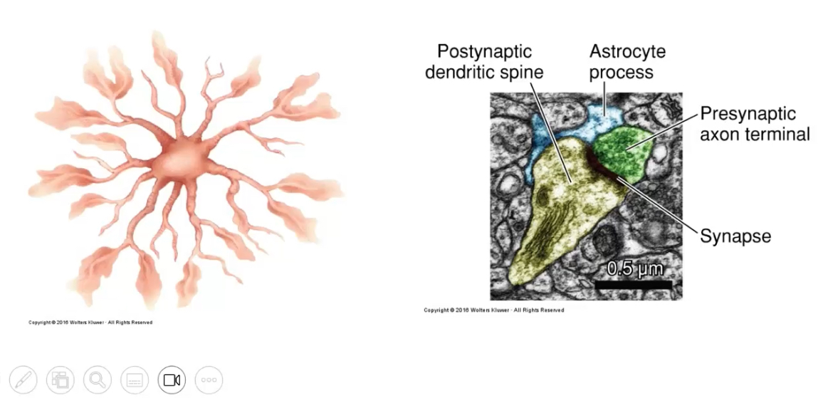
What are the main roles of astrocytes?
Most numerous glial cells.
Regulate extracellular ion balance.
Enclose synapses.
Remove neurotransmitters from synaptic clefts.
They form part of the blood-brain barrier and supply metabolic fuel to neurons via lactate shuttling.)
What do microglia do?
Main role: immune defence
Act as the resident immune cells of the CNS
Continuously survey the environment
Remove debris, dead cells, and pathogens by phagocytosis
Become activated during injury or disease
Think: immune system of the brain
They originate from yolk sac macrophages, not neural ectoderm.
What are the functions of ependymal cells?
Main role: cerebrospinal fluid (CSF)
Line the ventricles of the brain and central canal of the spinal cord
Produce and help circulate CSF via cilia
CSF cushions the brain and maintains a stable chemical environment
Think: CSF production and movement
What do oligodendrocytes and Schwann cells do?
Main role: myelination
Oligodendrocytes: myelinate axons in the CNS
One oligodendrocyte can myelinate multiple axons
Schwann cells: myelinate axons in the PNS
One Schwann cell myelinates one axon segment
Myelin increases conduction speed via saltatory conduction
Think: insulation
Schwann cells also support regeneration; oligodendrocytes do not → CNS regeneration is limited.)
Origin: Both from neural tube derivatives except microglia.