Lecture 20 - Vision I
1/32
Earn XP
Description and Tags
March 10th
Name | Mastery | Learn | Test | Matching | Spaced | Call with Kai |
|---|
No analytics yet
Send a link to your students to track their progress
33 Terms
What type of energy is transduced during hearing?
Mechanical
Which speech/language representations are in the correct order from smallest to largest?
Phonemes, Morphemes, Words, Sentences
Which type of aphasia is traditionally associated with damage to the inferior frontal gyrus?
Broca’s (nonfluent) aphasia
Visible light is a narrow band of electromagnetic radiation…
Wavelength = length between two peaks in a repeated stimulus such as a wave, light, or sound
Photon = quantum of electromagnetic energy in the range of wavelengths we call light
Light is an amalgamation of the two
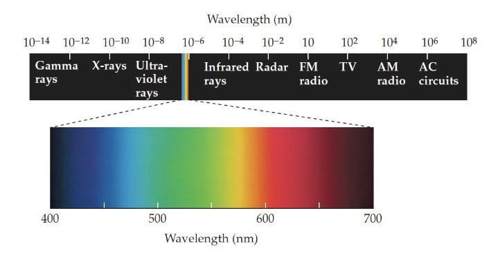
Why so narrow?
Higher energy past ultraviolet rays, high energy radiation penetrates through your skin. Leaves trails, we don’t see them because they’re too energetic for biological creatures to not be destroyed by them
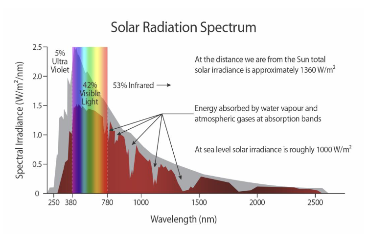
Visible light we see covers the most light that reaches the Earths surface from the Sun
Wavelengths longer than visible light would require much larger cellular structures to capture with the same acuity
We don’t get many gamma rays because the atmosphere gets rid of it, protected
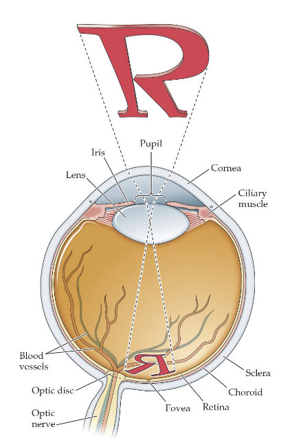
The structure of the Eye
Cornea = transparent outer layer of the eye, whose curvature is fixed
Pupil = the aperture, formed by the iris, that allows light to enter the eye
Iris = circular structure that provides an opening to form the pupil
Lens = structure that helps the focus an image on the retina
Ciliary muscle = controls the shape of the lens
Accommodation = process of focusing by the ciliary muscles and lens
Retina = the receptive surface inside the eye that contains photoreceptors and other neurons
Cornea
Transparent outer layer of the eye, whose curvature is fixed
First structure to focus the eye
Pupil
The aperture, formed by the iris, that allows light to enter the eye
Hole created by the iris, not empty space behind it, over the lens
Iris
Circular structure that provides an opening to form the pupil
Colored part of the eye, limits amount of light, makes pupil larger/smaller
Lens
Structure that helps the focus an image on the retina
Key to focus light on to back of the eye, somewhat malleable, near sighted or far sighted lens isn’t as flexible (focus of different distances)
Ciliary muscles
Controls the shape of the lens
Accommodation:
Process of focusing by the ciliary muscles and lens
Image projected to back of the eye is reversed, light gets bent by the lens. Upside down and mirror reversed
Retina
The respective surface inside the eye that contains photoreceptors and other neurons
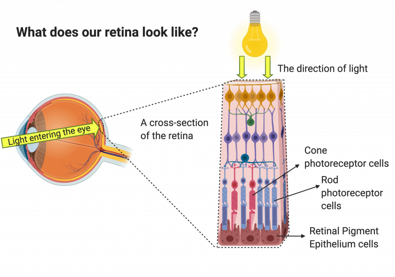
Anatomy of the Retina
Layered structure, very awesome, very complex, so many more photoreceptors than bits of information leaving the eye. SO much neural processing during the eye before it gets to the brain.
Pigmented Epithelium = cell layer that reduces scattered light in the eye
Rod and Cone cell layer = photoreceptors, neural cells that respond to light

Pigmented Epithelium
Cell layer that reduces scattered light in the eye
Back most of the eye, absorbs so that light doesn’t create crazy images
Bipolar Cells
Interneurons that receive information from the rods and cones
Form the middle layer
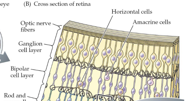
Horizontal Cells
Contact both the photoreceptors and bipolar cells creating lateral connections
Also form the middle layer, involves many different photoreceptors

Ganglion Cells
Class of cells in the retina whose axons form the optic nerve
Proper neurons, have axons that come out of eye and send info back into the brain
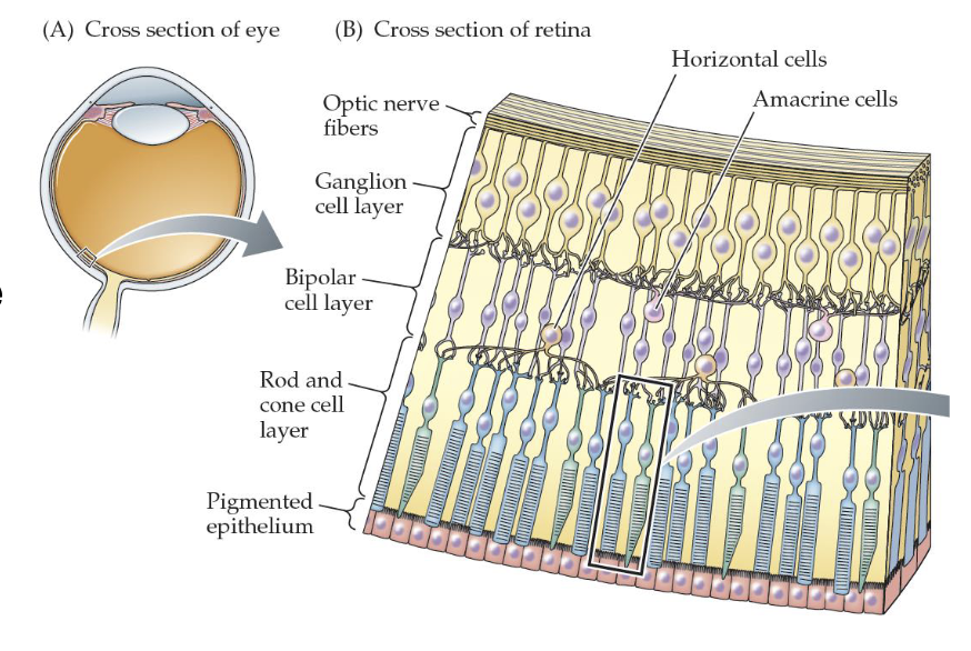
Amacrine Cells
Contact both bipolar and ganglion cells and are especially significant in (lateral) inhibitory interactions
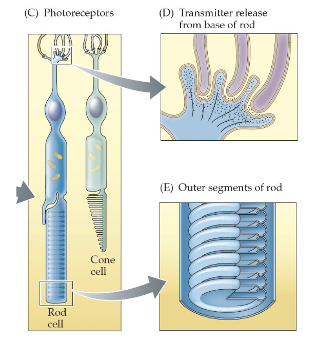
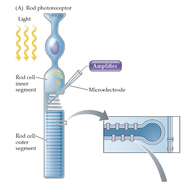
Photoreceptor Structure
We have two types of photoreceptor cells - rods and cones
Photoreceptors don’t send action potentials; they have graded potentials that regulate the amount of neurotransmitter (glutamate) they release
The discs in the outer segments contain the photopigment proteins
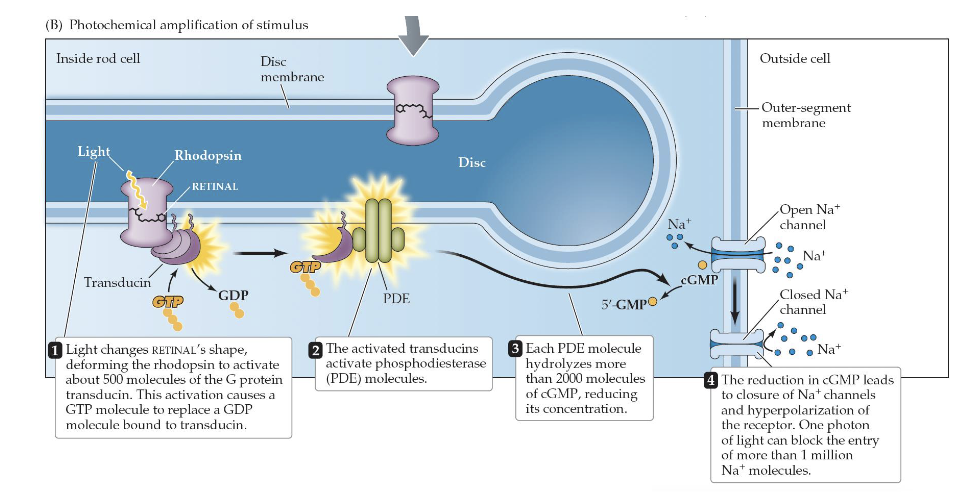
How is light transduced in rods?
Rods and cones contain photopigment receptor molecules
In rods, the photopigment is called rhodopsin (hit when light it changes shape and creates cascade of chemical reactions)
Cones have similar photopigments
Details about trandsuction in vision
Even small amounts of light can cause activation of a large amount of molecules - this allows weak signals to be amplified (500 molecules of different protein get release)
The main effect of all this is to close sodium channels and hyperpolarize the photoreceptor cell (they work in a counterintuitive way, when light enters cell, they close sodium channels)
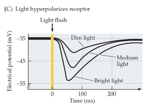
Light hyperpolarizes the photoreceptor cell
When light enters the cell, it causes the potential to becomes more negative than it already was (hyperpolarization)
This causes it to release less neurotransmitter than it was already releasing
The brighter the light, the greater the hyperpolarization, the less neurotransmitter released
Less glutamate gets released, greater sensitivity to light and more adaptability
Visual System Characteristics
The cascade of processes required to stimulate the visual receptors helps account for three characteristics of the visual system:
Its sensitivity, because weak stimuli are amplified to produce physiological effects (one photon blocks millions of Na+ ions)
The integration of the stimulus over time, which makes vision relatively slow (compared, for example, with audition) but increases its sensitivity
The adaptation of the visual system to a wide range of light intensities
Two Functional Systems of the Retina
Photopic system = high levels of light, sensitivity to color, involves cones
Scotopic system = low levels of light, involves rods
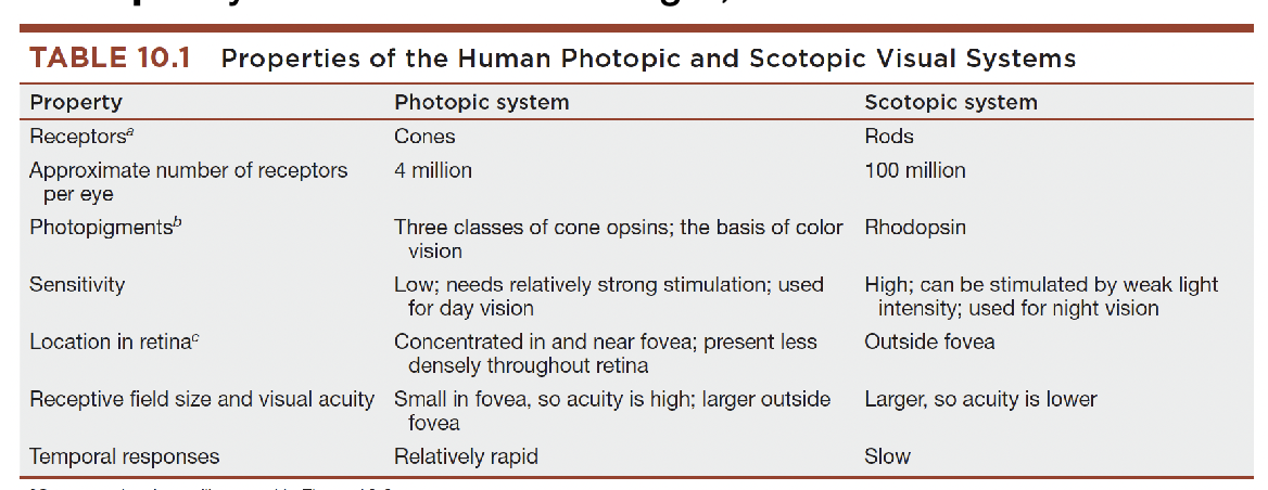
Photopic System
Receptors - Cones
Approximate # of Receptors per eye - 4 million
Photopigments - Three classes of cone opsins; the basis of color vision
Sensitivity - Low; needs relatively strong stimulation; used for day vision
Location in retina - Concentrated in and near fovea; present less densely throughout retina
Receptive field size and visual acuity - Small in fovea, so acuity is high; larger outside fovea
Temporal responses - Relatively rapid
Scotopic system
Receptors - Rods
Approximate # of Receptors per eye - 100 million
Photopigments - Rhodopsin
Sensitivity - High; can be stimulated by weak light intensity; used for night vision
Location in retina - Outside fovea
Receptive field size and visual acuity - Larger, so acuity lower
Temporal responses - Slow
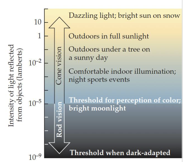
Photoreceptor adaptation
Tendency of rods and cones to adjust their light sensitivity to match the ambient levels of illumination
The visual system is concerned with differences, or changes, in brightness, not the overall level of illumination
This is part of why we can see over such wide ranges of brightness
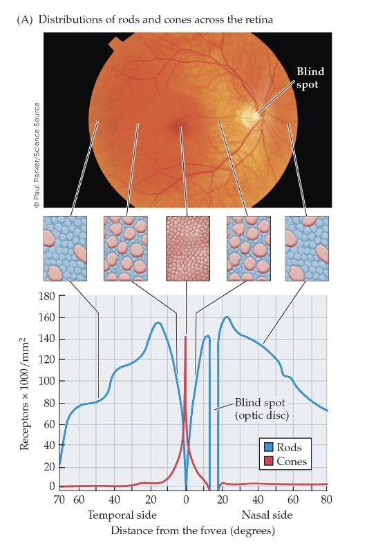
Distribution of rods and cones
Rods and cones are distributed unequally across the retina
Cones (in red) are concentrated at the fovea, the central portion of the retina
Optic Disc = region on the retina without any receptor cells because that’s where ganglion cell axons and blood vessels exit the eye
Blind Spot = portion of the visual field corresponding to the optic disc
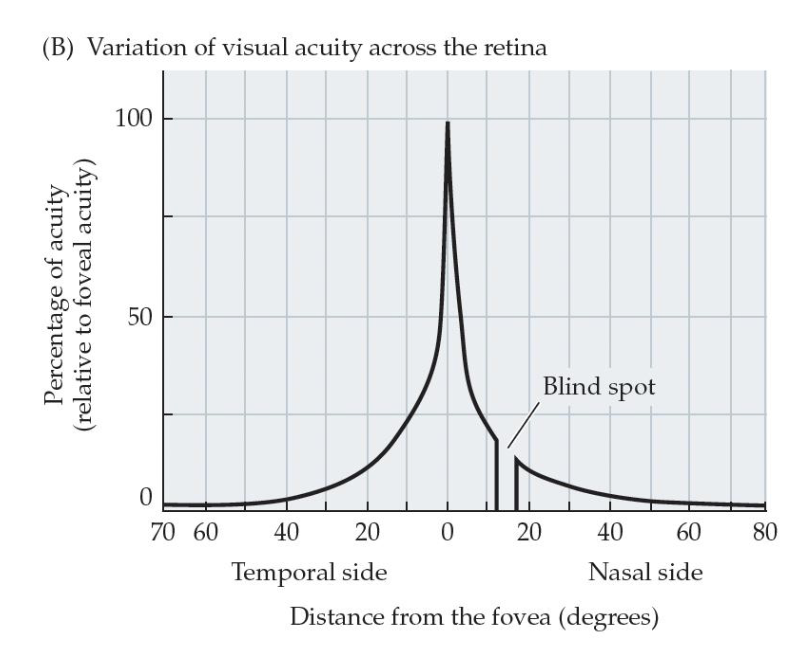
Visual acuity reflects the distribution of cones
Visual Field = the whole area you can see without moving your eyes
Visual Acuity = the sharpness of vision
Saccades = fast movements of the eyes that present various parts of the visual scene to the fovea
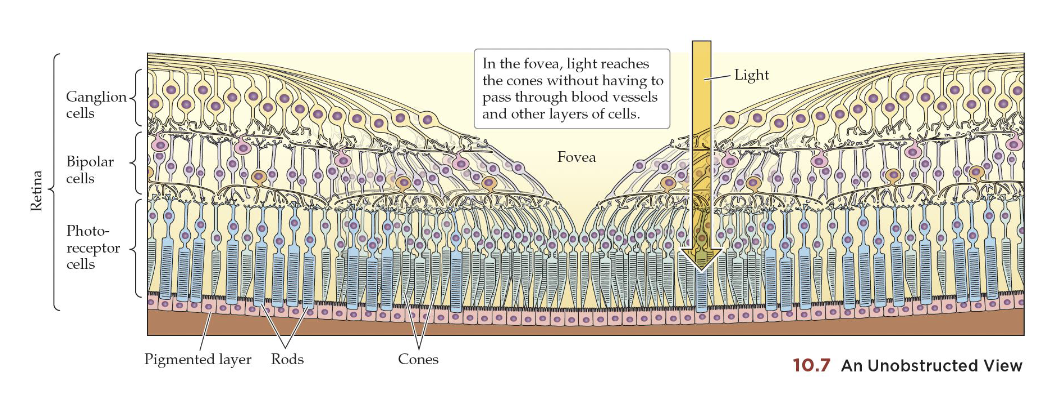
Structure of the Fovea
Spreading apart of bipolar, horizontal, amacrine, ganglion, to get to the photoreceptors cells
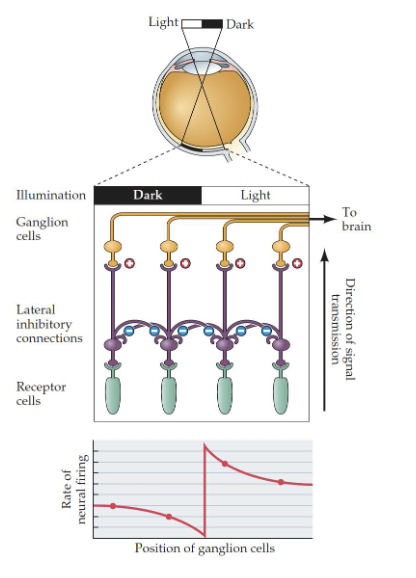
Perception of Brightness
Sharpness and differentiates things
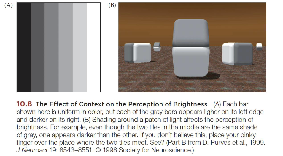
Lateral Inhibition
When neurons in a region are interconnected, either through their own axons or by means of interneurons (*cough* horizontal cells *cough*) and each neuron tends to inhibit its neighbors
This allows contrasting edges to be emphasized at the level of the retina
Right at the retina, to enhance
Emphasize on differences, no emphasize on sameness, need to see tiny differences