Biochemistry Class 8 - Endocrine, Cardiovascular, Immunology
1/69
There's no tags or description
Looks like no tags are added yet.
Name | Mastery | Learn | Test | Matching | Spaced |
|---|
No study sessions yet.
70 Terms
What are characteristics of endocrine glands?
Make hormones
Release into bloodstream
No duct needed
What are characteristics of exocrine glands?
Products: enzymes, bile, stomach acid, bicarbonate, saliva, tears, sebum, earwax, mucus, breastmilk, semen, etc.
Release outside the body or inside a body cavity
Requires ducts to get the product where it needs to be (except mucus cells)
What are they made of, receptors located, mechanism of action, speed of effects, and longevity of effects? Peptide hormones
Made from: amino acids
Receptors: cell surface
MoA: 2nd messenger systems
Speed: quick
Longevity: temporary
What are they made of, receptors located, mechanism of action, speed of effects, and longevity of effects? Steroid hormones
Made: derived from cholesterol
Receptors: intracellularly in cytoplasm or nucleus
MoA: binds to DNA, modifies transcription
Speed: slow
Longevity: more permanent
What is thyroid hormone?
Made of amino acids, but is hydrophobic enough to cross cell membranes
Looks like peptide hormone, acts like steroid hormone
How is hormone release controlled? 3
Neural, hormonal, humoral
How are hormones controlled neurally?
Action potential triggers release of hormone
Ex: sympathetic nervous system triggers release of epinephrine
How are hormones controlled hormonally?
Hormone triggers release of another
Tropic hormones do this
Ex: adrenocorticotropic hormone triggers release of hormones from adrenal cortex
How are hormones controlled humorally?
Something in blood that is not a hormone triggers hormone release
Ex: glucose regulates insulin and glucagon, calcium regulates parathyroid hormone and calcitonin
What is the pituitary gland?
Controlled by hypothalamus and sits below it
Anterior (adenohypophysis) and posterior (neurohypophysis)
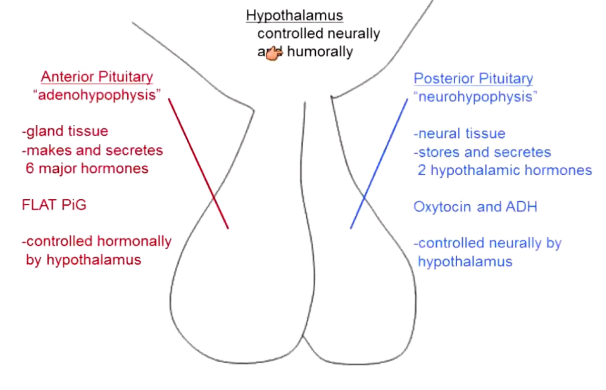
What does the anterior pituitary do?
Gland tissue that makes 6 hormones
FLAT PiG (FLAT are all tropic)
FSH, LH, ACTH, TSH, prolactin, growth hormone
Controlled hormonally by hypothalamus: all by releasing hormones (aka ___RH), except prolactin (by prolactin inhibitory hormone aka prolactin IH)
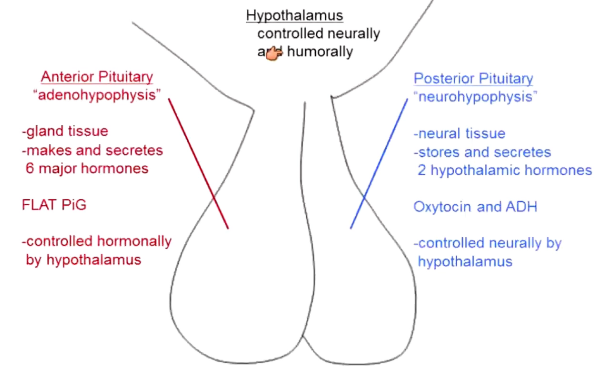
What does the posterior pituitary do?
Neural tissue
Stores and secretes 2 hypothalamic hormones
Oxytocin and ADH (antidiuretic hormone aka vasopresin)
Controlled neurally by hypothalamus
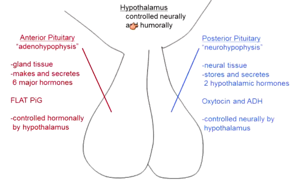
How is the hypothalamus controlled?
Neurally and humorally
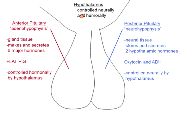
What is the anatomy of the posterior pituitary?
Neurons with cell body in the hypothalamus project down into it
These make and release the hormones and transport them down the axon
Released into capillary bed → bloodstream
What is the anatomy of the anterior pituitary?
Hormone making cells line capillaries → veins connect these capillaries to the capillaries in the hypothalamus (that are also lined by hormone making cells)
Called a portal system (connected by hypothalamic/hypophyseal portal veins)
How do portal systems work?
Hormone released in hypothalamus (ex: growth hormone releasing hormone) → travels to anterior pituitary → stimulates cells in anterior pituitary (ex: release growth hormone) → hormone (ex: growth) goes to body
Double negative feedback
Allow extremely targeted delivery
What is the liver portal system?
Liver capillaries connetc to intestinal capillaries via portal veins
What is the circulatory system? Basic parts of the
Loop
Artery: blood away from heart
Capillaries: site of nutrient exchange
Vein: blood towards heart
Heart: pump that controls all this
How do arteries function?
High pressure system
Blood moves via forward momentum (from high pressure at heart to low pressure in capillaries)
Muscular walls (regulate flow and diameter)
Elastic (can snap back to shape after stretching)
How do capillaries function?
Very large surface area
Relatively low pressure
Do nutrient/waste exchange
Fluid out due to pressure near the arteries → blood in due to osmosis near the veins → net outward flow of blood → must recover this via lymphatic system
How do veins function?
Low pressure
Blood moves when veins are squished (normal body movement)
Valves ensure unidirectional flow
Not muscular and not elastic
What does the lymphatic system do?
Structurally like vein (valves, non muscular, non elastic)
Pick up fluid from capillaries and returns it to veins (filters through lymph nodes)
What are lymph nodes?
Concentrated areas of white blood cells
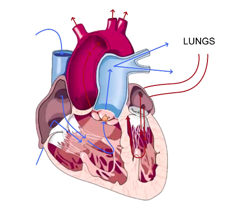
How does blood flow in the heart?
Blood from body superior vena cava → right atrium → contracts → right ventricle → contracts → pulmonary artery to lungs → pulmonary vein to left atrium → contract → left ventricle → contract → to body via aorta
Left ventricle is thicker walled
What drives blood flow?
Pressure gradient
Once pressure is greater than arterial pressure → blood flow to arteries
Valves prevent backflow between chambers/between arteries and ventricles
Blood passively drains into atrial until ventricle pressure is lower → goes to ventricles
What are atrioventricular valves?
Tricuspid AV valve: between right atrium and ventricle
Bicuspid (mitral) AV valve: between left atrium and ventricle
What are semilunar valves?
Pulmonary semilunar: between right ventricle and pulmonary artery
Aortic semilunar: between left ventricle and aorta
What do systole and diastole mean?
Systole: heart contracted
Diastole: heart relaxed
Usually talking about ventricles → MCAT will specify atrial if it wants that
What do the heart sounds mean?
Lub: closing of AV valves, begins systole
Dub: closing of semilunar valves, begins diastole
What is blood pressure?
120/80, 115/70
Systolic/diastolic
Arterial pressure when heart is contracted/when relaxed
What is blood pressure directly proportional to?
Cardiac output and peripheral resistance
What is cardiac output?
Volume of blood pumped per minute
Stroke volume (volume pumped per beat ) x heart rate (beats per minute)
What is the FS Law of Heart?
More blood in heart → muscles stretch → contract back with greater force → more blood out
Within parameters
What happens in congestive heart failure?
Heart is so stretched out, the muscle fibers no longer overlap → cannot pump effectively
How do you change stroke volume?
Change blood volume (ex: drunk water, ingest salt, donate blood, etc.; renal system regulates this primarily)
Change activity level (moving increases blood to heart)
Change posture (gravity makes less blood reach heart for a second after you stand)
What is peripheral resistance?
How hard it is to move blood through vessels
Vessels constrict → decrease in diameter → decrease in flow → increased resistance → increased blood pressure
Vessels dilate → increase diameter → increase flow → decrease resistance → decreased blood pressure
How do cardiac muscles reach action potential?
Connected via gap junctions
Rest at -80 mV → reaches threshold once its neighbor does → voltage gated Na+ channels open → inactivate at +40 mV → voltage gated K+ channels open → voltage gated Ca2+ channels open → action potential plateaus → Ca2+ channels close → hyperpolarization → K+ channels close → back to rest
200-300 milliseconds from threshold to rest (100x longer than nerve action potentials)
Prevents tetanic contractions (sustained contractions) because it is impossible to increase the frequency of action potentials enough for this
What are skeletal muscle action potentials like?
20-30 milliseconds
Each action potential triggers a twitch → add a bunch of them → tetanic contraction (sustained)
What are cardiac autorhythmic cells?
Initial cells that start cardiac contractionsW
hat is the action potential like for cardiac autorhythmic cells?
Rest at -40 mV → constantly drifting upward due to sodium leak channels → reach threshold → slow voltage gated Ca2+ channels → cause slow influx of calcium up to 20 mV → depolarizes → rises up again thanks to sodium leak channels
When it hits thresholds, it fires action potentials
These are connected to other cardiac cells → trigger action potentials in them
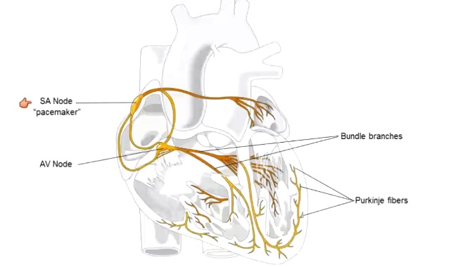
What is the cardiac conduction system?
Cardiac autorhythmic cells
SA (sinoatrial) node: leakiest to sodium, is first to reach action potential → controls pace of contractions
AV (atrioventricular) node → branches into left and right bundle branches → form Purkinje fibers
What is the pacemaker of the heart?
SA node
Has a resting rate of 100 beats per minute → brought to ~70 by parasympathetic nervous system
~70 is parasympathetic tone (aka vagal tone)
Epinephrine will increase the beats per minute past 100
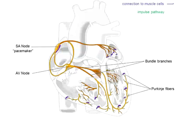
Where are heart muscle cells connected to autorhythmic cells?
SA node connects to right atrium → left connected at end of fibers that project from it
Purkinje fibers connect to ventricles
To the muscle cells, this signals contraction
What is the impulse pathway through the heart?
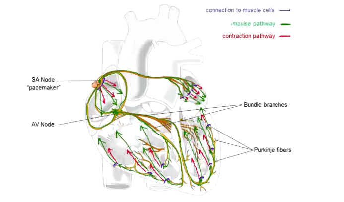
SA node in right atrium → left atrium → AV node → right and left bundle branches → Purkinje fibers
Atria contract downward and ventricles contract upward
Why does the heart conduction system run like it does?
Atria and ventricular muscle cells not electrically connected (valves separate them): atria cause ventricles to contract
AV node delays impulse, allowing atria to contract before ventricles do
Ventricles contract from bottom to top, pushing blood out through the aorta (attached at top of ventricles) → more efficient ejection
What are the three components of blood?
54% plasma: anything hydrophilic dissolved in blood
1% (variable depending on gender, if you are sick, age, weight) leukocytes: white blood cells and platelets
45% (variable, see above) hematocrit: red blood cells
How do oxygen and carbon dioxide travel through the blood?
Oxygen: 3% dissolved in plasma, 97% bound to hemoglobin
Carbon dioxide: 7% dissolved in plasma, 20% bound to hemoglobin, 73% as bicarbonate
How does oxygen bind to hemoglobin?
Cooperative binding: once one oxygen binds, it is easier for the others to
Released when the [oxygen] is released
![<p>Cooperative binding: once one oxygen binds, it is easier for the others to</p><p>Released when the [oxygen] is released</p>](https://knowt-user-attachments.s3.amazonaws.com/e38bcfda-71f7-4947-9660-8fa5c7810612.png)
How is CO2 carried as bicarbonate?
CO2 + H2O ←→ H2CO3 (carbonic acid) ←→ H+ + HCO3- (bicarbonate)
Tissues always producing CO2 → shifts equilibrium towards bicarbonate
Lungs are getting rid of CO2 → shifts equilibrium towards CO2
Affects blood pH
What are three categories of non-specific immune defense?
Barriers
Chemicals
Cells
What type of immune barriers do you have?
Skin, mucus, hair, earwax, etc.
What are chemicals for immune defense?
Mucus, lysozyme (saliva, tears), stomach acid, enzymes, complement system, etc.
What are general immune defense cells?
Macrophages, neutrophils, eosinophils, dendritic cells, basophils, etc.
What is an antigen?
Foreign protein that triggers an immune response
What are antibodies?
Highly-specific marker that binds to an antigen
What are pathogens?
Disease-causing organisms
What are the two types of specific immunity?
B cell “humoral” immunity
T cell “cell mediated” immunity
What is characteristic of B cell immunity?
B cells produce and secrete antibodies
Each B cell makes one type of antibody
Diversity generated via DNA rearrangement of genes that produce antibodies
Occurs in the bloodstream
What are antigen receptors?
Displayed on the surface of B cells
Bind to the antigen that B cell creates antibodies for → pulls it in → creates a bunch of antibodies to that antigen → cell clones itself so there is a greater response
What are types of T-cells? What does each do?
Killer T: bind to and kill own cells that have become abnormal
Helper T: secrete cytokines that allow B cells and killer T cells to proliferate
How do killer T cells work?
Has antigen and “self” receptors → bind to antigens displayed by MHC I
Self receptor allows it to identify cells that originate from the host
Both self and antigen receptors must be bound to create a response
What is MHC I?
Found on all cells
Allows cells to display cell contents on the cell surface → allows killer T cells to identify abnormal ones
How do helper T cells work?
Look for antigens displayed on MHC II
What is MHC II?
Found on macrophages and B cells
Allows cells to display stuff they have “eaten” on the cell surface
What is the primary immune response?
1st exposure to antigen → 7-10 days → antibodies generated and T cells activated → memory cells produced
Takes a super long time, relative to the viral lifecycle
What are memory cells?
Remember an antigen of a previous infection → allows faster response next time
Allows for long-term immunity
What is the secondary immune response?
2nd exposure → make antibodies, activate T cells, make more memory cells
Existing memory cells allow this to happen in <1 day
How do vaccines work?
Inject antigens of whatever you want immunity against → body produces antibodies, activates T cells, makes memory cells in 7-10 days
No active infection
Memory cells can now respond quickly if you are exposed to the actual infection
Boosters allow for stronger, faster responses
What is autoimmunity?
Body produces B and T cells that recognize a large number of antigens, including self-antigens → self reactive lymphocytes will attack our own antigens and cause autoimmune diseases
We must have a way to identify self-reactive lymphocytes and inactivate/destroy them
How are self-reactive lymphocytes inactivated/destroyed?
By B cells in the bone marrow and T cells in the thymus
B cells recognize soluble proteins → become anergic (inactive)
T cells recognize cell-surface proteins → apoptosis
Making them anergic/apoptosing them occurs only in the bone marrow and thymus
If the B and T cells do not recognize self things → released into circulation