NEUR1020 Module 2
0.0(0)
0.0(0)
Card Sorting
1/30
Earn XP
Study Analytics
Name | Mastery | Learn | Test | Matching | Spaced | Call with Kai |
|---|
No study sessions yet.
31 Terms
1
New cards
Some facts about the brain: weight percentage? number of neurons/synapses/pathways? blood and energy use?
* 2% total body weight
* contains 100 billion neurons and 1,000,000 billion synapses: giving rise to 10^1 million possible interconnected circuits. This large number gives the brain it’s capacity for information processing
* it receives 20% of the blood pumped from the heart and consumes 20% of the body’s energy
* contains 100 billion neurons and 1,000,000 billion synapses: giving rise to 10^1 million possible interconnected circuits. This large number gives the brain it’s capacity for information processing
* it receives 20% of the blood pumped from the heart and consumes 20% of the body’s energy
2
New cards
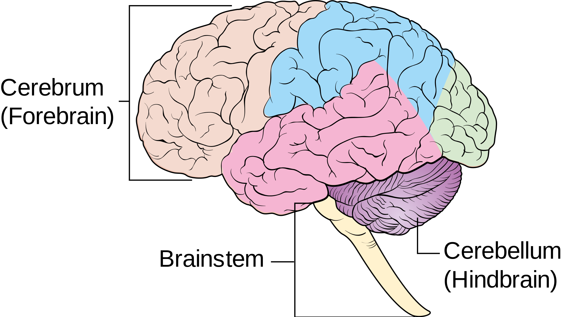
What are the 3 major regions of the brain?
1. Brainstem (interconnects between the spinal chord and cerebral hemispheres)
2. Cerebellum (aka hind brain: posterior, back of the brainstem)
3. cerebrum (including the cerebral hemispheres + forebrain)
3
New cards
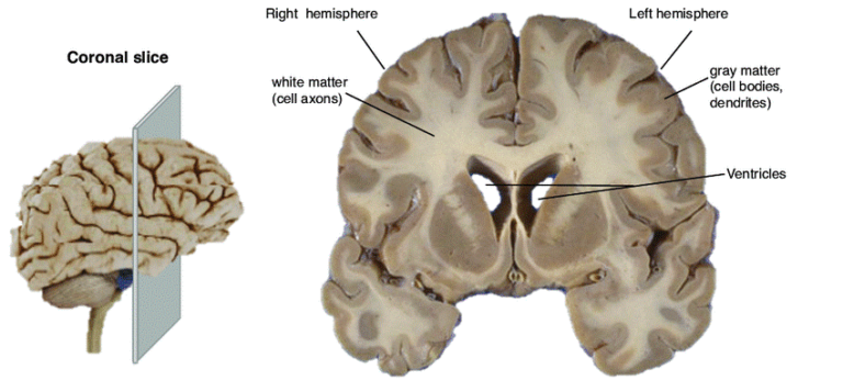
Cerebrum: made up of? cerebral cortex? white matter?
=cerebral hemispheres + forebrain
* 2 hemispheres= left and right brain, divided down the middle by the longitudinal fissure (aka the inter-hemispheric fissure)
* most the inside surface of the brain have no interconnections between the hemispheres, with connections existing deeper in the brain through the corpus collosum
* the **cerebral cortex** (aka grey matter)= outermost surface layer of the cerebrum.
* it contains the majority of the cell bodies of the brain’s neurons
* approx 2-4mm thick
* highly folded to maximise surface area (the amount of cortex that can fit inside the cell). Therefore more folding=more intelligence because it maximises the number of neuron and synapse connections that can exist in one space
* beneath the cerebral cortex is **white matter**= the “wiring” of the brain
* the axons of the neurons that interconnect with other neurons and go down the spinal chord all travel through white matter.
4
New cards
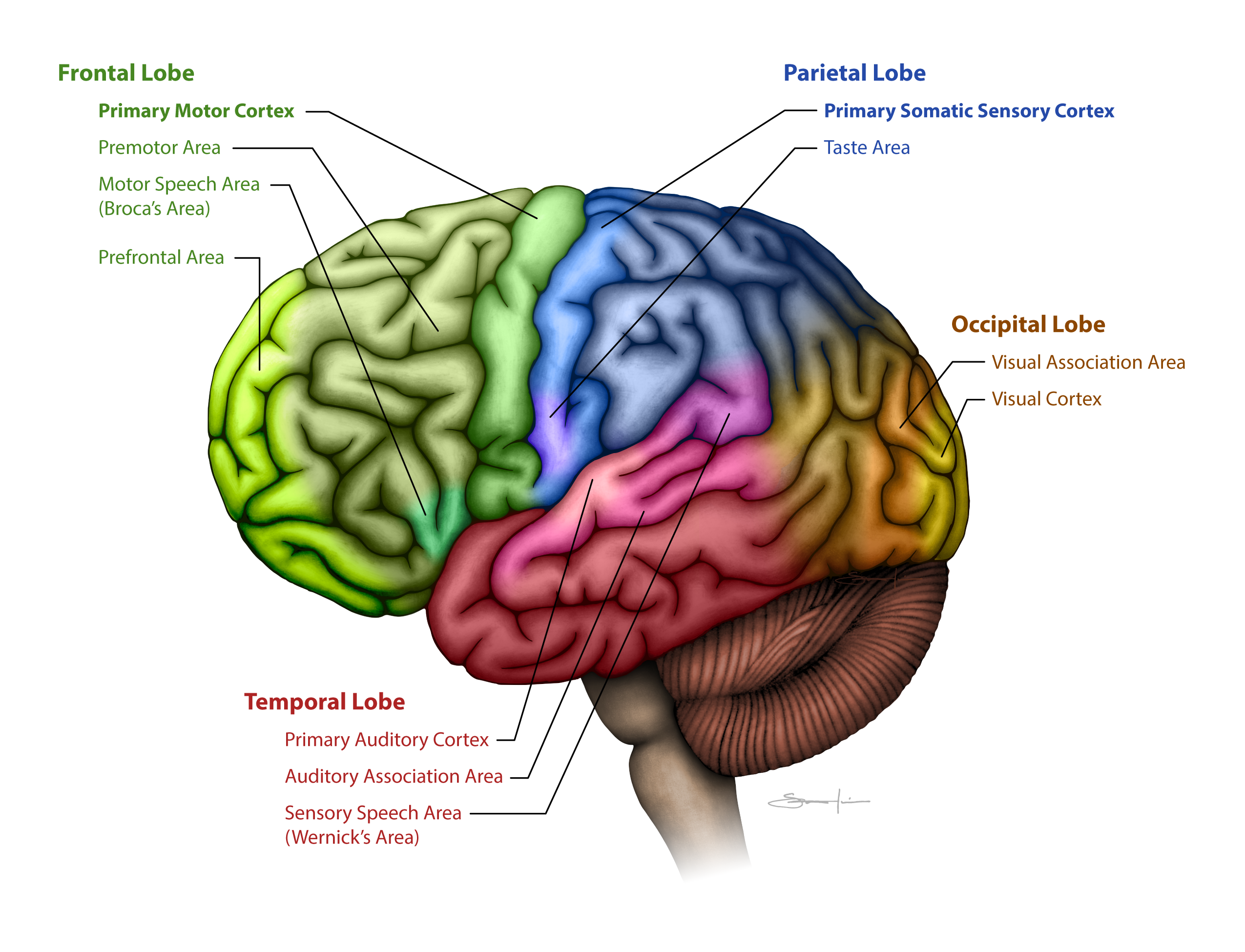
What are primary areas?
=the first site of input into the brain from our sensors OR the last stage of output before it goes down the spinal chord
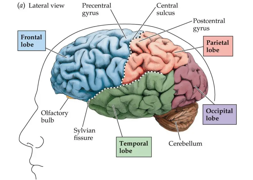
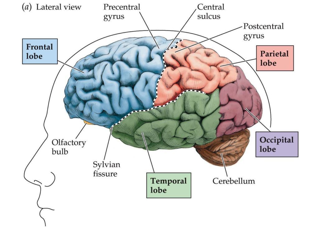
* Primary visual cortex= where input from our eyes first go. If there is any damage to this cortex it creates a blind spot in our vision field. Occipital lobe.
* Primary auditory cortex= receives first input from cochlea in the ears. Temporal lobe.
* Primary somatic sensory cortex= touch sensors on out skin relay signals up the spinal chord and to this cortex to generate sensation. Damage to this cortex=part of the body becomes numb. Runs down the centre of the brain/central sulcus from the parietal lobe.
* Primary motor cortex= the last place where neurons fire signals down the spinal chord to cause muscle contraction for voluntary movement. Damage to this region= paralysis. Located next to/anterior the central sulcus. Frontal lobe.
5
New cards
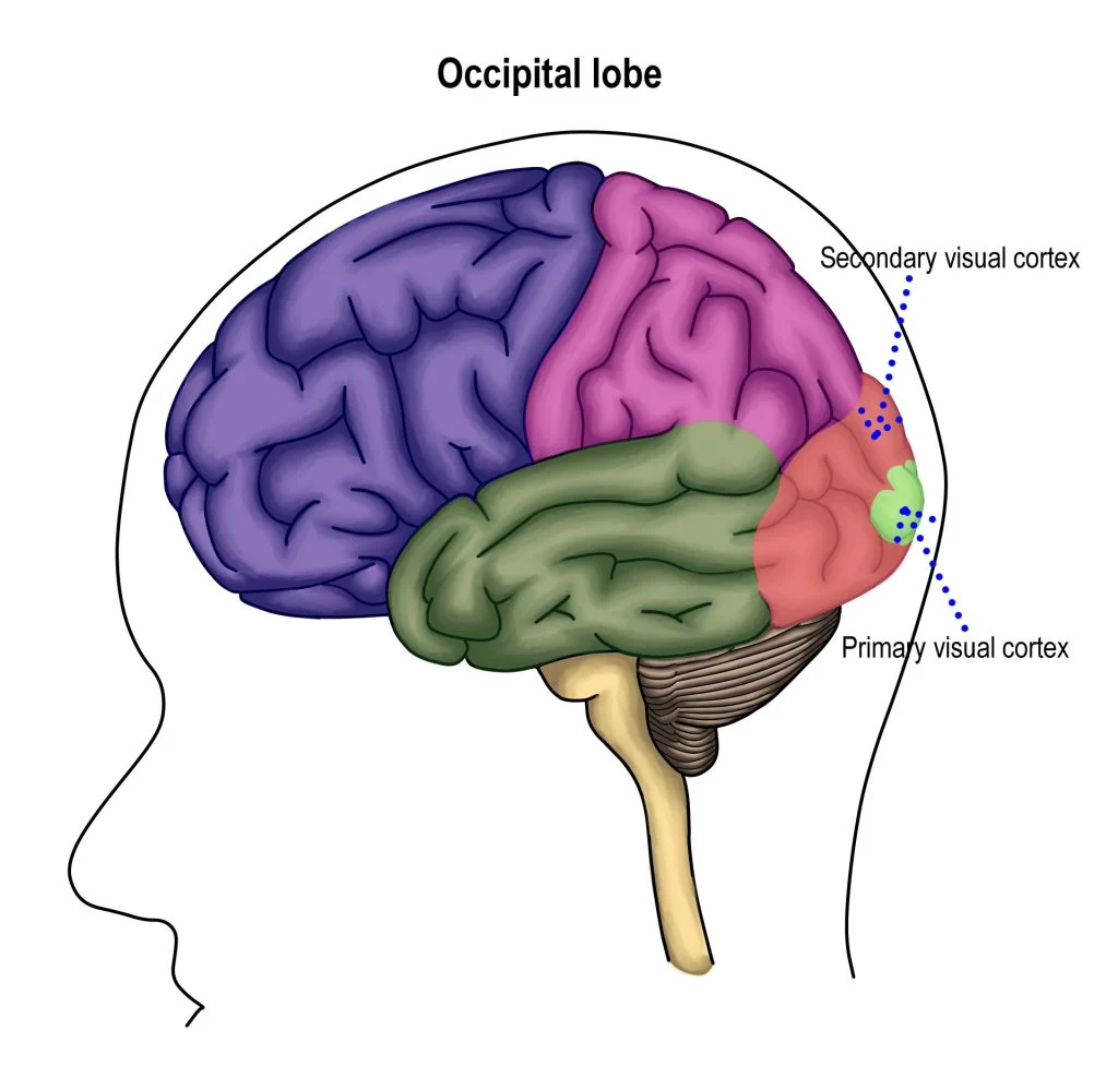
Occipital lobe
Responsible for all our sense of vision (where input is sent to FIRST)
* the primary visual cortex controls all visual perception: input from the retina + front of eye→cortex where it is processed through different modules
* particularly detects edges between dark and light in different orientations. An individual neuron signals edges in different locations each to build visual scenes
* and the visual association area + higher visuall areas
* different regions/neuron groups process shape, colour, orientation, motion etc
6
New cards
Temporal lobe
Major role in sense of audition, language and ability to comprehend speech/language
* the primary auditory cortex= perception of sound
* the auditory association area
* Wernick’s area (aka the sensory speech area)= comprehend language and speech.
* damage= comprehension deficit: can’t understand language, speech has no meaning but is fluent with normal rhythm and intonation
* **medial temporal lobe= limbic system** containing areas needed for learning and memory: the amygdala and hippocampus
* amygdala= alerts us to threats= fear and arousal response
* hippocampus= learning and memory= form new episodic memories. Damage causes anterograde amnesia (can’t make new memories)
7
New cards
Parietal Lobe
Controls how we act/interact within the world around us + builds a stable representation/sense of space of our surroundings and vision. It comprises a very large/major volume of the brain.
* contains the primary somatic sensory cortex:
* touch sensors on out skin relay signals up the spinal chord and to this cortex to generate sensation/perception of touch.
* gives a sense of stable world around us relative to our body position
* provides spatial attention: directing attention and eye movements to explore the visual world
* links vision to fine control of movement: understand the spatial location of objects around us to guide actions
* has the taste area

8
New cards
Frontal Lobe
Responsible for complex human behaviour
* Has the executive functions (prefrontal cortex):
* reasoning, planning, problem solving
* inhibitory control (Eg. regulate appropriate behaviour against other biological desires; selecting what is appropriate and inhibiting competing alternatives)
* working memory
* Has motor functions:
* premotor cortex= motor planning
* primary motor cortex=execution
* Responsible for speech production
* Broca’s area (aka motor speech area). Damage= suffer speech deficit called Broca’s aphasia, where speech is slow and non fluent, you have difficulty finding appropriate words but speech still carries meaning and you still comprehend
9
New cards
Corpus Callosum
Main area that interconnects the left and right hemispheres to allow communication between them with neuron connections
* site where the output arm of axons cross from one hemisphere to the other
* site where the output arm of axons cross from one hemisphere to the other
10
New cards
Brainstem + nervous system divisions
**Brainstem=** Part of the brain that connects between the cerebral hemispheres, the cerebellum and the spinal cord. A part of it called the Medulla/Medulla oblongata drives the automatic nervous system.
**The nervous system has 2 main divisions:** the central nervous system (brain + spinal cord) and the peripheral nervous system (nerves out to the rest of the body).
* The peripheral nervous system has two main sections:
* The somatic nervous system=nerves that go out to muscles that we have voluntary control over (motor and sensory muscles)
* the autonomic nervous system= controls involuntary actions (emotions, body actions) in two divisions:
* the sympathetic nervous system prepares the body to face danger: emotional arousal, stress, fear, fight or flight response, increased heart rate, body temp, respiration, perspiration, pupil dilation etc
* the parasympathetic nervous system brings everything back down to a rest and digest state in opposition of the SNS. Lowers heart rate, respiration, increases digestion.
A part of the brainstem called the Medulla/Medulla oblongata drives the automatic nervous system by controlling the involuntary actions it fluctuates.
* the medulla also has reflex centres for coughing, sneezing, swallowing and vomiting
11
New cards
Persistent Vegetative State and Locked in Syndrome
PVS= severe damage to upper brain (hemispheres and cortex), giving patients no conscious awareness (coma)
* if the brainstem isn’t damaged, the autonomic nervous system functions can remain (respiration, heart rate control, involuntary actions)
LIS= ALS or motor neuron disease= progressive loss of motor neurons from the cortex down by the spinal cord
* intact cerebrum and brainstem, but “disconnected” from the spinal cord. They can’t move/respond (can’t contract muscles) but have normal cognitive function, vision and hearing
* occurs due to brain injury (accident)
12
New cards
Cerebellum
Structure in the hind brain that is important for our sense of balance and coordination of complex movement (eg. centre of gravity)
* motor learning: gives fine adjustments to movements based on feedback
* we are constantly changing our movement: stand, walk, run etc. Cerebellum automatically makes postural adjustments with this by changing spine/core muscles to centre our mass with our feat. This is called a feedback loop
* Still many functions we don’t understand about it
13
New cards
Motor Programmes theory
Most movements are complex coordinated contractions of multiple muscles in a timed sequence. This is a theory from the 60s on complex movement execution.
Theory= Movements are planned and programmed by the brain before initiation: either creating the program just before movement or retreiving remembered programs for actions that have already been learnt (Eg. signature)
* visual and sensory feedback about a movement dictate the next one with fine adjustments in a feedback loop
* the brain computes the differences between planned actions and feedback during a performed action
Moreover, the brain automatically links sensory events with our own actions to infer causality. Eg. when someone else tickles you, you have no access to the motor plan and just feel the sensation. When you try and tickle yourself, you have the motor plan and don’t feel the sensation (?)
14
New cards
Define cognitive neuroscience
The study of the neural basis of behaviour and thought
15
New cards
Top-down and bottom-up. Evolution.
Behaviour is a consequence of forces driven “top-down” from the brain (controlled, conscious choice. selection and inhibition) and “bottom up” (automatic brain response, senses and learnt patterns, evolved for survival)
Most evolved parts of the brain from lowest level to highest:
1. Brainstem, cerebellum
* autonomic nervous system functions
2. Limbic system
* fear/threat and memory
3. Cerebral hemispheres
* planning, reasoning, problem solving (frontal lobe) + language (temporal) are the most highly evolved
* also includes visual perception and action (occipital and parietal)
16
New cards
Neurons composition
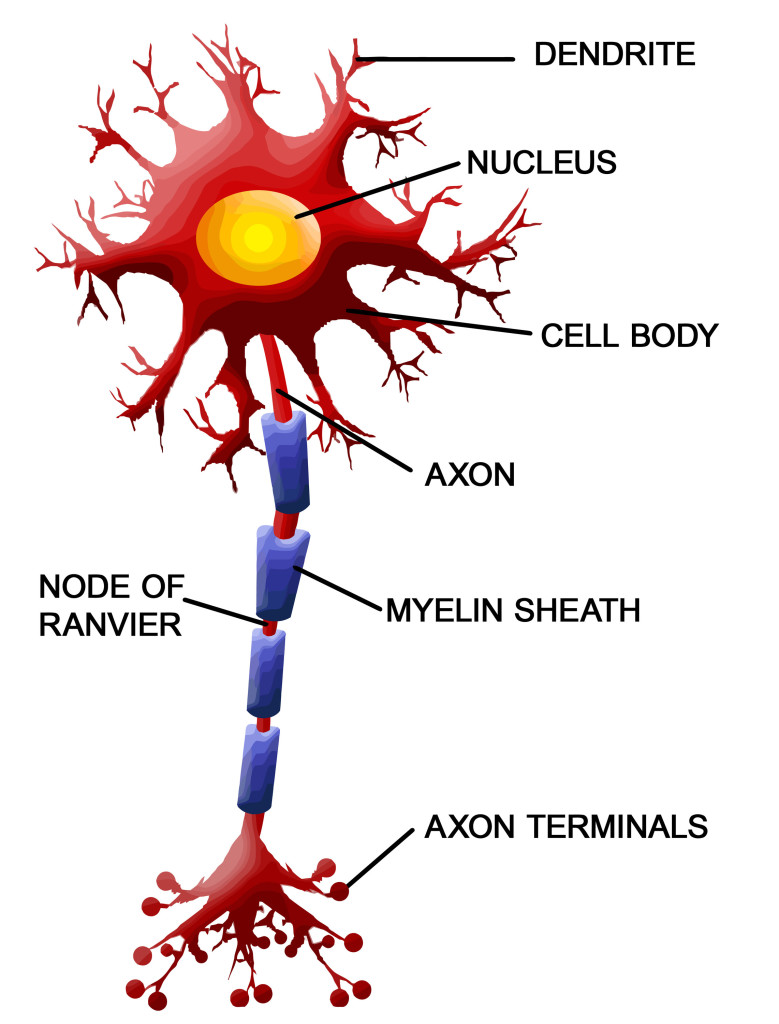
Neurons form synapses with one another and other cells to receive and send input from the dendrites/cell body, down the axon and released at the axon terminals.
* has a **cell body** with a nucleus and all structures necessary for normal cell functioning (DNA)
* **Dendrites** are unique to neurons. They receive signals from synapses, and there are many per neuron so that one neuron can receive thousands of inputs
* **axons** are unique to neurons. they send signals, output from the axon hillock at the cell body, to the axon terminals. There is only one per neuron= one signal sent at a time.
* wrapped in **myelin** to increase signal transmission efficiency along the axon
* can grow very long, forming the nerve pathways throughout the body (longest in human body is 1m from the spine to the big toe)
* **axon terminals**=terminal buttons that form synapses with other neurons. They secrete neurotransmitters to send signals across synapses to other neurons
* neural signals only ever go one way: from terminal → dendrites
* We define the neuron transmitting the signal as the pre-synaptic neuron and the other as the post-synaptic
\
There are 100 billion neurons that each have 10,000 synapses= 100 trillion connections
17
New cards
Glial cells
Supporting cells for neurons. 3 types:
1. **Oligodendrocytes.** Produce the myelin sheath that wraps around the axons
2. **Astrocytes**. Supply nutrients from blood→neurons through the blood brain barrier. This barrier blocks certain substances from the blood from entering the brain as protection for neurons and other brain cells
3. **Microglia**=brain’s immune system. Clean up foreign or toxic substances (because of the blood brain barrier, the body’s usual immune mechanisms can’t do this)
18
New cards
Action potentials- how they work at the membrane level
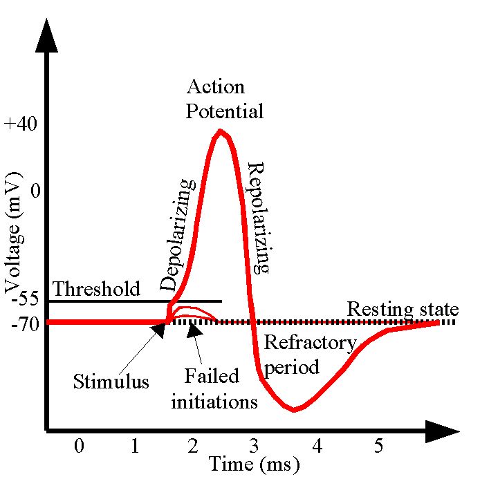
Action potentials= Electrical impulses that travel along the axons of neurons to allow communication between the brain and body
* the water surrounding and filling cells contains ions. Cations give each side of the membrane an electrical potential, and at equilibrium (resting potential) there are more cations outside the cell than in, giving an overall inner membrane potential of -70mV (relative to the extracellular domain)
* The cell membrane has a number of channels/gates that allow the flow of ions along their concentration gradient, changing the electrical potential difference. There are 3 crucial channels for an action potential:
1. Sodium Potassium pump: 3Na+ out for every 2K+ in: maintaining negative resting membrane potential
* activating the pump consumes 25% of the body’s total energy and 70% of brain energy
Inputs coming into the neuron through synapses increase the membrane potential. If this voltage exceeds a threshold of approximately -55mV, an action potential is triggered. This is done by a rapid depolarization of the membrane potential back to 0 (equal ion balance) followed by a repolarization back to resting potential
2. Voltage gated sodium channels:
* At rest these are closed, but when the threshold is reached and an action potential is triggered, they open and sodium flows into the cell along it’s concentration gradient, causing the rapid depolarization.
3. Voltage gated potassium channels
* When the membrane potential peaks, sodium channels close and potassium channels open. K+ ions flow outside the cell along their concentration gradient: causing repolarization back to resting potential.
* repolarization undershoots below resting potential, giving a refractory period where membrane potential is further from the threshold and therefore action potential is more difficult to trigger
* action potentials are ‘all or nothing’: if the threshold level is reached, no matter by how much force, the action potential is of a fixed size for that neuron.
* The strength of a neuron signal is determined by the rate of repeated signal releases. Not strong enough=doesn’t reach threshold=action potential won’t occur
19
New cards
Action potentials- how they conduct along an axon
* starts at the axon hillock: the membrane here has the lowest threshold to trigger an action potential
* depolarisation spreads to neighbouring regions of cell membrane by causing it to pass the threshold and triggering an action potential. Meanwhile, repolarisation is occurring as each segment finishes it’s action potential as the signal travels along the axon.
* depolarisation spreads to neighbouring regions of cell membrane by causing it to pass the threshold and triggering an action potential. Meanwhile, repolarisation is occurring as each segment finishes it’s action potential as the signal travels along the axon.

20
New cards
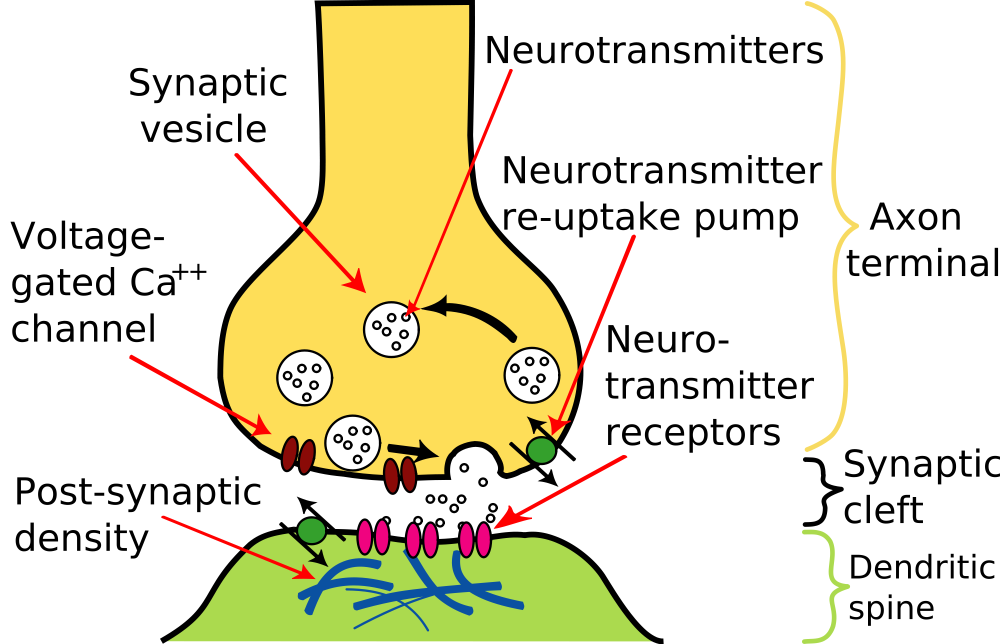
Synapses- how they work
Transmit signals from the presynaptic neuron’s axon terminal to the postsynaptic’s dendrites.
* when an action potential depolarises the axon terminal, it triggers the release of neurotransmitters (chemical messengers, eg. dopamine, seratonin) into the synaptic cleft
* the neurotransmitters are released from synaptic vesicles by exocytosis.
* in the cleft, it can join to receptors on the dendrite spine. These receptors are gates that, when joined to neurotransmitters, open ion channels on the postsynaptic neuron.
* opening ion channels changes membrane potential, causing an action potential if the threshold is reached
* each receptor will only bind to a specific type of neurotransmitter determined by it’s molecular shape: must have stereoisomerism (lock and key model). This allows drugs to replace specific neurotransmitters/act on specific receptors without having unintended impacts
* To recycle neurotransmitters when the signal needs to be turned off, they get taken back into the presynaptic vesicles via endocytosis by the reuptake pump.
* There are also enzymes that break neurotransmitters down: changing their molecular shape so it can’t bind with receptors
21
New cards
Dopamine and Serotonin neurotransmitter roles + treatment for diseases that target them
Dopamine
* an important neurotransmitter for motor function/control and pleasure (as it acts to reinforce behaviours that have positive outcomes)
* Parkinsons involves a loss of neurons in the basal ganglia (a brain structure), resulting in a loss of dopamine in the brain. This slow and inhibits movements
* Treatment: L-DOPA turns into dopamine when it crosses the blood brain barrier and replaces dopamine lost to the brain.
Serotonin
* important for mood regulation- it is in low levels for people with depression
* 2 treatment drugs: SSRIs and MAOIs act on processes that break down serotonin in the synaptic cleft to keep it there for longer: increasing serotonin signalling. SSRIs supress the re-uptake pump while MAOIs inhibit action of breakdown enzymes.
22
New cards
Excitatory and inhibitory signals
Excitatory signals= receptor opens channels that cause depolarisation, bringing the potential closer to the threshold for action potential.
* EPSP= excitatory postsynaptic potential
Inhibitory signals= recpetors open channels that cause hyperpolarisation, taking the potential further from the threshold. IPSP.
\
The effect of neurotransmitters on a post synaptic cells can be strong or weak, giving graded potentials, based on the sum of excitatory and inhibitory signals. The graded potential needs to be strong enough to make a change in membrane potential that hits the threshold for action potential to occur.
* the timing of inputs is also important: if enough excitatory inputs occur in close intervals, the threshold can be incrementally reached
* The sum of all inputs/signals (excitatory, inhibitory, strong and weak) coming from many synapses is called neural integration
23
New cards
Brain Lesion Studies
* explains normal brain function by examining what changes when part of the brain is damaged
* studies brain lesions (caused by strokes, brain injury etc) and induced lesions in animals (electrical and chemical)
Assumption=whatever changes in behaviour/cognition must rely on that part of the brain that is damaged. We use this to explain normal brain function
* ie. if a brain injury is sustained and a particular behaviour/mechanical/etc function is lost, that part of the brain must have been essential for that function
24
New cards
Single Neuron Recording: what does it do and how does it work? Pros and cons? example?
* place a thin electrode into an animal’s brain and record action potentials firing from a single neuron
* allows us to determine what that individual neuron is encoding (what stimulus, action or thought it represents) + what causes it to fire
Pros: best at detecting localisation and timing of brain function by directly measuring action potentials from individual neurons
Cons: Highly invasive, animals only (although some recent studies use human volunteers during brain surgery)
\
eg. Visual Cortex study on Monkeys
* Monkey stares straight ahead at a fixation cross in the centre of a screen. A bar of light is moved around the screen and visual cortex neurons are recorded
* Found neurons fire particularly when the light is slightly in the upper right area of the monkey’s peripheral vision; suggesting this particular neuron encodes this specific location and other neurons in the primary cortex encode other locations in the periphery to make up our visual space.
25
New cards
EEG (Electroencephalography) and Event related potentials: what does it do and how does it work? Pros and cons? example?
The summed activity from action potentials of neurons in the cortex cause electrical potential changes on the scalp. EEG measures this via a cap fitted with approximately 64 electrode sensors.
* each sensor measures the summed electrical activity coming from action potentials around it’s area
* brain activity shows constant oscillations (waves) that change in frequency depending on levels of alertness, sleep, arousal etc. There are 2 particularly important frequency bands in EEG:
1. Alpha activity (8-12 Hz)=idling activity of the brain. Becomes very large when people are relaxed/sleepy/closed eyes; indicating alertness and cognitive load
2. Delta frequency waves (1-3 Hz) is a slow wave that indicates very deep sleep. Can use to measure sleep stages/patterns + coma states
EEG research is interested in Event-related potentials=brain activity related to a specific event, stimulus or event-related potentials
* 100+ short time windows of EEG activity called epochs, each epoch following the same stimulus event, are averaged. Each time window occurs one after the other to get an idea of how the brain processes the information over time. There will be different peaks of activity that occur, representing different stages of stimulus processing as neurons send signals to different levels of the brain.
* ERPs can show the precise time of information processing
* eg. in the graph for face recognition stimulus, a peak of brain activity occurs 170ms after seeing the face. This is early processing; absorbing brightness, colours, edges etc. Then lighter peaks are exhibited as higher order cognitive processing occurs. All of this occurs in the visual cortex.
Pros of ERPS: show precise timing, direct measure of neuron firing
Cons: difficult to accurately localise activity to specific brain areas due to measuring being based on scalp areas.
26
New cards
Functional brain imaging (fMRI): what does it do and how does it work? Pros and cons? typical MRI session layout?
Measures the change in blood oxygen levels in the brain, giving images of brain anatomy (MRI, anatomical data) and a statistical map of where there is a change in blood oxygen level, appearing as colourful activation spots that change with particular tasks (fMRI, functional data)
* when neurons become active they consume more energy and therefore more oxygen (cellular respiration). Oxygen is delivered to active neurons through the blood, so change in blood oxygen levels (called BOLD signals) indicates changes in brain activity.
* increased brain activity→ increased blood flow to deliver more oxygen→ increased fMRI signal
We can measure the changes in brain activity in response to different stimuli using this method to determine what brain regions are responsible for processing of certain stimuli and map the process of this.
Pros: good localization of brain activity
Cons: indirect measure of brain activity (BOLD signal is not precise to the timing of neural activity), very expensive
\
General MRI scanner session:
* it places participants inside a superconducting magnet that is always on with a head coil to capture brain images and investigate changes in blood O2 levels as brain activity increases.
* They hold a button to respond to tasks, headphones to hear tasks and there is a mirror on the head coil to see external stimuli (Eg. a screen)
27
New cards
PET- Position emission tomography- what is it?
* an early brain imaging technique (80s-90s) where radioactive substances are injected into the bloodstream
* this radioactive label is used to map the location of blood flow through the brain
* today, this idea is used to map neurotransmitters or receptors in the brain
* this radioactive label is used to map the location of blood flow through the brain
* today, this idea is used to map neurotransmitters or receptors in the brain
28
New cards
Brain measurement and mind reading
You cannot infer what people are thinking, doing or feeling based on measurement of their brain activity.
* a good experimental design manipulates an independent variable (Eg. task or behaviour) to measure the effect on a dependent variable (eg. brain activity)
* reverse inference is impossible: you can’t look at brain activity (dependent variable) to determine what the independent variable was
* think of it this way: fluctuations in height in a height experiment could be due to many independent variables, such as height, age, genetics, posture, etc.
\
Current research, however, is investigating using MRI to read the contents of consciousness using brain decoding methods: attempting to reconstruct what people are seeing based on activity in the visual cortex
29
New cards
What is neuroplasticity?
The capability of the brain to alter its functional organisation as a result of experience and learning
* aka brain plasticity
2 main processes= neurogenesis and synaptogenesis, involving generating new neurons and changes in synapse connections
1. **Neurogenesis=** new neurons are formed, allowing the brain’s performance to be tweaked and optimised.
* Neurons do not regenerate when they are damaged or die, however, new neurons are born throughout all of life through stem cells that occur within the brain.
* stem cells=undifferentiated cells that can become any kind of cells that occur naturally in the brain
* stem cells can become neurons in 2 areas: the hippocampus (learning and memory) and the subventricular zone for olfactory bulb
2. **Synaptogenesis=** generation of new synapses (brain connections), or strengthening/weakening existing ones
* new synapses are constantly formed and strengthened with experience and learning
* environmental enrichment (opportunity for sensory and motor exploration) allows the growth of dendrites (so more synapses can exist) and more extensive synaptic connections through the brain.
* **long-term potentiation, LTP,** is the strengthening of synapse connections through a range of chemical and molecular processes with learning
* changes in the structure of synapses are made to give stronger signals from presynaptic to postsynaptic neurons (eg. more postsynaptic receptors for stronger graded potentials)
* graded potentials depend on the strength of synapse connections, where strong stimulus= large change in membrane potential and weak=small change. Synapse connections will strengthen every time those neurons fire together (pass an action potential down to each other) so that, in future, activation of the presynaptic neuron is more likely to cause the postsynaptic neuron to also fire (via the greater graded potential).
* This allows the brain to learn through associations as neurons are fired and wired together. This process is called **Hebbian Learning**
30
New cards
Spreading Activation Model Theory
Neurons represent a specific concepts (Eg. grandma, Jenifer Aniston) and share connections with neurons that represent related concepts (eg. I could have a connection between Nana cells and schnitzel imagery cells).
* The more associated things are encountered together=the stronger the connection between them becomes, and activating/firing one neuron is more likely to create a graded potential that spreads activation to the connected neurons.
* Making and strengthening concepts allows learning and memory
* The more associated things are encountered together=the stronger the connection between them becomes, and activating/firing one neuron is more likely to create a graded potential that spreads activation to the connected neurons.
* Making and strengthening concepts allows learning and memory
31
New cards
Brain reorganization
* the mapping connection between our visual world and our actions via motor programmes and specific muscle contractions is automatic but can be relearned/remapped at any time
* the primary senory and primary motor cortexes have homunculus organisation where the size of the area on the cortex represents the sensitivity we have with that part of the body on the motor side, aka our level of fine motor control. This size and capability is learned and changed with experience.
Some studies of large scale brain reorganisation with experience and learning (key takeaway= bold part):
1. The sensory cortex was measures of people who play string instruments: measuring activity for index and pinky finger sensation. Found that string players have larger areas on the primary sensory cortex for the left hand than non string player
* **Overall, the sensory cortex finger areas expand with use and experience**
2. Researchers lesioned the motor cortex of monkeys to impair hand movement to study brain reorganisation following brain injury. With no rehabilitation (hand isn’t used at all), the motor cortex area became smaller=maladaptive plasticity. With rehabilitation (movement training) the motor cortex area for the hand expanded beyond previous areas as other areas took over the function in compensation.
* **Overall, the motor cortex can reorganise with use to recover functions, expanding its area as new areas take over**
3. Studied brain activity in blind vs sighted people who knew braille while reading braille. Thesensory cortex was active in sighted participants as it should be, as braille involves touching dots. However, the visual cortex was only activated in blind participants. This cortex lacks input from them but still has neurons, so the brain reorganises touch-reading functions there.
* **Overall, brain areas lacking their normal input can take new functions with use.**